Understand the concepts of 3D imaging Clinical applications of 3D echocardiography 3D image acquisition and optimization - Iowa ACC Chapter
←
→
Page content transcription
If your browser does not render page correctly, please read the page content below
4/19/2021

                    Aiman Smer, MBBCh, FACC, FASE
                    Associate Professor of Medicine
                Creighton University School of Medicine
                            No Disclosure

Understand the concepts of 3D imaging

Clinical applications of 3D echocardiography

3D image acquisition and optimization

                                                                 1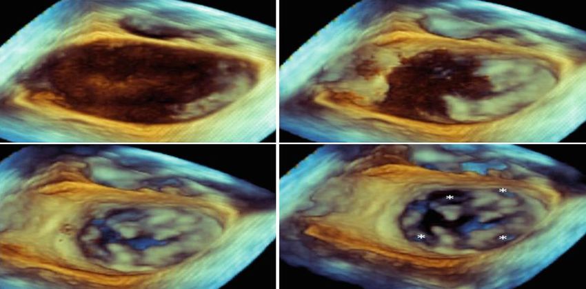
4/19/2021

                                                                                                   3D-mode

                                            2D-mode

                                   M-mode

                          B-mode

                 A-mode

1st Cardiac US
     1960

                                                      From Edler I, Ultrasound cardiography. Acta Med Scand Suppl 370 1961; 170:39

                                                                                                                                     2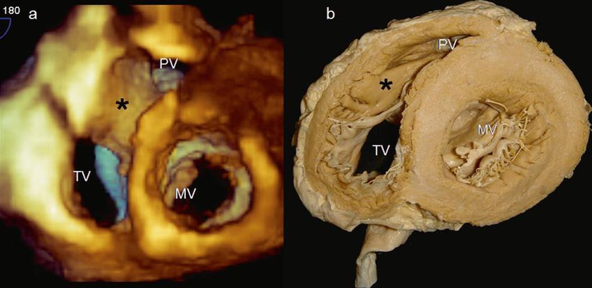
4/19/2021

New generation Piezoelectric crystals (PureWave Crystals)

Microelectronics

New generation Piezoelectric crystals (PureWave Crystals)

Microelectronics

            Modified from Bulwer BE, Rivero JM, editors. Echocardiography Pocket Guide: The Transthoracic Examination. Burlington, Mass: Jones & Bartlett Learning, 2011, 2013, p 208

                                                                                                                                                                                        3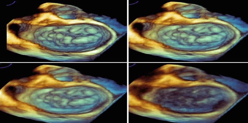
4/19/2021

The 3D probe provides all conventional modalities, such as 2D imaging, M-mode, pulse
and continuous wave Doppler, and color Doppler imaging

X-Plane technology

Chamber quantification
Valvular heart disease
  Native and prosthetic valves
Intracardiac masses
Congenital heart defects
  Anatomic assessment and procedural guidance
Interventional cardiac procedures
  TAVR, MitraClip, LAA occlusion devices
Stress echo

                                                                                         4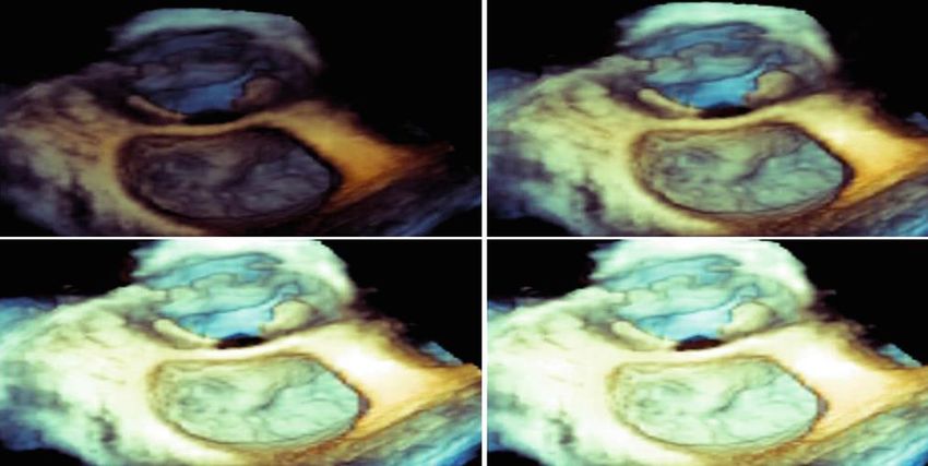
4/19/2021

2D method                                        Heart Model
   Subjective
   Experience dependent
   Large inter- and intra-observer variability

3D method
   No geometric assumption
   Avoid foreshortening
   Reproducible
   Excellent correlation with CMR

                                                        Mor-Avi V, Lang RM et al., Circulation 2004. 110: 1814-1818
                                                        Jacobs LD, et al. Eur Heart J 2005; 27:460-8

Assessment of mitral valve anatomy

LVOT and aortic annulus sizing

Valve area measurement via direct
planimeter in mitral and aortic valve
stenosis

Regurgitant orifice measurement via
vena contracta in regurgitant lesions

Evaluation of prosthetic valve
malfunction and paravalvular leak

                                                                                                                      5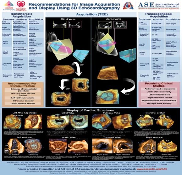
4/19/2021

       6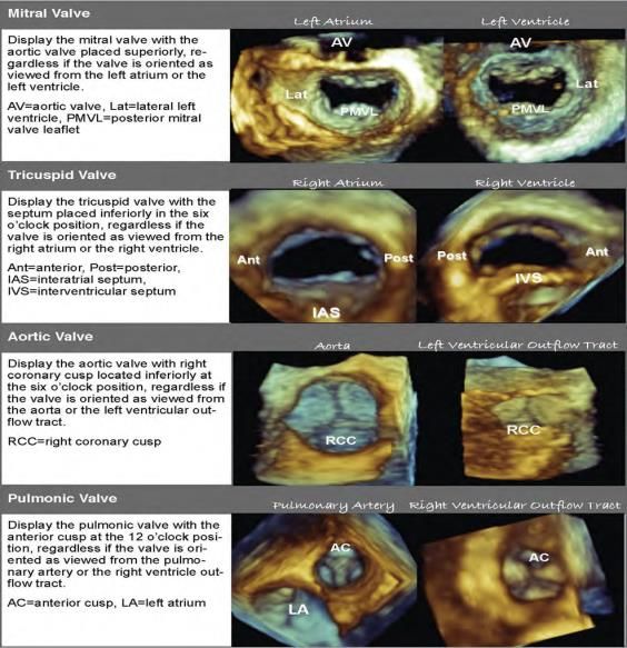
4/19/2021
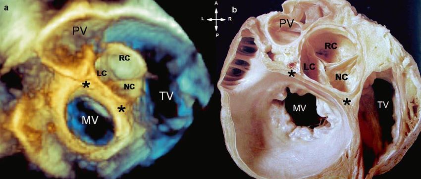
LA en face view of MV
7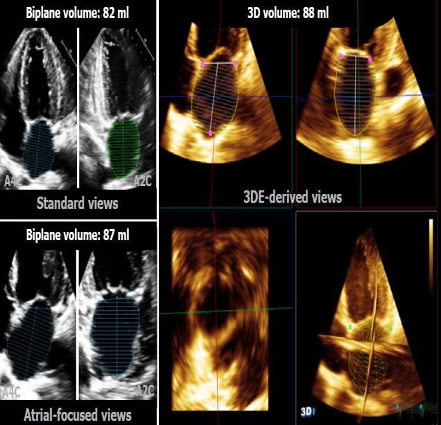
4/19/2021

       8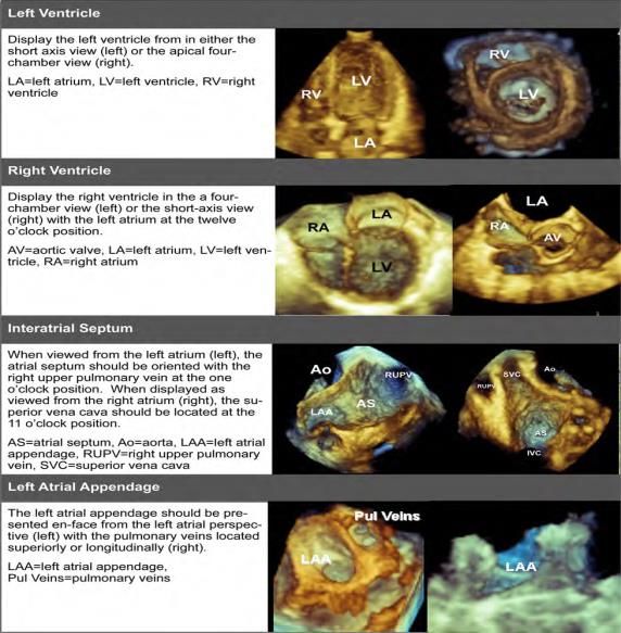
4/19/2021

Lambl's excrescences     Papillary fibroelastoma

 MitraClip             LAA occlusion device

                                                          9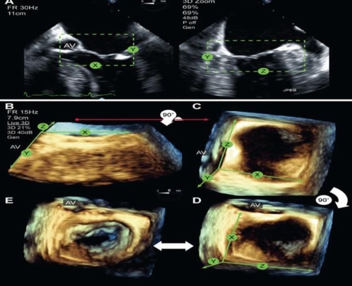
4/19/2021

1. 2D Image
                                                       Live 3D
                                                       3D Zoom
                             2. Mode of Acquisition    Full Volume
                                                       Single vs multi-beat
                                                       Color Doppler
              3. Rendering

                                         4. Image Acquisition and Display
                                                       Cropping

                                   5. Final Image

                                                                                    10
4/19/2021
Real-time (live) acquisition
Live 3D narrow volume
3D zoom
Full-volume (ECG-gated)
Real-time (live) acquisition
Live 3D narrow volume (50x30)
3D zoom
114/19/2021
Full-Volume
A pyramidal volume of 60 x 60 up to 100 x 100
Large cardiac volume
Require ECG gating
Full volume is constructed by merging 2-6 narrow
segments of the pyramidal database
Full-Volume 3 D with Color Doppler
Structural or
LV, LA or RV Interventional
congenital heart
quantification Guidance
defects
Live 3D + Color
Live 3D
Full Volume 3D Zoom,
3D Zoom
Full Volume
124/19/2021
1) Initial view in 3D zoom is a
biplane preview
2) X and Y planes and elevation
width (Z axis) to include entire
structure of interest
3) Press 3D zoom again will
generates a pyramid of 3D
volume (X,Y, Z)
4) En Face MV view, tilt 3D
volume down 90 degree, then
rotate clockwise or counter-
clock to position the aortic
valve at 12 clock
5) Decrease gain
Saric M et al. J Am Soc Echocardiogr
2010;23:1128-35
134/19/2021
J Am Soc Echocardiogr 2012;25:3-46
144/19/2021
Optimize 2 D image
Adjust the gain
Zoom on the ROI
Adjust focus depth
Choose small sector
Quality 3D images always start wit a good 2D image
Cropping
Gain
Compression
Brightness
Smoothing
Color-Map vision
154/19/2021
Removes noise from structures
High values of compression add “soft” echos, making objects appear more opaque and
larger
Lower compression values are preferred, 2-4 on scale of 0-10
164/19/2021
To avoid under-illumination or over-illumination, a medium level of brightness ranging
from 40-50 (scan 0-100) should be used
Removes subtle roughness of the surface
Medium values 6-8 on a scale 0-10 allows precise definition of structure
174/19/2021
Grayscale shades or dual-color mapping for depth perception
Evaluation of cardiac chamber volumes and mass
Assessment of regional left ventricular wall motion and
quantification of systolic dyssynchrony
Presentation of realistic views of heart valves
Volumetric evaluation of regurgitant lesions and shunts with
3DE color Doppler imaging
184/19/2021
Time consuming
Requires training in 3D analysis
Accuracy varies with expertise and vendors
Temporal resolution
Relies on good 2D image
194/19/2021
20You can also read


