Tissue Engineering: From 3D culture models to regenerative medicine | Von Ambrustschützen zum Virennachweis: Die Abteilung Infektionsdiagnostik
←
→
Page content transcription
If your browser does not render page correctly, please read the page content below

Periodisches Informationsblatt des Departementes Biomedizin
    Universität Basel, Universitätsspital Basel und
    Universitäts-Kinderspital beider Basel

Tissue Engineering: From 3D culture models to regenerative medicine | Von
 Ambrustschützen zum Virennachweis: Die Abteilung Infektionsdiagnostik
    im ehemaligen Stachelschützenhaus | Berlin – creative and dynamic
                                                                  1 16
Editorial
                                                                                                   1
                                                                 UniBasel immunologists retreat to
                                                                 the mountains         8
                                                                 SweetySpot-Software development
                                                                 in the DBM Flow Cytometry Facility
                                                                 for droplet based Cell Sorters   15
                                                                 Tag der Biomedizin

                       2
                                                                                                  20
                                                                 Publikationen
                                                                                                  22
                                                                 Art
Tissue Engineering: From 3D                                                                       35
culture models to regenerative                                   Mitarbeitende/Colleagues
medicine
                                                                                                  36
from Ivan Martin
                                                                 Auszeichnungen/Congratulations
                                                                                                9/37
                                                                 DBM IT
                                                                                                  46
                                                                 Das DBM stellt sich vor
                                                                                                  48

                    10                           17
                                                                  IMPRESSUM

                                                                  Redaktion
                                                                  Heidi Hoyermann
                                                                  Übersetzungen
                                                                  Paula Cullen
Von Ambrustschützen zum          The Anatomical Museum Basel
                                                                  Layout
Virennachweis: Die Abteilung     from Magdalena Müller-Gerbl
                                                                  Eric Spaety, Morf Bimo Print AG, Binningen
Infektionsdiagnostik im ehema-
ligen Stachelschützenhaus                                         IT-Unterstützung
von Rainer Gosert                                                 Niklaus Vogt
                                                                  Administration
                                                                  Manuela Bernasconi
                                                                  Fotos
                                                                  www.sublim.ch (20)
                                                                  Frank Neumann (2)
                                                                  Titelfoto: shutterstock
                                                                  Druck
                                                                  Morf Bimo Print AG, Binningen

                    38                           42
                                                                  Anschrift
                                                                  Redaktion DBM Facts
                                                                  Departement Biomedizin
                                                                  Hebelstrasse 20
                                                                  4031 Basel
                                                                  heidi.hoyermann@usb.ch

Persian New Year (Norouz)        Berlin – creative and dynamic
from Zeinab Barekati             from Mathias Schmaler

DBM Facts 1|2016                                                                  Departement Biomedizin
EDITORIAL

                                                                                  Radek Skoda
                                                                                    Leiter DBM

   Liebe Leserinnen und Leser
   Der Frühling ist die Zeit der Pläne, der Vorsätze. Auch die Mitarbeitenden des DBM haben sich Einiges vorgenommen
   und so Manches bereits umgesetzt:
   Am 9. April fand der Tag der Biomedizin statt. Über 2000 Interessierte aus den umliegenden Kantonen und dem grenz-
   nahen Ausland kamen, um sich von den DBM-Forschungsgruppen in die Welt der Wissenschaft entführen zu lassen
   (siehe Seite 20).
   Nicola Aceto und Eline Pecho-Vrieseling haben eine SNF-Förderprofessur erhalten. Nicola widmet sich in seinem Projekt
   den grundlegenden molekularen Mechanismen, die der Entstehung von Krebsmetastasen zugrunde liegen, mit einem
   besonderen Schwerpunkt auf der Analyse von zirkulierenden Tumorzell-Clustern beim Menschen. Zusätzlich zur SNF
   Professur erhielt er letztes Jahr auch einen renommierten ERC Starting Grant. Eline untersucht in ihrem Projekt am Bei-
   spiel der Huntington-Krankheit die Rolle von falsch gefalteten Proteinen. Ähnliche Vorgänge kann man auch bei ande-
   ren neurodegenerativen Erkrankungen – etwa der Alzheimer-Krankheit – finden. Das DBM wünscht beiden viel Erfolg!
   Ivan Martin, Leiter der Forschungsgruppe “Tissue Engineering”, koordiniert das EU-Projekt “BIO-CHIP”, das vom EU-För-
   derprogramm Horizon 2020 mit insgesamt 5.1 Millionen Euro unterstützt wird. Sieben Institutionen aus der Schweiz,
   Deutschland, Italien und Kroatien sind daran beteiligt. Mehr über seine Forschung erfahren Sie ab Seite 2.
   Wie vielseitig das DBM ist, zeigt Magdalena Müller-Gerbl in ihrem Artikel über das Anatomische Museum (Seite 16) und
   Rainer Gosert, der uns an den alltäglichen Herausforderungen im Dienstleistungsbereich der Medizinischen Mikrobiolo-
   gie teilhaben lässt (Seite 10). Die aktuellsten Publikationen aus dem DBM folgen ab Seite 22.
   Mit Zeinab Barekati können wir das iranische Neujahrsfest feiern (Seite 34) und Mathias Schmaler nimmt uns mit in
   seine Heimatstadt Berlin (Seite 38). Und noch vieles andere mehr erwartet Sie in der neuesten Ausgabe. Viel Spass bei
   der Lektüre!

   Dear Readers
   Spring is the time for plans and new resolutions. Even the employees of the DBM have taken this to heart and have started
   to make many changes:
   The “Tag der Biomedizin” took place on the 9th of April. More than 2000 interested individuals from the surrounding cantons
   and the neighbouring countries came to be introduced to the world of science by the DBM research groups (see page 20).
   Nicola Aceto and Eline Pecho-Vrieseling have both received SNF professorships. Nicola has applied himself to his research on
   the fundamental molecular mechanisms that drive the formation of cancer metastasis, with particular focus on the analysis
   of circulating tumor cell clusters in humans. In addition to his SNF professorship he also received a renowned ERC Starting
   Grant. Eline’s research investigates the role of misfolded proteins in Huntington’s Disease. Similar findings have also been
   made in respect of other neurodegenerative diseases such as Alzheimer’s. We at the DBM wish them both every success!
   Ivan Martin, leader of the research group “Tissue Engineering”, is coordinating the EU-Project BIO-CHIP which is being
   funded by the EU Framework Programme Horizon 2020 for a total of 5.1 Million Euro. Seven institutions from Switzerland,
   Germany, Italy and Croatia are taking part in the project. You can read more about Ivan’s research from page 2.
   The versatility of the DBM is highlighted by Magdalena Müller-Gerbl in her article on the Anatomical Museum (page 16) and
   by Rainer Gosert who lets us take a look at the daily challenges incurred by the services sector of medical microbiology
   (page 10). The latest publications from the DBM can be found from page 22 onward.
   We celebrate the Iranian new year with Zeinab Barekati (page 34) and Mathias Schmaler introduces us to his hometown,
   Berlin (page 38). All that and more awaits you in this latest edition.
   Happy reading!

DBM Facts 1|2016                                                                                     Department of Biomedicine
2    WISSENSCHAF T | SCIENCE                                                                         DBM HEBEL STR ASSE

Tissue Engineering:From 3D culture models
to regenerative medicine
The Tissue Engineering group was started in 1999 as an        group and extension to new research areas. This short
initiative of the surgical units at the University Hospital   and hopefully “light” article will describe the current
Basel, coordinated by the recently retired Prof. Michael      scientific and translational programs of the Tissue Engi-
Heberer and with the goal to develop biological cartilage     neering group, and provide a few hints on the structural
implants based on adult human cells. The program was          and functional organization of a team now including ap-
initially strictly focused, as required by fact that only 4   proximately 30 people (Figure 1).
working hands were available in the lab, namely those
of a young assistant surgeon (now Prof. Marcel Jakob,         Objectives and organization of the group
head of trauma and orthopaedic surgery) and mine
(yes, I could indeed handle a pipetboy). The first suc-       The common denominator of the research projects
cessful SNF and European grant applications, the collab-      in the group is related to the establishment of 3D cell
oration with newly established surgical professorships        culture systems, combining interdisciplinary efforts in
with strong interest in regenerative medicine (Prof.          cell biology, engineering technologies and materials
Dirk Schaefer, Prof. Hans-Florian Zeilhofer, Prof. Stefan     science. These systems are used as models to investi-
Schären) and the embedding in the positively develop-         gate fundamental aspects of tissue development, and
ing DBM, enabled a progressive consolidation of the           as grafts to induce tissue regeneration. Several collab-
                                                                               orative programs have been estab-
                                                                               lished within the DBM to employ the
                                                                               developed tools for 3D culture of tu-
                                                                               mor cells (Prof. Giulio Spagnoli, Prof.
                                                                               Gandomenica Iezzi), endothelial cells
                                                                               (PD Dr. Andrea Banfi), thymic epithelial
                                                                               cells (Prof. Georg Holländer), glial cells
                                                                               (Prof. Raphael Guzman), pluripotent
                                                                               stem cells (Prof. Christian De Geyter)
                                                                               and cardiac cells (PD Dr. Anna Marsa-
                                                                               no). However, our main focus has been
                                                                               maintained around the development
                                                                               of cartilage and bone tissues, includ-
                                                                               ing bone marrow. Figure 2 illustrates in
                                                                               a simplified way the priority research
                                                                               programs, the areas embedding our
                                                                               scientific questions and translational
                                                                               targets, and the horizontal platforms,
                                                                               necessary to provide the engineering
                                                                               background to the different fields and
                                                                               the regulatory perspective for clinical
                                                                               translation. The latter platforms are
                                                                               those linking the group with its sec-
Figure 1: The Tissue Engineering group in February 2016                        ondary affiliations within the Medical

DBM Facts 1|2016                                                                                Departement Biomedizin
DBM HEBEL STR ASSE                                                                                  WISSENSCHAF T | SCIENCE    3

                                                                                       from the damaged joint, which display
                                                                                       a large and uncontrollable inter- and in-
                                                                                       tra-donor variability. We have been first
                                                                                       to identify that chondrocytes from the
                                                                                       nasal septum, as compared to those
                                                                                       from articular cartilage, have a superior
                                                                                       and more reproducible chondrogenic
                                                                                       capacity following in vitro expansion, in
                                                                                       addition to being available in an autolo-
                                                                                       gous setting, under minimally invasive
                                                                                       conditions. The possibility of generat-
                                                                                       ing cartilage tissues from nasal chon-
                                                                                       drocytes (Figure 4) has inspired plastic
                                                                                       surgeons to use the materials as autol-
                                                                                       ogous grafts for the reconstruction of
Figure 2: Schematic representation of the research programs of the                     the alar lobule of the nose after skin tu-
group and of their structural organization
                                                                     mor resection, a procedure for which normally cartilage
                                                                     from ears, ribs or large parts of the nasal septum from
Faculty, namely the Department of Biomedical Engi-                   the same patient would be used. The results of a first-
neering (DBE) and the Department of Clinical Research                in-man clinical trial (Clinical-trials.gov NCT 01242618,
(DKF), respectively. A tight interaction with the clinic has         Swissmedic TpP-I-2010-002) have demonstrated that
been pivotal for the development of the group; more                  the engineered grafts could satisfy structural, function-
than 20 trained surgeons (Figure 3) have worked in the               al and aesthetic needs, bypassing additional morbidity
past years full time in the lab. Thanks to their profes-             of native cartilage tissue harvest (1). These results are
sional development after their time in research, they                now opening the way to implementation into more
have enabled clinical translation of our concepts and                challenging and larger facial defects.
strategies. The variety of topics and the size of the
group can now only be managed thanks to the profes-                  But going back to the original clinical target, are nasal
sional help of staff scientists employed in the group on a           chondrocyte-derived grafts compatible with implan-
stable basis, coordinating the cartilage-related projects            tation in a joint? After demonstrating that nasal chon-
(PD Dr. Andrea Barbero), the bone-related projects (PD               drocytes can respond to physical forces resembling
Dr. Arnaud Scherberich), the engineering technologies                joint loading and can recover after exposure to the in-
(Dr. David Wendt) and the regulatory aspects (Dr. Sylvie             flammatory factors typical of joint injury, we addressed
Miot and Anke Wixmerten). The following sections have                their genetic compatibility with an articular cartilage
been sketched by these “scientific pillars” and are meant            environment. We found that nasal chondrocytes – of
to provide a flavor of the different targets / questions             neural crest origin – display a distinct profile of HOX
currently addressed.                                                 gene expression from articular chondrocytes – of me-
                                                                     soderm origin –, independently of their in vitro manipu-
Nasal chondrocytes and environmental plasticity                      lation. However, upon implantation in a joint, they can
(A. Barbero)                                                         be reprogrammed by the recipient site and acquire the
                                                                     HOX “signature” typical of articular chondrocytes (2).
The reproducible and durable repair of articular carti-              These results have led to pre-clinical goat studies and
lage following traumatic injuries is still an unmet clini-           ultimately to the treatment of 17 patients with trau-
cal need. The most advanced cell-based therapies are                 matic cartilage injuries in the knee (Clinical-trials.gov
based on the use of autologous chondrocytes harvested                NCT01605201, Swissmedic TpP-I-2012-001). Clinical

DBM Facts 1|2016                                                                                     Department of Biomedicine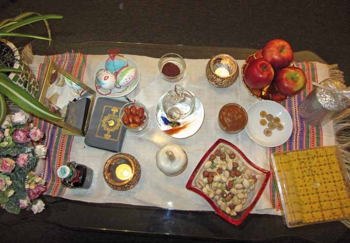
4    WISSENSCHAF T | SCIENCE                                                                                    DBM HEBEL STR ASSE

                                                                                            embryonic development. The strategy
                                                                                            has brought, in collaboration with the
                                                                                            team of Prof. Rolf Zeller, to the genera-
                                                                                            tion of intermediate templates con-
                                                                                            sisting of hypertrophic cartilage that
                                                                                            – upon in vivo implantation – have the
                                                                                            capacity to autonomously remodel
                                                                                            into bone tissue and thereby recapitu-
                                                                                            late the events occurring during endo-
                                                                                            chondral ossification (3). Remarkably,
                                                                                            the same processes can be triggered
                                                                                            by the same tissues if suitably decel-
                                                                                            lularized, thus highlighting a pivotal
                                                                                            role of extracellular matrices embed-
                                                                                            ding the cocktail of factors necessary
Figure 3: The clinical partners of the Tissue Engineering group in February 2016
                                                                                            and sufficient to prime regenerative
                                                                                            processes (4). Moreover, the bone or-
outcomes still need to be consolidated at longer terms,                   gan developed by this approach is capable to host fully
but assessments by MRI (stability, integrity and qual-                    functional hematopoietic stem cells (5) and is thus be-
ity of the grafts and repair tissue) and improvements                     ing investigated in collaboration with the groups of Prof.
in the clinical scores assessed by the patient are highly                 Radek Skoda and Prof. Claudia Lengerke as a humanized
promising. These studies have also been instrumental                      environment to study the interaction of leukemic cells
to receive funding within the Horizon2020 program of a                    with a 3D stromal niche (6).
phase II clinical study, which is coordinated by our team
and will include treatment of a total of 108 patients                     Mesenchymal stromal cells are also found within the
(Project BIO-CHIP, http://biochip-h2020.eu). In parallel,                 Stromal Vascular Fraction (SVF) of adipose tissue, in
pre-clinical studies are ongoing to test the feasibility of               conjunction with endothelial lineage cells. The dem-
the procedure for the treatment of more challenging in-                   onstrated capacity of these different cell types to self-
dications, with features typically associated with osteo-                 assemble into osteogenic and vasculogenic structures
arthritic degeneration.                                                   upon ectopic or orthotopic implantation (7) represents
                                                                          an opportunity for the engineering of osteogenic and
Bone, Bone marrow and Vascularized bone                                   simultaneously vasculogenic grafts to enhance fracture
(A. Scherberich with A. Todorov)                                          healing. Given the abundance of this cellular material,
                                                                          these grafts can be manufactured without cell expan-
The golden standard for the treatment of critical bone                    sion or in vitro culture, thus in a mode which is com-
defects is the use of autologous bone, typically harvest-                 patible with intra-operative procedures. This concept
ed from the iliac crest, which is associated with large                   has led to the clinical treatment of 8 elderly individuals
morbidity at the donor site. Mesenchymal stromal/                         with osteoporotic fractures in the humerus (ClinicalTri-
stem cells from the bone marrow have previously been                      als.gov NCT01532076), where the standard procedure
proposed to generate osteogenic grafts as a surrogate                     of plate fixation has been augmented by the implanta-
of autologous bone, but the efficiency and reproducibil-                  tion of SVF-based grafts generated during the operation
ity of the procedure has not yet been convincing. The                     (Figure 5). Despite the limited number of patients, biop-
working hypothesis of the group is that the robustness                    sies taken at the repair site upon plate removal indicate
of bone formation by engineered tissues could be in-                      the formation of de novo bone tissue throughout the
creased if these could mimic processes typical of bone                    implant site, as opposed to osteoconduction from the

DBM Facts 1|2016                                                                                           Departement Biomedizin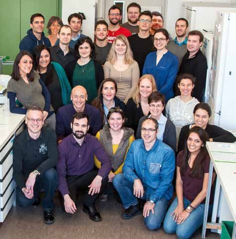
DBM HEBEL STR ASSE                                                                                      WISSENSCHAF T | SCIENCE      5

                                                                                           3D, predominantly because – unable to
                                                                                           vascularize – tissues in vitro have to be
                                                                                           supplied with oxygen and nutrients in
                                                                                           a different way. We have pioneered the
                                                                                           use of direct perfusion of fluid through
                                                                                           the matrix of engineered tissues to mim-
                                                                                           ic interstitial fluid flow within custom-
                                                                                           designed bioreactor systems in order
                                                                                           to generate uniform tissue structures
                                                                                           (Figure 6A). More recently, we have also
                                                                                           validated a microfluidic system allowing
                                                                                           the spontaneous formation of 3D cellu-
                                                                                           lar structures and their exposure to dif-
                                                                                           ferent combinations and concentrations
                                                                                           of regulatory molecules. The system is
                                                                                           in use to investigate in a relatively higher
Figure 4: Engineering of autologous nasal cartilage grafts. (A) Collection of a nasal car- throughput the signals supporting the
tilage biopsy from a patient, this procedure is performed under local anaesthesia and
results in minimal donor site morbidity. (B) Biopsy of nasal cartilage septum. (C-D) Tis-  growth and differentiation of mesen-
sue engineered cartilage graft: macroscopic (C) and histological appearance (Safranin- chymal progenitors (8) (Figure 6B).
O staining specific for sulfated glycosaminoglycans (D).

existing bone, thus strongly suggesting osteogenesis                   Despite the compelling clinical need, advances in
by the grafted cells. Studies are ongoing to combine the               the field of tissue engineering have yet to result in vi-
strategy of engineered and decellularized matrices pre-                able products with widespread therapeutic adoption.
sented in the previous section with the use of SVF cells               Among the challenges to be addressed, overcoming
to intraoperatively “re-activate” such constructs in or-               manufacturing-related limitations will be key for a suc-
der to enhance bone and vascular development.                          cessful translation. Since the production of tissue grafts
                                                                       is typically based on conventional manual cell and tis-
Engineering platforms (D. Wendt)                                       sue culture techniques, these labor-intensive processes
                                                                       are challenging to standardize, difficult to scale-up, and
Conventional 2D culture systems (Petri dishes, culture                 incur high costs in the long-term. While the clinical tri-
flasks) cannot adequately recapitulate the complex mi-                 als conducted by our laboratory have demonstrated the
croenvironment experienced by cells in vivo. 3D model                  safety, feasibility, and preliminary evidence of the effica-
systems, which mimic specific aspects of this micro-                   cy of functional cartilage tissue grafts, the manufactur-
environment, are vital to gain a greater understanding                 ing processes used to produce the engineered tissues
of basic cell and tissue functions within the native mi-               will ultimately pose significant regulatory and economic
lieu. However, challenges in biology and engineering                   challenges for routine clinical practice. Bioreactors have
have to be overcome to establish and maintain cells in                 the potential to overcome the limitations of manual

                                                                                          Figure 5: Intraoperative engineering of
                                                                                          autologous Stromal Vascular Fraction-based
                                                                                          grafts for the clinical treatment of humerus
                                                                                          fractures in osteoporotic individuals

DBM Facts 1|2016                                                                                          Department of Biomedicine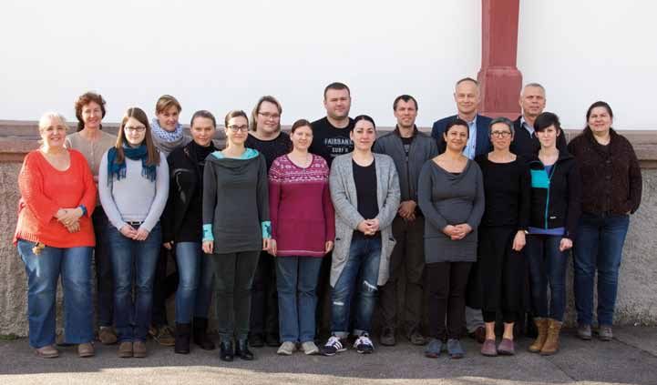
6 WISSENSCHAF T | SCIENCE DBM HEBEL STR ASSE
Cellec Biotek AG (www.cellecbiotek.
A
com).
Regulatory platform
(S. Miot, A. Wixmerten)
From a regulatory standpoint, Tis-
sue Engineered Products belong to
a recently developed, new category
of medicinal products, the Advanced
Therapy Medicinal Products (ATMPs).
Manufacturing and use of these prod-
B ucts require the establishment of a
Quality Management System, regulat-
ing processes in compliance with Good
Manufacturing Practice (GMP), Good
Clinical Practice (GCP) and Good Distri-
bution Practice (GDP). For us as experi-
enced basic and pre-clinical research-
ers, entering the new field of clinical
Figure 6: (A) Direct perfusion system to support efficient cell seeding into 3D porous scaf-
trials and subsequent regulatory as-
folds and uniform in vitro tissue development. (B) Microfluidic-based system to allow
Figure 6: (A) Direct perfusion system to support efficient cell seeding into 3D porous scaffolds and uniform pectsin was comparable to a jump in at
high-throughput test of compounds within culture chambers, where cells can organize
vitro tissue development. (B) Microfluidic-based system to allow high-throughput test of compounds within
into microtissues and monitored by fluorescence microscopy online.
culture chambers, where cells can organize into microtissues and monitored by fluorescence microscopythe deep end. Fortunately we could
online.
benefit from the knowledge of the col-
Despite the compelling clinical need, advances in the field of tissue engineering have yet to result in
manufacturing
viable products with methods by: i.)therapeutic
widespread providingadoption.
a controlled leagues attothe
Among the challenges be Hospital
addressed,Pharmacy to establish a quality
physico-chemical culture environment
overcoming manufacturing-related limitationsthat
willtightly
be keyreg- management
for a successful translation.system,
Since theincluding a document manage-
production of tissue grafts is typically
ulates the bioprocesses, minimizing process and prod- based on conventional manual cell and tissue culture
ment system and several folders of SOPs (Standard oper-
techniques, these labor-intensive processes are challenging to standardize, difficult to scale-up, and
uct variability, ii.) including non-invasive sensor-based ating procedures) for the GMP-compliant production of
incur high costs in the long-term. While the clinical trials conducted by our laboratory have
monitoring
demonstratedwhich offer
the safety, a high level
feasibility, of traceability
and preliminary evidence and grafts.ofWe
of the efficacy quicklycartilage
functional discovered that the translation from
facilitate compliance
tissue grafts, to regulatory
the manufacturing processes guidelines,
used to produce engineeredlab
andtheiii.) protocol
tissues to SOP includes
will ultimately pose not only a change in format
significant regulatory and economic challenges for routine clinical practice. Bioreactors have the
automating the various bioprocesses to standardize and addition of detail, but also the need to use different
potential to overcome the limitations of manual manufacturing methods by: i.) providing a controlled
production
physico-chemicalmethods
cultureand maximize
environment that prospective
tightly regulatesscale- reagents
the bioprocesses, (GMP-grade,
minimizing process andfor human use and not derived
up and variability,
product cost-effectiveness
ii.) includingin non-invasive
the long-term (9). Based
sensor-based monitoringfrom
whichanimals)
offer a highandlevel
to document
of every procedural step
traceability and facilitate compliance to
on the solid foundation established by the past work inregulatory guidelines, and iii.) automating the various
in manufacturing protocols. Furthermore, the person-
bioprocesses to standardize production methods and maximize prospective scale-up and cost-
the group, we now aim to conduct a Phase I clinical trial nel had to adapt to working in a clean room, which re-
effectiveness in the long-term (9). Based on the solid foundation established by the past work in the
based on cartilage grafts produced in a streamlined
group, we now aim to conduct a Phase I clinical trial based on cartilage bio- quiresgrafts
extensive
producedprotective
in a clothing and monitoring of
reactor system
streamlined withinsystem
bioreactor the DBM GMP
within thefacility.
DBM GMP This clinical the environmental
facility. This clinical trial will provide conditions.
an Following further efforts
unprecedented proof-of-concept of bioreactor-based,
trial will provide an unprecedented proof-of-concept of fully streamlined manufacturing of an engineered
to formally document the design and course of the trial
tissue for clinical use.
bioreactor-based, fully streamlined manufacturing of (study protocol), manufacturing and characteristics of
an engineered tissue for clinical use. the medicinal product (Investigator’s brochure, Investi-
gational medicinal product dossier) as well as data col-
A crucial step to industrialize the 3D perfusion culture lection (Case Report Form, database) and Patient docu-
model described above, as well as to develop a clinical- ments (Patient information and informed consent), we
grade system for the engineering of implantable cellular received approval by the ethical committee (EKNZ) and
grafts, was the founding of the lab’s spin-off company Swissmedic to carry out clinical studies and manufac-
DBM Facts 1|2016 Departement Biomedizin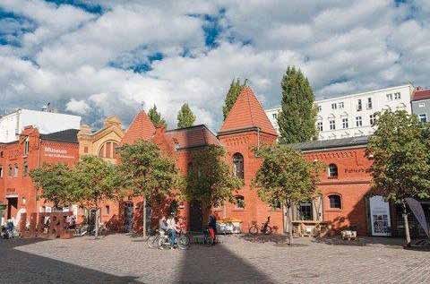
DBM HEBEL STR ASSE                                                                              WISSENSCHAF T | SCIENCE           7

ture cell-based transplant products in the established
                                                              Most relevant publications cited in the text
infrastructure (now managed in tight cooperation with
                                                              1. Pelttari K, Pippenger B, Mumme M, Feliciano S, Scotti C,
PD Dr. Werner Krenger, head of the DBM-GMP facility).
                                                                 Mainil-Varlet P, Procino A, von Rechenberg B, Schwamborn
                                                                 T, Jakob M, Cillo C, Barbero A, Martin I. Adult human neural
A conclusive, personal note from a PhD student                   crest-derived cells for articular cartilage repair. Sci Transl
                                                                 Med. 6:251ra119 (2014)
(S. Pigeot)
                                                              2. Fulco I, Miot S, Haug MD, Barbero A, Wixmerten A, Feliciano
                                                                 S, Wolf F, Jundt G, Marsano A, Farhadi J, Heberer M, Jakob
How is life in the Tissue Engineering Group? How can I           M, Schaefer DJ, Martin I Engineered autologous cartilage
capture the main impressions of being part of this team?         tissue for nasal reconstruction after tumour resection:
                                                                 an observational first-in-human trial. Lancet 384:337-346
The management of the group is obviously challenging,            (2014)
due to the large size and diversity of topics investigat-
                                                              3. Scotti C, Tonnarelli B, Papadimitropoulos A, Scherberich A,
ed. But the established structure and coordinated ef-            Schaeren S, Schauerte A, Lopez-Rios J, Zeller R, Barbero A,
forts by Ivan and the senior scientists allow efficient and      Martin I. Recapitulation of endochondral bone formation
                                                                 using human adult mesenchymal stem cells as a paradigm
frequent interactions, such that I feel well supervised          for developmental engineering. Proc. Natl Acad Sci USA
while maintaining my independence and responsibili-              107:7251-7256 (2010)
ties. What I find of high value are the daily “after lunch    4. Bourgine PE, Scotti C, Pigeot S, Tchang LA, Todorov A,
meetings” which in rotation Ivan has with each of the            Martin I. Osteoinductivity of engineered cartilaginous tem-
                                                                 plates devitalized by inducible apoptosis. Proc Natl Acad
group members, and which give us the opportunity to              Sci USA 111:17426-17431 (2014)
periodically assess our progress and ensure a general
                                                              5. Scotti C, Piccinini E, Takizawa H, Todorov A, Bourgine P,
cohesion between the different project lines. The sci-           Papadimitropoulos A, Barbero A, Manz MG, Martin I. Engi-
entific interactions within the lab are stimulated in our        neering of a functional bone organ through endochondral
                                                                 ossification. Proc Natl Acad Sci USA 110:3997-4002 (2013)
“Progress report” series, where every week a member
of the group presents the own results and/or plans to         6. Di Maggio N, Piccinini E, Jaworski M, Trumpp A, Wendt D,
the rest of the team, and in the “Regenerative medicine          Martin I. Toward modeling the bone marrow niche using
                                                                 scaffold-based 3D culture systems. Biomaterials 32:321-
journal club”, which is organized every two weeks with           329 (2011)
the goal to get inspired by outstanding achievements
                                                              7. Gueven S, Mehrkens A, Saxer F, Schaefer DJ, Martinetti R,
by other groups. On a more practical standpoint, the             Martin I, Scherberich A. Engineering of large osteogenic
working organization also needs to be well orchestrated          grafts with rapid engraftment capacity using mesenchy-
                                                                 mal and endothelial progenitors from human adipose
and all the lab members obviously need to help out. In           tissue. Biomaterials 32:5801-5809 (2011)
addition to the work carried out by Francine Wolf and
                                                              8. Occhetta P, Centola M, Tonnarelli B, Redaelli A, Martin I,
Sandra Feliciano, our technical assistants, each group           Rasponi M. High-throughput microfluidic platform for
member is responsible for a few tasks in the lab such            3D cultures of mesenchymal cells, towards engineering
                                                                 developmental processes. Sci Reports 5:10288 (2015)
as general ordering, preparing general aliquots, buf-
fers, etc. Teams are generated to clean the cell culture      9. Martin I, Simmons PJ, Williams DF. Manufacturing challeng-
                                                                 es in regenerative medicine. Sci Transl Med 6:232fs16 (2014)
rooms weekly and the incubators monthly. It goes with-
out saying that such an organization sometimes incurs
into implementation challenges, but overall the spirit of
mutual assistance prevails and the daily life in the lab
carries on in a friendly and enjoyable atmosphere.

                                              Ivan Martin

DBM Facts 1|2016                                                                                  Department of Biomedicine
8    WISSENSCHAF T | SCIENCE                                                                                  DBM

UniBasel immunologists
retreat to the mountains

The immunologists at the University of Basel have or-     Medicine and Surgery at the USB. Major activities include
ganized themselves into a community, which fosters        a well-attended weekly research seminar and a seminar
scientific exchange and collaborations. The UniBasel      series where distinguished immunologists from around
Immunology Community (UBICO) was formed in the            the world are invited to Basel to present their work and
spring of 2014 and is comprised of basic scientists       interact with UBICO Faculty members. Immunology PhD
and clinicians with a research focus in Immunology.       students organize a Journal Club where a paper from
The UBICO grew out of the Immunology Schwerpunkt          the visiting immunologist’s lab is informally discussed.
at the DBM, but additionally includes scientists at the   Discussions between the PhD students and the visiting
Biocenter, UKBB and the BSSE. The UBICO is generously     immunologists have been very lively.
supported by the DBM and the Departments of Internal
                                                                            Additionally, the UBICO sponsors a
                                                                            research meeting exclusively for PhD
                                                                            students where they present and dis-
                                                                            cuss their work. The “Basler Studien-
                                                                            stiftung” also sponsors UBICO activi-
                                                                            ties for promoting the education of
                                                                            young MD and PhD students in Im-
                                                                            munology.

                                                                            We had our first retreat in Engelberg
                                                                            on November 2-3, 2015. On an early
                                                                            Monday morning 100 students, post-
                                                                            docs and faculty began their journey
                                                                            to the conference site – Hotel Edel-
                                                                            weiss, a very cozy and charming ho-

DBM Facts 1|2016                                                                           Departement Biomedizin
DBM WISSENSCHAF T | SCIENCE 9
tel in Engelberg built in 1901 in the middle of “Angel
Mountains”. Susanne and Peter Kuhn, the owners of the
hotel, welcomed us with an extraordinarily warm and
personal hospitality. Everything was perfectly organized
and the hotel was technically well equipped. Our scien-
tific program with oral presentations started shortly af-
ter our arrival and ended the next day in the afternoon.
The program covered basic concepts, models and tech-
nologies, recent news from genes to proteins regulating
development and effect functions of immune cells and
translation into therapies. After an UBICO group leader
meeting on the first day, we had a poster session with groups presented their work and the atmosphere was
an apero until late in the night. Twenty-four research very interactive and friendly.
Daniela Finke
Feedback from the students:
“I found it extremely useful both in terms of getting myself many useful comments and advices on how to proceed
in-depth information about novel technical approaches in my project not only from scientific point of view but also
immunology and as well in interacting with experienced tips on my protocols which I’m trying to improve.”
clinical and non-clinical immunologist.”
“The presentations and poster session were full of un-
“I learned the main topics of each group in Basel, which biased interaction between all participants, regardless
gave me the opportunity to think about possible collabo- whether they are students or professors. Definitely the
rations or even the available equipment we have in the retreat enhanced the cohesion of our growing Uni Basel
city. I also could train my networking with not only other Immunology Community and I think it would be a great
PhD students but also Postdocs and PIs (which I find use- success to enforce such kind of retreat once a year.”
ful for future career). As I was presenting the poster I got
Auszeichnungen
Bürgi-Preis an Anna Rickli
Anna Rickli von der Forchungsgruppe Psychopharma- Anouk Blatter hat für ihre Diplomarbeit “Kritisches
cology Research (Departement Biomedizin Hebelstras- Hinterfragen des aktuellen Immunflureszenztests für
se) wurde während der Tagung der Schweizerischen Flaviviren” in der Forschungsgruppe “Molecular Viro-
Gesellschaft für Pharmakologie und Toxikologie mit logy” (Departement Biomedizin Petersplatz) von der
dem Bürgi-Preis ausgezeichnet. Der Bürgi-Preis wird Schweizerischen Union für Labormedizin die Auszeich-
alle zwei Jahre für die beste Dissertation mit pharmako- nung für die beste Arbeit erhalten.
logischem Inhalt innerhalb der Schweiz verliehen.
Das DBM gratuliert ganz herzlich!
DBM Facts 1|2016 Department of Biomedicine10 WISSENSCHAF T | SCIENCE DBM Petersplatz
Von Armbrustschützen zum Virennachweis:
Die Abteilung Infektionsdiagnostik im ehemaligen
Stachelschützenhaus
Im 16. Jahrhundert beherbergte das Stachelschützen- (Durchfall) vor. Er verneint Antibiotikaeinnahme und
haus mit den städtischen Armbrustschützen, deren extensiven Alkoholkonsum, welche zu einer Diarrhoe
Aufgabe es war, die Stadt zu verteidigen, wenig aka- führen können. Da er im Moment seinen Militärdienst
demisches Personal. Dies änderte sich Anfang des 18. ableistet und somit engem menschlichen Kontakt aus-
Jahrhunderts, als Bernoulli im neugebauten Südflügel, gesetzt ist, kommen Adenoviren als Auslöser der Di-
dem „physikalischen laboratorio“, seine Experimente arrhoe in Frage und eine Stuhlprobe geht zur Abklärung
durchführte. Schließlich fand im 20. Jahrhundert die ins Labor Virusisolierung (LabVI) am Haus Petersplatz.
neugegründete Hygienische Anstalt ihr Zuhause am Pe-
tersplatz. Stuhlproben sind aufgrund enthaltener Inhibitoren und
unterschiedlicher Konsistenz ein eher problematisches
Heute bietet das Haus Petersplatz Raum für mehre- Material. Davids Stuhlprobe wird aufgeschwemmt, d. h.
re Forschungsgruppen des Departements Biomedizin eine definierte Menge wird in einen Puffer überführt,
(DBM) der Universität Basel sowie für die Abteilung In- suspendiert und zur Abtrennung der festen Bestand-
fektionsdiagnostik (AbtID). Die AbtID besteht aus vier teile zentrifugiert. Nach der Aufarbeitung wird im Lab-
Labors, der Molekularen Diagnostik, Serologie, Genoty- VI innerhalb von 15 min ein Schnelltest auf Adeno- und
pisierung und Resistenz sowie dem Labor Virusisolie- Rotaviren durchgeführt, der negativ ausfällt.
rung, welche für das Universitätsspital Basel (USB), das
Universitätskinderspital beider Basel (UKBB) und weitere Da mit dem Schnelltest kein Erreger in Davids Probe
Spitäler und Arztpraxen in der Nordwestschweiz Analy- identifiziert werden konnte, wird die Suche auf wei-
sen durchführen. Die AbtID ist gemäß QM (Qualitätsma- tere gastroenteritische Erreger ausgedehnt. Dazu ver-
nagement) nach ISO/IEC 17025 zertifiziert und dies seit wendet das LabVI das xTAG Gastrointestinal Pathogen
1999, daher gilt sie als eines der ersten Diagnostiklabors Panel (GPP), eine Next-Generation Multiplex Analyse
der Schweiz. Darüber hinaus beherbergt die AbtID das basierend auf Bead-Technologie, die gleichzeitig 15 der
Bestätigungslabor für HIV/AIDS Diagnosen und das Nati- wichtigsten Pathogene (Viren, Bakterien und Parasiten)
onale WHO-Referenzlabor für Poliomyelitis. Das Leitbild nachweist und das Resultat innerhalb 4 Stunden nach
der AbtID sieht den Patienten im Mittelpunkt ihrer Tä- Extraktion der Probe liefert. Damit ist der Laborleiter
tigkeiten. Dies wird durch eine breite Angebotspalette,
dem Einsatz neuester Untersuchungsmethoden, einer
schnellen Resultatübermittlung und qualitativ hochste-
hender Beratung erreicht. In der AbtID werden jährlich
ca. 35’000 Analysen durchgeführt, wovon etwa knapp
zwei Drittel aus dem USB eingehen. Im vorliegenden
Beitrag möchte ich Sie zu einer kurzen Reise durch die
verschiedenen Labors der AbtID einladen.
Ein nicht ganz alltäglicher Fall der AbtID
David stellt sich im Notfall des USB mit Kopfschmerzen,
Beatrice Hess, Labor Virusisolierung, beim Pipettieren der Zellen
Müdigkeit, Schwindel, trockenem Mund und Diarrhoe für einen Neutralisationstest.
DBM Facts 1|2016 Departement BiomedizinDBM Petersplatz WISSENSCHAF T | SCIENCE 11
Im LabVI ist außerdem das Natio-
nale WHO-Referenzlabor für Polio-
myelitis beheimatet. Entsprechend
werden Stuhlproben aus der gesam-
ten Schweiz auf Polioviren (PV) unter-
sucht. Obwohl im Jahr 2015 mit bisher
weltweit 74 Fällen an akuter schlaffer
Lähmung (engl. Acute Flaccid Paraly-
sis, AFP), verursacht durch Wild PV, ein
Allzeittief erreicht wurde, hat die welt-
weite Überwachung der Poliomyelitis,
durch die Flüchtlingskrise in Syrien,
nichts an Brisanz eingebüßt. Noch im
Jahr 2013 wurde Wild PV aus Pakistan
Rita Reuter, Labor Molekulare Diagnostik, bei der Auswertung einer Real-Time PCR.
nach Syrien eingeschleppt und es kam
in der Lage gleichentags den behandelnden Arzt tele- zu 23 Poliomyelitis Fällen. Bereits ein einzelner bestä-
fonisch über ein positives Resultat zu informieren. Das tigter Poliomyelitis-Fall in der Schweiz wäre wie ein Aus-
LabVI kann in Davids Stuhlprobe keinen gastroenteri- bruchsereignis einzustufen, da weniger als 1% der In-
tischen Erreger nachweisen. fektionen unter dem Bild der AFP verlaufen.
Neben den beschriebenen Techniken führt das LabVI Vi- Nach Rücksprache mit dem behandelnden Arzt wird
rusisolierungen durch. Diese sind geeignet, wenn es sich nun eine Blutprobe von David ans Labor Molekulare
um relativ rasch wachsende, lytische Viren handelt. Das Diagnostik (LabMD) zur weiteren Abklärung geschickt.
Probenmaterial wird auf Zellkulturen gegeben und die Im LabMD wird Probenmaterial auf pathogene Erreger
im Material enthaltenen Viren infizieren die Zellen und mittels Nukleinsäure-Amplifikations-Testung (NAT) oder
vermehren sich. Die massenhafte Vermehrung verur- auch Polymerase-Ketten-Reaktion (PCR) analysiert.
sacht einen sogenannten cytopathischen Effekt (CPE), Dazu wird DNA und RNA aus Vollblut, Plasma, Serum,
ein Abrunden und Absterben der infizierten Wirtszellen, Körperflüssigkeiten, Biopsien, Punktaten oder Liquor
der mikroskopisch beobachtet werden kann. Die Virusi- (Rückenmarks-Flüssigkeit) gewonnen und teilweise die
solierung hat den Vorteil, dass infektiöse Partikel nachge- direkt anschließende PCR-Reaktion pipettiert. Dieses
wiesen werden und gilt deshalb als Goldstandard. Aller- Labor ist am meisten automatisiert und liefert die quan-
dings ist eine nachgeschaltete Analyse, z. B. in Form einer titativen Viruslast-Resultate für Humanes Immundefizi-
Immunfluoreszenz mit spezifischen, markierten Antikör- enz Virus (HIV), Hepatitis B, Hepatitis C und CMV täglich
pern (AK), zur genauen Identifizierung des Virusisolats mit Hilfe eines Vollautomaten. Analysen für andere Erre-
nötig. Auf diese Weise werden im LabVI Herpes-Simplex- ger (z. B. Epstein-Barr-Virus oder HSV 1/2) erfolgen auf
Virus 1/2 (HSV 1/2), Cytomegalievirus (CMV), Varizella- Real-Time PCR Cyclern, welche in Echtzeit die Menge an
Zoster-Virus (VZV), Adenovirus, Enteroviren, Influenza- vervielfältigter Nukleinsäure mittels Fluoreszenzsignal
viren und Respiratory Syncytial Virus (RSV) identifiziert. messen. Durch die parallele Analyse international de-
finierter WHO-Quantifizierungs-Standards erlaubt das
Aktuell läuft auch ein Projekt zur routinemäßigen HSV Verfahren eine Quantifizierung der Nukleinsäure in der
1/2 Resistenztestung für antivirale Substanzen wie Probe. Die einzelnen Schritte der Analyse sind räumlich
Aciclovir mittels automatisierter Interpretation des CPE. getrennt um das Kontaminationsrisiko zu minimieren.
Diese Innovation zeigt, dass trotz des Schwerpunkts auf
molekulare Verfahren, bestimmte phänotypische Tests Das LabMD bietet neben Dengue- und Chikungunya-
weiterhin sinnvoll sein können. seit kurzem einen Zikavirus Nukleinsäurenachweis an.
DBM Facts 1|2016 Department of Biomedicine12 WISSENSCHAF T | SCIENCE DBM Petersplatz
Fällen kontaktiert die LabortechnikerIn den Auftragge-
ber und klärt ab, ob zusätzliches Probenmaterial vor-
handen ist. Wenn nicht, nimmt der Laborleiter Kontakt
mit dem behandelnden Arzt auf. Er klärt im direkten Ge-
spräch, ob eine Priorisierung der Analysen sinnvoll und
machbar ist oder das Material verdünnt werden soll,
was die analytische Sensitivität der Nachweismethode
herabsetzt.
Bei respiratorischen Proben ist die Qualität des Mate-
rials von entscheidender Bedeutung. Respiratorische
Christine Bauer, Labor Genotypisierung/Resistenz, beim Anset- Erreger der unteren Atemwege lassen sich am besten
zen einer Sequenzier-PCR.
aus broncheoalveolärer Lavage nachweisen, Erreger
Zikavirus gehört zur Gattung der Flaviviren, deren Ver- der oberen Atemwege hingegen werden in Nasen-, Ra-
treter, durch Stechmücken übertragen, schwere Erkran- chenabstrich, Sputum oder Bronchialsekret detektiert.
kungen, wie hämorrhagische Fieber oder Infektionen Neben Schnelltesten für Influenzaviren und RSV mit ei-
des ZNS verursachen. Zikavirus, erstmals 1952 beim ner Bearbeitungszeit von nur 15–60 min, verwendet das
Menschen beschrieben, wurde bisher mit eher mil- LabMD für eine breitere Suche nach respiratorischen
den Krankheitsverläufen in Verbindung gebracht. Seit Erregern eine Next-Generation Multiplex Analyse, die
Ende 2015 besteht im Zusammenhang mit der aktuell auf Fluoreszenz-Bead-Technologie basiert und 96 Pro-
in Lateinamerika laufenden Epidemie der Verdacht auf ben auf einmal analysieren kann. Der RPP detektiert
einen Zusammenhang zwischen Zikavirus-Infektionen gleichzeitig 22 respiratorische Erreger und liefert das
bei Schwangeren und Mikrozephalie (signifikant ver- Resultat innerhalb 3 Stunden nach Extraktion der Pro-
minderter Kopfumfang) bei Föten und Neugeborenen. be. Dies erlaubt dem Laborleiter den behandelnden Arzt
Allerdings gibt es noch keine verlässlichen Daten über gleichentags telefonisch über ein positives Resultat zu
die Häufigkeit von Mikrozephalie in Lateinamerika vor informieren und somit eine schnelle Isolierung des Pa-
der Zikavirus-Epidemie. Zudem wurde für andere virale tienten zu ermöglichen. Während der Influenza-Saison
Erreger, wie Rubellavirus (Erreger der Röteln) und CMV werden vom LabMD täglich bis zu 30 RPP-Analysen
oder das Protozoon Toxoplasma gondii, ein Zusammen- durchgeführt. Die wöchentlich aktualisierte Statistik für
hang zwischen Mikrozephalie und Infektionen während respiratorische Erreger ist unter www.zid.ch > Infekti-
der frühen Phase der Schwangerschaft erbracht. In der onsstatistik einsehbar.
ersten Woche nach Symptombeginn, in der virämischen
Phase, kann die RNA des Virus durch eine Zikavirus- Das LabMD findet in Davids Blutprobe 350’000 Kopien
spezifische Reverse-Transkriptase PCR (RT-PCR) im Blut HIV pro mL. Die hohe Viruslast deutet auf eine HIV-
nachgewiesen werden. Analysen auf andere Flaviviren, Neuinfektion hin und muss mit Hilfe einer zweiten,
wie Dengue- oder das Alphavirus Chikungunyavirus, unabhängigen Blutprobe bestätigt werden. Ein Bestäti-
welche ähnliche Symptome verursachen können, sowie gungstest ist vom Gesetzgeber vorgeschrieben um eine
die Reiseanamnese, sollten in die Gesamtbeurteilung falsch-positive Diagnose auszuschließen. Dazu wird
eingehen. vom USB eine neue Blutprobe Davids ans Labor Serolo-
gie (LabS) geschickt. Das LabS fungiert als HIV-Referenz-
Um eine hohe analytische Sensitivität zu erreichen, labor und erhält Aufträge zur HIV-Testung und -bestäti-
muss vom Auftraggeber ausreichend Probenmaterial gung aus der ganzen Schweiz.
eingesandt werden. Dies ist öfter nicht der Fall bei Mate-
rialien, welche von Natur aus in geringen Mengen vorlie- Der Nachweis von Antigenen (Ag) erlaubt einerseits die
gen, wie Vorderkammerpunktat oder Liquor. In solchen Identifizierung von Ag-tragenden Krankheitserregern,
DBM Facts 1|2016 Departement BiomedizinDBM Petersplatz WISSENSCHAF T | SCIENCE 13
andererseits können durch den Nachweis bestimmter
Antikörper (AK) im Blut Krankheiten diagnostiziert
werden. Beim HIV-Bestätigungstest sind auf einer Trä-
germembran die unterschiedlichen HIV-Proteine (Ag)
nebeneinander aufgebracht. Von der LabortechnikerIn
wird Davids Serumprobe auf die Membran pipettiert.
Sind AK gegen HIV vorhanden, heften sich diese an die
Virusproteine auf dem Streifen. Nach weiteren Arbeits-
schritten werden dunkle Striche auf dem Teststreifen
sichtbar. Damit ist der Bestätigungstest positiv ausge-
fallen. Jetzt muss David darüber informiert werden,
dass er HIV-1-positiv ist. Angelika Aebli, Labor Serologie, bei der Validierung der Masern /
Mumps Serologie.
In der Schweiz nehmen die Fälle sexual übertragener den, damit der Patient isoliert und eine Therapie einge-
Krankheiten seit einigen Jahren zu. Als Folge erhält das leitet werden kann.
LabS viele Untersuchungen zum Nachweis von Trepo-
nema pallidum, dem Erreger der Lues (Syphilis). Da T. Im Jahr 2013 wurden schweizweit mehr als 22 Millionen
pallidum nur mit Spezialmethoden gezüchtet werden Auslandsreisen unternommen. Entsprechend hoch ist
kann, kommt der Serologie eine besondere Bedeutung die Zahl an Reiserückkehrern mit Verdacht auf tropische
zu. Als Suchtest dient die Treponema-pallidum-Parti- Erkrankungen. Von den Ärzten werden deshalb ver-
kel-Agglutination (TPPA). Als Ag werden Lysate von T. mehrt Aufträge erteilt, Flavivirus-Serologien (Dengue-,
pallidum verwendet, die an Gelatinepartikel gekoppelt West-Nil- und Zikavirus) durchzuführen. Seit März bietet
sind. Sind AK im Serum vorhanden, reagieren sie mit das Haus Petersplatz eine Zikavirus-Serologie an.
den Ag der «beladenen» Gelatinepartikel und es kommt
zur Verklumpung (Agglutination) der Partikel. Ein po- David wird vom Arzt im USB darüber informiert, dass er
sitives TPPA Resultat wird mit einem Bestätigungstest eine HIV-Erkrankung hat. Der Arzt erklärt ihm, dass die
überprüft. Zur Beurteilung des Krankheitsverlaufs, bzw. von David beschriebenen Symptome und die Diarrhoe
Behandlungserfolges, wird die Reaktion des Serums mit kompatibel mit einer HIV-1 Infektion sind. HIV-1 ist die
unspezifischen Lipoid-Ak durch den RPR-Test (Rapid- Ursache für die Entstehung des Acquired Immune De-
Plasma-Reagin) benutzt. ficiency Syndrome (AIDS). Das Virus schädigt das Im-
munsystem, da CD4 T-Helfer-Lymphozyten zerstört
werden. Der Arzt wird David die Therapiemöglichkeiten
In der Schweiz erkranken jährlich ca. 550 Menschen erläutern. Bei einer wirksamen kombinierten antiretro-
an Tuberkulose, wobei eine Verschiebung von älteren viralen Therapie (cART) wird die Vermehrung von HIV-1
Schweizern zu jungen Migranten zu beobachten ist. durch Medikamente unterdrückt und es ist kein Virus im
Zum spezifischen Nachweis einer Infektion mit Myco- Blut des Patienten nachweisbar. Steigt die Virusmenge
bacterium tuberculosis verwendet die Serologie das In- im Verlauf der cART trotz Einhaltung der Medikation an,
terferon-Gamma-Release Assay (IGRA). T-Lymphozyten kann dies ein Hinweis auf eine Mutation des HIV-1 und
des Patienten werden mit Ag von M. tuberculosis über damit auf eine Resistenz sein. Um zu klären, ob David
Nacht stimuliert. Ist eine Infektion vorhanden, kann die sich bereits mit einem resistenten HIV-Stamm infiziert
Sekretion von Interferon-Gamma nachgewiesen wer- hat oder das Virus gegen die verschiedenen Medikamen-
den. Als Testformate werden das Immunoassay (Quan- tenklassen empfindlich ist, wird der Arzt eine Resistenz-
tiFERON-TB) oder der ELISPOT (T SPOT.TB) angeboten. bestimmung veranlassen. Dazu wird Davids Blut ans La-
Bei beiden Tests kann das Ergebnis bereits nach 1 Tag bor Genotypisierung/Resistenz (LabGR) geschickt.
abgelesen und der behandelnde Arzt informiert wer-
DBM Facts 1|2016 Department of Biomedicine14 WISSENSCHAF T | SCIENCE DBM Petersplatz
Im LabGR werden die Genabschnitte Protease, Reverse ganmanifestationen auftreten. Bei Reaktivierung stehen
Transkriptase und Integrase der viralen RNA mit Hilfe dem Arzt effektive Medikamente zur Verfügung, deren
der RT-PCR vermehrt und sequenziert. Die Nukleotidab- Wirksamkeit durch das Auftreten von Resistenzen stark
folge von Davids HIV-1 wird mit der Referenzsequenz in herabgesetzt werden kann. Die Mitarbeiter des LabGR
der SmartGene Datenbank abgeglichen, es erfolgt eine suchen in so einem Fall nach Mutationen im UL97 (Phos-
genotypische Resistenzbestimmung. Das HIV-1 in Da- photransferase) und UL54 (DNA-Polymerase) Gen. Die
vids Blut weist keine relevanten Punktmutationen auf Genotypisierung deckt Resistenzen gegen die Medika-
und somit ist klar, dass kein resistentes HIV-1 übertra- mente Ganciclovir, Foscarnet und Cidofovir ab, was dem
gen wurde. In Davids Fall sollten alle Medikamentenklas- Arzt erlaubt auf eine Alternativtherapie zu wechseln.
sen die Vermehrung von HIV-1 effizient unterdrücken.
Als Spezialuntersuchung gilt die Bestimmung des HIV-1 Fazit
Tropismus gegenüber der zellulären Rezeptoren CCR5 Die Arbeit in der AbtID ist spannend und dank der sich
und CXCR4. Die Untersuchungen werden innerhalb we- täglich mit der Klinik ändernden Herausforderungen
niger Tage vom LabGR durchgeführt und der Arzt erhält durch die Auftraggeber sehr anspruchsvoll. Die Kom-
einen Bericht, der die Resistenz gegen die unterschied- bination von Routinediagnostik und Forschung, Ent-
lichen Medikamentenklassen übersichtlich graphisch wicklung sowie Einführung neuer diagnostischer Tests
darstellt. erfordert flexible und interessierte Mitarbeiter, die in
einem hochmotivierten Team gute Entfaltungsmöglich-
CMV führt wie alle Herpesviren zu einer persistierenden keiten erhalten.
Infektion. Unter Immunsuppression kann es zur Reak- Rainer Gosert
tivierung kommen und es können unterschiedliche Or-
Mitarbeiter der Abteilung Infektionsdiagnostik. Vordere Reihe (links nach rechts): Christel Widia, Anna Sophie Bahlmann (Assistentin),
Klaudia Bagienski (Leiterin LabGR), Bettina Oettli, Zeliha Özkan, Hülya Atici, Sibylle Stauffer, Aline Weber, Frauke Mekolli; hintere Reihe
(links nach rechts): Angelika Aebli, Christine Bauer, Rita Reuter, Sylvie Goepfert, Sasa Maksimovic, Rainer Gosert (Leiter LabVI und WHO
Nationales Referenzlabor für Poliomyelitis), Hans H. Hirsch (Leiter AbtID), Thomas Klimkait (Stv Leiter AbtID, Leiter LabS)
DBM Facts 1|2016 Departement BiomedizinDBM WISSENSCHAF T | SCIENCE 15
SweetySpot-Software development in the DBM Flow
Cytometry Facility for droplet based Cell Sorters
For years, BD Influx users have jealously been watching the user How it works
friendly BD Aria Diva Software with Sweet Spot software. Now my A camera is used to observe the break off point. The images are cap-
new software development for droplet-based cell sorters closes the tured as a so called array. (picture 102 from left first image). In the
gap because it can do the same as Sweet Spot but is now available for next step, the array image is transformed to an 8-bit greyscale image.
the BD Influx and BD FACS Jazz. In theory it could be used for every The standard camera on the machine has a resolution about 640x480
droplet-based cell sorter with a camera watching the break-off point pixels, so we have 307 200 pixels. Each pixel is represented in our
and an accessible communication protocol. My software observes greyscale image by an integer value in the range of 0 to 255 (8-bit
and automatically readjusts the break-off point by setting new val- deep greyscale image -->28=256 possible values, but we use only val-
ues for the amplitude. Droplet based cell sorters work with liquid ues between 0 to 255). Now we set a threshold to our grey image, so
which is pressed through a nozzle with a specific diameter. Because that we can more easily calculate where the break off point is. The
of a transducer with a piezoelectric crystal that vibrates in a specific library I use for thresholding has different methods and types for set-
frequency, the pressed stream is broken up into single drops. This ting a threshold. To get the best result and an almost noise free im-
break-off point is very important and has to be stable through the age, I use adaptive thresholding but I implemented every type of the
whole time of sorting. Before sorting this break-off point has to be thresholding method so that the user can decide what is the best for
defined and the cell sorter calibrated. The operator has to calculate their needs. You can see the threshold image in picture 102 (from left
the time a particle needs from the measurement point to this break- the second image). Thereby the values in the range of 0 to 255 are re-
off point, the so-called drop delay. Obviously this point needs to be placed with almost two possible values around 0 (black) and around
stable during the sort because it defines which drop with your par- 255 (white). In figure 102 (from left picture 3 and 4), I extracted the
ticle inside will be deflected and collected in a tube. But only if the pixel intensity values from our threshold image and exported them
drop delay was calculated correctly. Otherwise the charged drop is to Microsoft Excel (picture 3) to make it more clear. Picture 4 shows a
empty or a particle is inside you don’t want to be sorted. You can see cut-out of the excel sheet after all values close to zero were marked
this process of building the break off point in picture 101. black (picture 4). With the pixel intensity values of 0 and 255 from
the threshold image it is easier to calculate where the break off point
is. With my own search algorithmic with simple math, I can calculate
the stream position and the gap between the stream position and the
first satellite drop, which are the necessary values to keep the break-
off point stable. You can see the final processed image in picture 102
from left, image 5. These two parameters are calculated for every
camera frame (around 25–30 frames per second).
The advantages of SweetySpot
My Software is 100% written in Python. Python is one of the most
powerful Programming languages because it is relative easy to read.
Like Java, Python is an interpreter language which means that you
don’t have to compile your software for a specific operating system.
The Python interpreter translates it to machine code. The interpreter
is available for almost every operating system (Windows, Mac, Linux).
SweetySpot is written completely in python for maximum flexibility
and fast update. The key functions like image processing and calcu-
picture 101 lation are written in C with python bindings for maximum perfor-
DBM Facts 1|2016 Department of Biomedicine16 WISSENSCHAF T | SCIENCE DBM
on the influx will stay original and un-
touched. For downgrading just discon-
nect the two tubing’s and reconnect
them to the originally installed valve.
Don’t forget, ”In theory it could be
used for every droplet-based cell sort-
er, not just the BD influx.”
This year I was invited by BD to the Eu-
ropean Technology Center in Heidelberg
Germany. They gave me the possibility
to test SweetySpot on the BD FACS Jazz
cell sorter. It has been rumoured that BD
picture 102 produces software with this purpose for
mance. It is very comfortable because it works out of the box (pos- the Influx, but in Europe nobody has seen this software, even in Hei-
sible in one folder or one file), meaning no software installation is delberg. For me it would be very interesting to compare my Software
necessary. Another great advantage is that my software is completely with the Software from BD. But it’s sure that the version from BD
independent from the company operating software (for example the won’t have a kind of “secure mode” that mine has and it’s also sure
BD Sortware Software for the BD Influx Sorter). The purpose of this that for the BD FACS Jazz cell sorter no software with this purpose ex-
software is to keep the break off point stable and to protect your ists from BD at the time of writing. (picture 103 from left to right) Dr
sample and collection tubes. Jens Fleischer and Toni Krebs after successfully testing SweetySpot
on the FACSJazz Cell Sorter from BD in Heidelberg.
How to bring the Influx closer to state of the art?
Generally, the fluidics system of almost all Cell-Sorters can be operat- To look ahead
ed through software. Unfortunately, with Influx it isn’t possible. I did In my opinion SweetySpot is a great development but there is no mar-
a little hardware modification to make it possible on our influx one. I ket for selling or licensing it. BD will come with his own version for the
installed a simple relay, connected it to the fluidic operating circuits Influx. I hope I will get the ok to start an open source project under,
of the influx and to a network (also allows connecting through Eth- probably the General Public Licence (GPL) or Berkeley Software Distri-
ernet, w-lan, microcontroller, computer etc.). Now we have an influx bution (BDS) license and make it available for free, for everyone who
with software which observes the break off point and automatically wants to use it under the terms of the GPL/BDS. As long as I get the
readjusts it by setting new values if the break-off point changes and ok from the University Hospital, I will create a website with the source
with the hardware modification, we can additionally operate the flu- code, instruction to build the dependencies for SweetySpot and user
idic system with SweetySpot. With the access to the fluidic system of manuals. There will also be information available about other proj-
the Influx it was also possible to program a “secure mode” in Sweet- ects in flow cytometry.
ySpot. My software recognizes a partial or full nozzle clock and tries Toni Krebs
to protect your sample and collection tubes. It automatically stops
the fluidic main stream of the machine and changes the position of
the WDU (robotic arm with holder of the collecting tubes) to avoid
contamination of your collection tubes. At the same time, it stops the
sample run to avoid losing any microliter of your sample. At the last
step it could send an email or give a sms or a call to inform the opera-
tor that something went wrong. The future plan for it is to change
two magnetic valves on the Influx, so that I can program an automatic
fluidics system, such as automatic start-up, automatic debubbling,
automatic washing steps and so on. But don’t be afraid. Everything picture 103
DBM Facts 1|2016 Departement BiomedizinYou can also read