Cytogenetic and Molecular Diagnosis in Gestational Disorders - Katherine Geiersbach, M.D.
←
→
Page content transcription
If your browser does not render page correctly, please read the page content below
Cytogenetic and Molecular
Diagnosis in Gestational Disorders

              Katherine Geiersbach, M.D.
      Assistant Professor, Department of Pathology
          University of Utah School of Medicine
   Medical Director, Cytogenetics & Molecular Oncology
                    ARUP Laboratories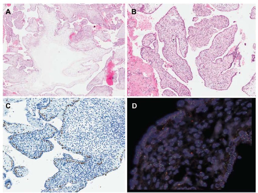
Overview
• Discuss the role of molecular genetic testing in
  evaluating pathologic conditions related to
  pregnancy
   – Viable pregnancy: prenatal screening and invasive
     diagnostic testing for genetic abnormalities
   – Pregnancy loss
   – Molar pregnancy

• Review laboratory testing strategies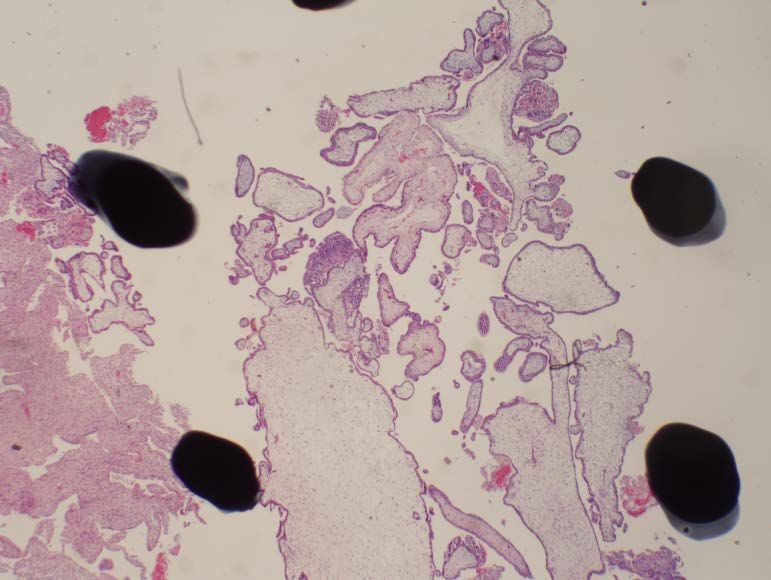
1. PRENATAL SCREENING AND INVASIVE
DIAGNOSTIC TESTING FOR GENETIC
ABNORMALITIES
The incidence of aneusomy increases dramatically with maternal age

     Maternal age and trisomy. Fig 6 in Hassold T, Hunt P. To err (meiotically) is human:
     the genesis of human aneuploidy. Nat Rev Genet 2001;2:280-91.
Meiotic ‘timelines’ for humans. Fig 1 in Hassold T, Hunt P. To err (meiotically) is human:
the genesis of human aneuploidy. Nat Rev Genet 2001;2:280-91.
Meiotic errors are correlated with the
number and location of recombination events

      Fig. 2-2 Chromosome behavior in meiosis I. In:
      Gersen SL, Keagle MB. The Principles of Clinical
      Cytogenetics. New Jersey: Humana Press; 2005.
Screening tests for genetic
   abnormalities of the embryo/ fetus
• Noninvasive tests for common trisomies (21, 18, (13))
   – Ultrasound
      • First trimester – nuchal translucency
      • Second trimester – anatomy scan (18-20 weeks)
   – Maternal serum screening
      • Biochemical markers of aneuploidy (PAPP-A, beta-hCG, AFP, uE3,
        inhibin A)
      • Cell free fetal DNA
• Ethnicity-based (parental carrier) screening for single
  gene disorders
   – Cystic fibrosis (Caucasian, Ashkenazi Jewish)
   – Tay Sachs disease and others (Ashkenazi Jewish)
   – Hemoglobinopathies (Mediterranean descent)
Non-invasive prenatal testing (NIPT)
• Cell free fetal DNA
   – Short fragments of DNA circulating in maternal serum,
     small proportion (
Non-invasive prenatal testing (NIPT):
         current controversies
• Variably regarded as “diagnostic” vs. “screening” test
• Concern over direct-to-consumer marketing
• Large preclinical trials have been performed, but more
  clinical validation studies are needed
• Lower sensitivity and specificity for trisomy 13
• Changing landscape of prenatal genetic testing
   – No longer restricted to women > 35
   – Shift toward increased patient autonomy
• Destigmatization of Down syndrome
• State-led initiatives to restrict abortions, concern over
  potential increase in pregnancy termination rate
Definitive (diagnostic) tests for genetic
 abnormalities of the embryo/ fetus
• Direct fetal sample is obtained via invasive procedure:
   – Chorionic villus sampling (11 - 13 weeks)
   – Amniocentesis (15 – 20 weeks)
• Offered to all pregnant women, along with genetic
  counseling, regardless of age (ACOG Practice Bulletin No.
  77, 2007)
• Preferred method of follow up for abnormal ultrasound
  and/ or abnormal prenatal screening (risk > 1:250 – 1:300)
• Cytogenomic testing methodologies:
   – Karyotype (chromosome analysis, cytogenetics)
       • All indications, including maternal preference for diagnostic testing
   – Genomic microarray for copy number variations, including
     microdeletion syndromes
       • Highest diagnostic yield (Wapner et al, NEJM 367 (23): 2175-84, 2012).
Definitive (diagnostic) tests for genetic
abnormalities of the embryo/ fetus
• Targeted molecular genetic testing
– FISH or microsatellite genotyping for aneuploidy
– Single gene / gene panel testing for Mendelian
disorders in the fetusChromosomal microarray
• DNA-based testing for copy number variations (CNVs) throughout the genome
• Size of detectable CNVs depends on array design and probe coverage
Affymetrix
CytoScan SNP array log2 ratio
(copy#)
“smooth
signal”
(copy#)
allele
difference
track (SNP
probes)Microarray showing Trisomy 21
Microarray - 1q21 microdeletion (346.8 kb within 2 Mb common microdeletion region)
2. PREGNANCY LOSS
Pregnancy loss is common in humans
• 15-20% of clinically recognized pregnancies
   – Recurrent miscarriage (≥2): 1%
   – Most losses (90%) occur during the first trimester
• 30-40% of chemically detected pregnancies
   • In a 1988 NEJM study*, women who were trying to achieve pregnancy
     were monitored by daily urine beta hCG levels. A total of 31% of
     detected pregnancies were lost.
     • 2/3 of the lost pregnancies were undetectable clinically

• Unknown % of pre-implantation pregnancies
   • Cytogenetic studies of IVF pre-embryos at day 3 has shown that about
     50% are genetically abnormal
• Probably
Etiology • An etiology for the loss can be determined in only about half of unintended pregnancy losses • Genetic abnormality • Immunologic factors • Infections • Endocrine • Environmental agents • Uterine anatomic abnormalities • Cytogenetic abnormalities are identified in up to 50% of first trimester losses
Value of genetic testing on pregnancy loss:
             for patient care

           Provides closure for families after a loss

           Identifies abnormalities associated with a risk for
           recurrence

           Identification of a genetic abnormality prevents additional
           costly workup for infertility
Fig. from Paxton CN et al, Prenatal Diagnosis 32, 1–7 [Epub 8 NOV 2012]
Distribution of abnormalities by
           mechanism

            6%         Nondisjunction in meiosis
       3%

 16%                   Abnormal fertilization

                       Error in zygote cleavage

                 75%   Breakage, aberrant
                       recombination, and/or
                       malsegregation
Cytogenetic Findings in POCs
• Aneusomy for virtually every chromosome
• 2-3% of trisomies associated with a potentially
heritable translocation
• Secondary aneusomy in polyploidy
– Triploidy: 9% have secondary trisomy or monosomy
– Tetraploidy: 14% have secondary trisomy or
monosomy
• Mosaicism (17% among all abnormal karyotypes)
– Eliminating cases of likely maternal cell
contamination, the true rate of mosaicism is probably
6%Anomaly POCs at Spontaneous Live births**
ARUP* abortions**
Trisomy 13 1.56% 1.10% 0.01%
Trisomy 18 1.19% 0.84% 0.02%
Trisomy 21 2.86% 2.00% 0.11%
Trisomy 16 3.04% 5.58% 0.00%
Other 9.52% 11.81% 0.00%
trisomies
Monosomy X 5.47% 8.35% 0.01%
Sex 0.07% 0.33% 0.15%
chromosome
trisomies
Triploids 5.50% 5.79% 0.00%
Tetraploids 0.98% 2.39% 0.00%
# Karyotyped 2,763* 3,353** 31,521**
*ARUP data estimated over 32 months of data
**data from Kline, J. and Stein, Z. (1986) The epidemiology of spontaneous abortion.
In Early Pregnancy Failure (Huisjes, H.J.and Lind, T. eds.), Churchill Livingstone, New York, pp. 240–256.When Is a Karyotype Insufficient? • Suspected molar pregnancy • Nonviable tissue – Culture failure rate for fresh tissue: ~14% – Only paraffin embedded tissue available • Uncharacterized structural abnormality – Marker chromosome – Additional material of unknown origin – Submicroscopic copy number variants (CNVs)
68,XX
Chromosome 1-22: 4 allele tracks, copy number 2, consistent with triploidy
X chromosome shows 3 tracks, as for 46,XX, but copy number approx. 1.3
Chromosome 9 shows copy number approx. 1.7

                                    …consistent with mosaicism for -9
                                    68,XX (55%)/67,XX,-9 (45%)
Characterization of a marker chromosome
45,XY,-21[8]/46,XX[11]
Chromosome 9 gain (copy number = 3)
Chromosome 21 mosaic loss (copy number approx. 1.85)

          (Prevalence of cell line with monosomy 21 is about 15% in this sample)
Chromosome X shows copy number = 1, Y copy number = 1 (not shown), consistent with pure male population (direct sample)
Fetal demise – alobar holoprosencephaly

2 Mb loss on 2p21: arr 2p21(43,461,488-45,412,846x1) (hg19)

                                                              SIX3
Metaphase FISH on
POC material
(used as positive control)
RP11-489K22 BAC (Empire Genomics)
Mother carries same deletion by FISHAbsence of heterozygosity affecting 22.5% of genome
Stretches of homozygosity

• May indicate:
  – Uniparental disomy if present on a single
    chromosome
  – Identity by descent if present in numerous
    independent regions of the genome
  – Complete hydatidiform mole if 100% homozygous
    (monospermy) or ~50% homozygous (dispermy)
• Associated with increased risk for autosomal
  recessive condition
  – In pregnancy loss / fetal demise, consider potentially
    lethal AR condition
Microarray for pregnancy loss:
summary
• Chromosomal microarray can be successfully
applied to pregnancy loss samples, may yield
different information than that provided by the
karyotype, and is more sensitive and specific for
characterizing clinically relevant genomic
imbalances
• Karyotype remains the most versatile method for
detecting mosaicism and some polyploidy
(tetraploidy); microarray is most valuable as a
reflex test for POCs with a normal karyotype or
POCs which fail to grow in culture3. MOLAR PREGNANCY
Hydatidiform Mole
• Associated risk for persistent trophoblastic
  disease
• Complete mole is often detected clinically,
  whereas partial mole is more often detected by
  the pathologist
   – BUT: histologic diagnosis is unreliable
• 3 major ancillary diagnostic tests (FFPE):
   – p57 immunohistochemistry (complete mole)
   – Flow cytometry (partial mole)
   – Microsatellite genotyping (complete and partial mole)
Complete hydatidiform mole

                23                                        23

                                                               23

         46,XX                                      46,XX or 46,XY
  Monospermy with endoreduplication                 Dispermy

       •Diploid
       •Complete uniparental disomy across entire genome
       •No maternal DNA
       •15% risk for persistent gestational trophoblastic disease
46,XX
5 STR loci: comparison of genotypes of villi (“fetus”) and decidua

Fig. 1 from Furtado et al, Diagnostic Utility of Microsatellite Genotyping for Molar Pregnancy Testing.
Archives of Pathology and Laboratory Medicine [In Press]
Partial hydatidiform mole

               23

                     23

          dispermy                               monospermy with endoreduplication
                                                 (theoretical mechanism)

       •Triploid
       •2:1 paternal: maternal DNA contribution
       •0.5% risk for persistent gestational trophoblastic disease
            •not a risk factor for choriocarcinoma?
69,XXY
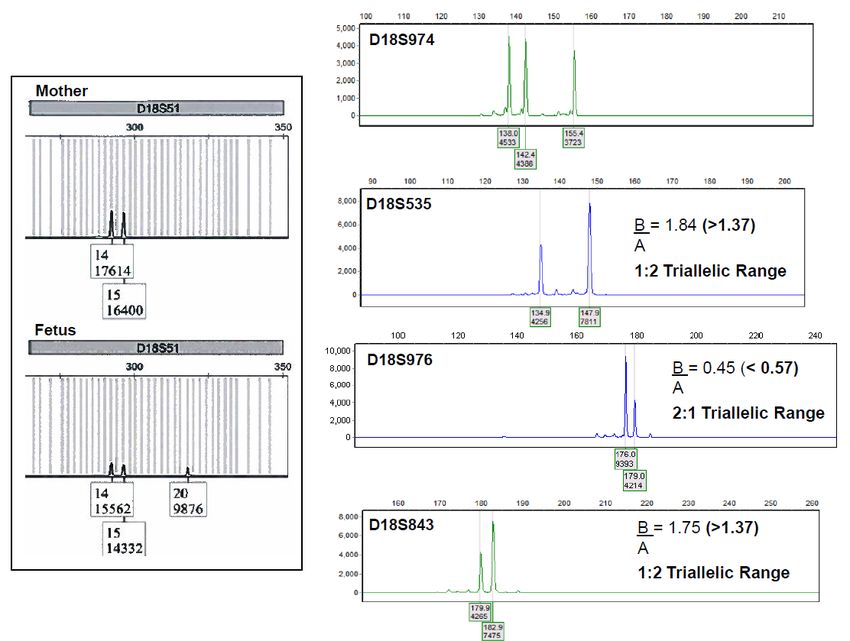
4 STR loci: comparison of genotypes of villi (“fetus”) and decidua

Fig. 2 from Furtado et al, Diagnostic Utility of Microsatellite Genotyping for Molar Pregnancy Testing.
Archives of Pathology and Laboratory Medicine [In Press]
Maternally derived triploidy (digyny): diploid egg

                23                                     23

      69,XXX or 69,XXY                       69,XXX or 69,XXY

       •Triploid
       •1:2 paternal: maternal DNA contribution
       •Fetal anomalies
       •NO risk for persistent gestational trophoblastic disease
Nonmolar hydropic abortion (HA)
23
* Autosomal trisomy, sex chromosome aneuploidy,
Mendelian disorders, non-genetic causes
May simulate molar pregnancy by histopathology
Trophoblastic proliferation
particularly +7, +15, +21, or +22
trophoblastic hypoplasia may also be seen (e.g. trisomy 18)
* Villous edema / hydropsIncidental Trisomy Detection (10/54 non-molar cases; not reported)
Unusual Case of Chimerism (Androgenetic / Biparental)
Fig. 4 from Furtado et al, Diagnostic Utility of Microsatellite Genotyping for Molar Pregnancy Testing.
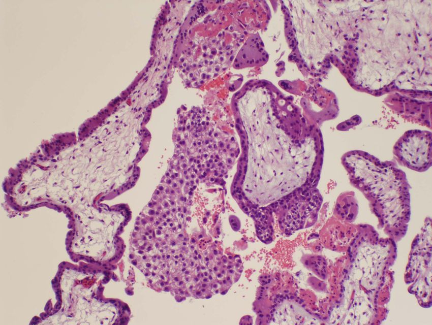
Archives of Pathology and Laboratory Medicine [In Press]p57 immunohistochemistry FISH with X and Y centromere probes
Fig. 5 from Furtado et al, Diagnostic Utility of Microsatellite Genotyping for Molar Pregnancy Testing.
Archives of Pathology and Laboratory Medicine [In Press]Conclusions
• New technology has introduced new options and
new testing algorithms for prenatal screening and
diagnosis
• Cytogenetic analysis is the most versatile method
for whole genome analysis of pregnancy loss
samples
– Microarray is a useful adjunct method, largely because
it can yield results in samples that fail to grow in
culture
• Accurate diagnosis of hydatidiform mole relies
upon ancillary testingYou can also read