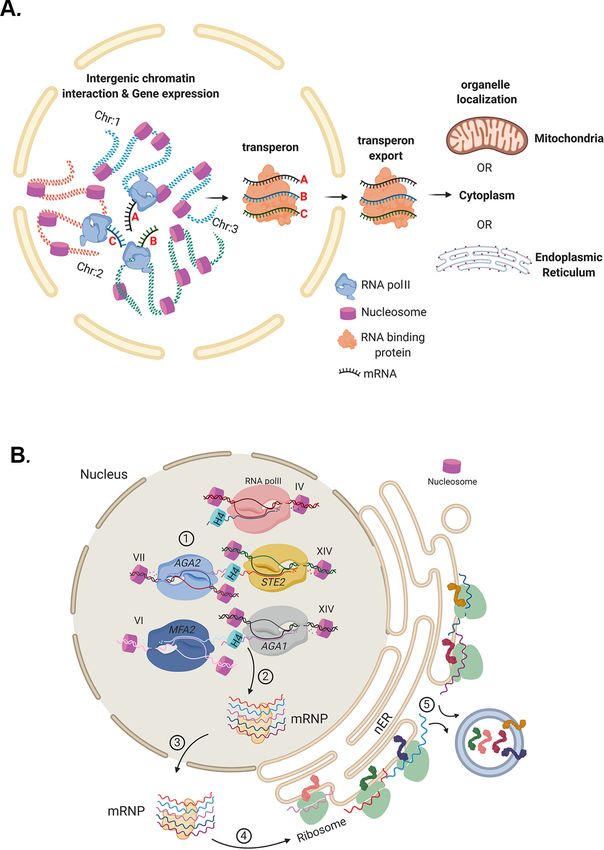
Multiplexed mRNA assembly into ribonucleoprotein particles plays an operon-like role in the control of yeast cell physiology
←
→
Page content transcription
If your browser does not render page correctly, please read the page content below
RESEARCH ARTICLE

                                    Multiplexed mRNA assembly into
                                    ribonucleoprotein particles plays an
                                    operon-like role in the control of yeast
                                    cell physiology
                                    Rohini R Nair1†, Dmitry Zabezhinsky1†, Rita Gelin-Licht1, Brian J Haas2,
                                    Michael CA Dyhr1, Hannah S Sperber1, Chad Nusbaum2‡, Jeffrey E Gerst1*
                                    1
                                        Department of Molecular Genetics, Weizmann Institute of Science, Rehovot, Israel;
                                    2
                                        Broad Institute of MIT and Harvard, Cambridge, United States

                                    Abstract Prokaryotes utilize polycistronic messages (operons) to co-translate proteins involved
                                    in the same biological processes. Whether eukaryotes achieve similar regulation by selectively
                                    assembling and translating monocistronic messages derived from different chromosomes is
                                    unknown. We employed transcript-specific RNA pulldowns and RNA-seq/RT-PCR to identify yeast
                                    mRNAs that co-precipitate as ribonucleoprotein (RNP) complexes. Consistent with the hypothesis
                                    of eukaryotic RNA operons, mRNAs encoding components of the mating pathway, heat shock
                                    proteins, and mitochondrial outer membrane proteins multiplex in trans, forming discrete
                                    messenger ribonucleoprotein (mRNP) complexes (called transperons). Chromatin capture and allele
                                    tagging experiments reveal that genes encoding multiplexed mRNAs physically interact; thus, RNA
                                    assembly may result from co-regulated gene expression. Transperon assembly and function
                                    depends upon histone H4, and its depletion leads to defects in RNA multiplexing, decreased
                                    pheromone responsiveness and mating, and increased heat shock sensitivity. We propose that
*For correspondence:                intergenic associations and non-canonical histone H4 functions contribute to transperon formation
jeffrey.gerst@weizmann.ac.il        in eukaryotic cells and regulate cell physiology.
†
 These authors contributed
equally to this work

Present address: ‡Cellarity Inc,
Cambridge, United States            Introduction
                                    Prokaryotic organisms can rely on polycistronic transcription (operons), which allows for the expres-
Competing interest: See
                                    sion of needed mRNAs from a single promoter and enables a rapid and robust response to corre-
page 28
                                    sponding stimuli (e.g., lactose operon) (Jacob and Monod, 1961). However, this mode of action has
Funding: See page 28                not been reported in eukaryotes, except for a few limited examples (e.g., in Caenorhabditis
Received: 23 December 2020          elegans [C. elegans]) (Spieth et al., 1993), and eukaryotic operons have been observed to give rise
Accepted: 02 May 2021               to dicistronic messages (Blumenthal, 2004). However, it is intriguing to postulate whether eukar-
Published: 04 May 2021              yotes have devised functional alternatives to operons. One possibility is that eukaryotic messenger
Reviewing editor: Karsten Weis,
                                    ribonucleoprotein (mRNP) particles, which are composed of multiple RNAs and RNA-binding pro-
ETH Zurich, Switzerland             teins (RBPs) (Mitchell and Parker, 2014), effectively confer combinatorial gene expression networks
                                    similar to prokaryotic operons (Keene and Tenenbaum, 2002). In this case, however, the mRNP par-
   Copyright Nair et al. This
                                    ticles contain individual mRNAs that undergo co-translational control and may encode proteins
article is distributed under the
                                    involved in the same biological process or molecular complex. The idea that mRNPs might essen-
terms of the Creative Commons
Attribution License, which          tially constitute RNA operons (Keene, 2007) or transperons, as termed here, implies several fea-
permits unrestricted use and        tures. First, although mRNAs derived from different chromosomes can reside in the same mRNP,
redistribution provided that the    they should have common regulatory elements not only for transcriptional and translational control,
original author and source are      but also for interacting at the post-transcriptional level with other mRNAs within the particle. The lat-
credited.                           ter includes motifs/structures to facilitate interactions with shared RBPs, as well as elements that

Nair, Zabezhinsky, et al. eLife 2021;10:e66050. DOI: https://doi.org/10.7554/eLife.66050                                              1 of 32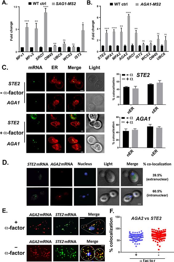
Research article                                                                                                   Cell Biology

                                    might facilitate RNA-RNA interactions (e.g., base-pairing). Second, some mechanism must confer
                                    the recruitment and assembly, in trans (i.e., multiplexing), of the individual mRNAs into single mRNP
                                    particles. Third, in order to be functionally relevant, transperons should contain mRNAs encoding
                                    proteins involved in the same functional context (e.g., organelle biogenesis, macromolecular com-
                                    plex, or biological process). Although the mechanism remains unknown, studies have shown that
                                    genomic DNA folds create sites of transcriptional hot spots (Lieberman-Aiden et al., 2009;
                                    Rao et al., 2014) that could facilitate multiplexing by co-localizing messages during transcription
                                    and coordinating the subsquent association of RBPs. Moreover, RNA-RNA interactome studies show
                                    that extensive interactions can occur directly between RNAs in living cells (Aw et al., 2016;
                                    Engreitz et al., 2014; Kudla et al., 2011; Nguyen et al., 2016; Sharma et al., 2016). Thus, the mul-
                                    tiplexing of mRNAs into mRNPs to yield transperons should be directly testable.
                                        Although individual yeast RBPs have been shown to bind to numerous (e.g., 10s-1000s) mRNAs
                                    (Hogan et al., 2008), these RNA-binding studies are based primarily upon crosslinking and RBP pull-
                                    downs that are unable to define the minimal number of mRNA species in a single mRNP particle. To
                                    test the idea of RNA multiplexing and define the composition of such particles, we employed a sin-
                                    gle mRNA species pulldown procedure (RaPID) (Slobodin and Gerst, 2010; Slobodin and Gerst,
                                    2011). This method employs MS2 aptamer tagging of endogenously expressed messages in yeast
                                    (Haim et al., 2007) and their precipitation from cell lysates via the MS2 coat protein (MCP) to iden-
                                    tify bound non-tagged transcripts using RNA-seq (RaPID-seq). We have previously demonstrated
                                    that RaPID-seq identifies MS2-tagged target mRNAs (Haimovich et al., 2016) and now show here
                                    that additional messages associate with these transcripts.
                                        By employing MATa yeast in RaPID-seq pulldown experiments, we found that select MS2-labeled
                                    target mRNAs (e.g., SRO7, EXO70, OM45) co-precipitated untagged mRNAs encoding secreted
                                    proteins involved in cell mating (e.g., STE3, SAG1, MFa1, MFa2). Since a corresponding complex (e.
                                    g., STE2, AGA1, AGA2, MFA1, MFA2) could also be precipitated from MATa cells, it suggested to
                                    us that mRNAs encoding secreted proteins involved in mating (e.g., pheromones, pheromone recep-
                                    tors, agglutinins, and proteases) multiplex into a functionally selective mRNP complex – the mating
                                    transperon. To identify potential factors involved in mating transperon assembly, we employed pull-
                                    downs of these mRNAs followed by mass spectrometry (RaPID-MS). We found that the yeast histone
                                    H4 paralogs, Hhf1 and Hhf2 (Dollard et al., 1994), interact with these mRNAs to regulate complex
                                    assembly. Furthermore, both pheromone responsiveness (e.g., shmooing) and mating were inhibited
                                    by either histone H4 depletion or a block in H4 acetylation. When mutated, no other histones had
                                    this effect. Moreover, we could identify conserved cis elements in the mating mRNAs and demon-
                                    strate that mutations therein had deleterious effects on mRNA multiplexing, pheromone responsive-
                                    ness, and mating, which were similar to those seen upon histone H4 mutation.
                                        To help elucidate the mechanism by which mRNA multiplexing might occur, we performed chro-
                                    matin conformation capture (Lieberman-Aiden et al., 2009), as well as genomic locus tagging
                                    experiments, to look for evidence of direct allelic interactions. Importantly, a specific interaction
                                    between the STE2 and AGA2 genes was identified in MATa cells, which suggests that interallelic
                                    coupling may give rise to RNA multiplexing. Furthermore, coupling appeared to occur in both a
                                    transcription- and histone H4-dependent manner. To help validate this hypothesis, we examined
                                    whether mRNAs encoding heat shock proteins (HSPs), whose genes were previously shown to
                                    undergo intergenic interactions during heat shock (Chowdhary et al., 2019), also undergo multi-
                                    plexing. Importantly, HSP mRNAs were found to multiplex in trans during heat shock to form RNP
                                    complexes, and histone H4 was required for both RNA multiplexing and robust post-heat shock cell
                                    recovery. Parallel studies revealed the existence of additional RNA multiplexes for mitochondrial
                                    outer membrane proteins and MAP kinase pathway proteins in yeast. Together, these results sug-
                                    gest that histone H4-mediated chromatin interactions may lead to the formation of functionally
                                    selective transperons in eukaryotic cells and possibly act as a means to co-regulate gene expression
                                    in place of polycistronicity.

Nair, Zabezhinsky, et al. eLife 2021;10:e66050. DOI: https://doi.org/10.7554/eLife.66050                                           2 of 32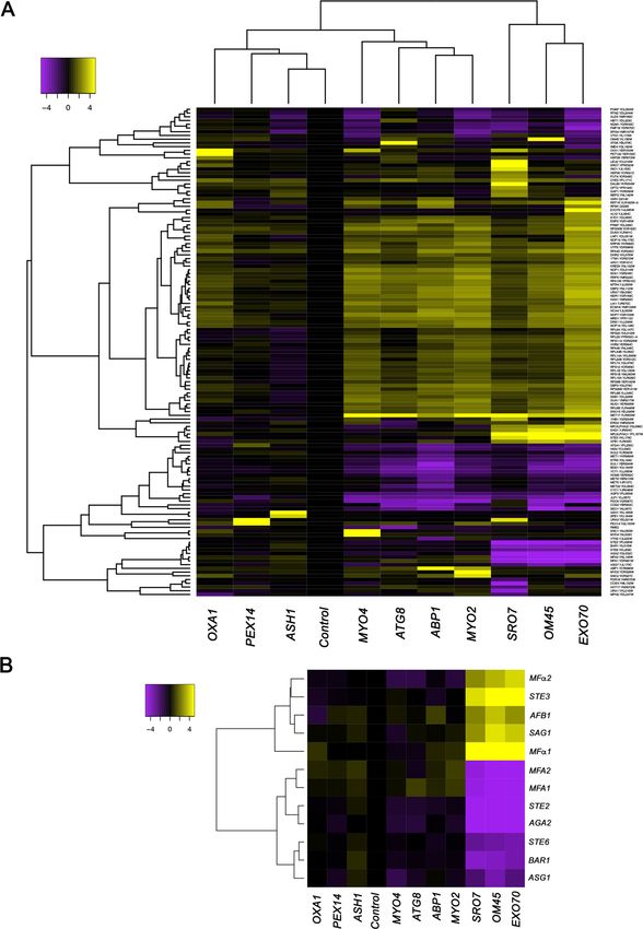
Research article                                                                                                    Cell Biology

                                    Results
                                    mRNAs encoding yeast mating pathway components co-precipitate in
                                    mRNPs
                                    We developed the RaPID procedure (Slobodin and Gerst, 2010; Slobodin and Gerst, 2011) to
                                    identify RBPs and mRNAs that bind to specific RNAs of interest. Unlike CLIP or PAR-CLIP, which col-
                                    lects information on all RBP-RNA interactions in the cell and is biased toward highly expressed
                                    mRNAs with long poly-A tails (Hafner et al., 2010), RaPID allows for a biochemical view of the pro-
                                    tein and RNA constituents of mRNPs at the single transcript level. RaPID employs the precipitation
                                    of MS2 aptamer-tagged messages using the MCP fused to streptavidin-binding peptide, followed
                                    by elution from immobilized streptavidin with free biotin, and then mass spectrometry
                                    (Slobodin and Gerst, 2010; Slobodin and Gerst, 2011) or RNA-seq (Haimovich et al., 2016) to
                                    identify bound proteins and RNAs, respectively. In order to address issues regarding whether the
                                    MS2 system might affect the stability of MS2 aptamer-tagged messages, we performed RaPID and
                                    RNA-seq (RaPID-Seq) on 11 endogenously expressed MS2 aptamer-tagged messages in MATa yeast
                                    (Haimovich et al., 2016). These included representative transcripts having a wide range of intracellu-
                                    lar patterns of localization (e.g., mitochondria: OXA1 and OM45; cortical endoplasmic reticulum
                                    [cER]/asymmetrically localized mRNAs: ABP1, SRO7, EXO70, ASH1, MYO2, and MYO4; peripheral
                                    nuclear endoplasmic reticulum [nER]: ATG8; and peroxisomes: PEX14) (Gadir et al., 2011;
                                    Haim et al., 2007; Zipor et al., 2009). In addition to identifying the target mRNA in each pulldown,
                                    we identified non-tagged RNAs that co-precipitated with the targets (Figure 1A, Figure 1—figure
                                    supplement 1, and see Supplementary file 1 for RNA-seq data). These included retrotransposable
                                    elements (e.g., Ty elements), tRNAs, ribosomal RNAs, telomere and small nuclear RNAs (Figure 1—
                                    figure supplement 1), and coding RNAs (Figure 1, Figure 1—figure supplement 1, and
                                    Supplementary file 1). Notably, five mRNAs encoding secreted and membrane proteins (mSMPs) of
                                    the mating process were found associated with a subset of target RNAs. These target transcripts
                                    consisted of two cER-localized mRNAs that encode polarity factors (mPOLs; SRO7 and EXO70)
                                    (Aronov et al., 2007) and OM45, an mRNA that encodes a mitochondrial outer membrane protein
                                    (MOMP), which we have shown to localize to both mitochondria and ER (Gadir et al., 2011). Co-pre-
                                    cipitated non-tagged mRNAs included STE3, which encodes the a-pheromone receptors, MFa1 and
                                    MFa2, which both encode a-mating factor, SAG1, which encodes a-agglutinin, and AFB1, which
                                    encodes the a-pheromone blocker (Figure 1B). This result is consistent with the idea that specific
                                    RNAs can undergo multiplexing.
                                       Importantly, the corresponding MATa mating-type RNAs (e.g., STE2, MFA1, MFA2, AGA2, and
                                    BAR1), as well as other MATa RNAs involved in mating (e.g., STE6, ASG7), were negatively enriched
                                    (depleted) in these pulldowns (Figure 1A and B). While this result was predicted, due to their lack of
                                    expression in MATa cells, nonetheless, it validates the specific pulldown of the MATa mating
                                    mRNAs.
                                       We verified the RaPID-Seq results using RaPID followed by qRT-PCR (RaPID-qPCR) and found
                                    that the pulldown of SAG1 mRNA co-precipitated cohort mRNAs of the mating pathway (e.g.,
                                    MFa1, MFa2), but not unrelated mRNAs (e.g., UBC6, WSC2) (Figure 2A). We noted that the original
                                    target mRNAs (SRO7, OM45) were present in this pulldown, though OM45 was lowly represented in
                                    the qRT-PCR data as those mRNAs directly related to mating. We note that IST2 mRNA was signifi-
                                    cantly enriched in the pulldown from MATa cells (Figure 2A), but not in pulldowns from MATa cells
                                    (Figure 2B).
                                       By employing RaPID-qPCR, we examined whether a corresponding set of mating pathway
                                    mRNAs (i.e., STE2 [a-factor receptor], AGA1 and AGA2 [agglutinins], and MFA1 and MFA2 [a-fac-
                                    tor]) could co-precipitate in MATa cells. Indeed, the precipitation of AGA1 mRNA using RaPID led
                                    to co-precipitation of the cohort mating pathway mRNAs (e.g., STE2, AGA2, MFA1, MFA2;
                                    Figure 2B), along with SRO7. Thus, we demonstrated that mRNAs encoding secreted proteins
                                    involved in mating undergo multiplexing in either yeast haplotype.

Nair, Zabezhinsky, et al. eLife 2021;10:e66050. DOI: https://doi.org/10.7554/eLife.66050                                            3 of 32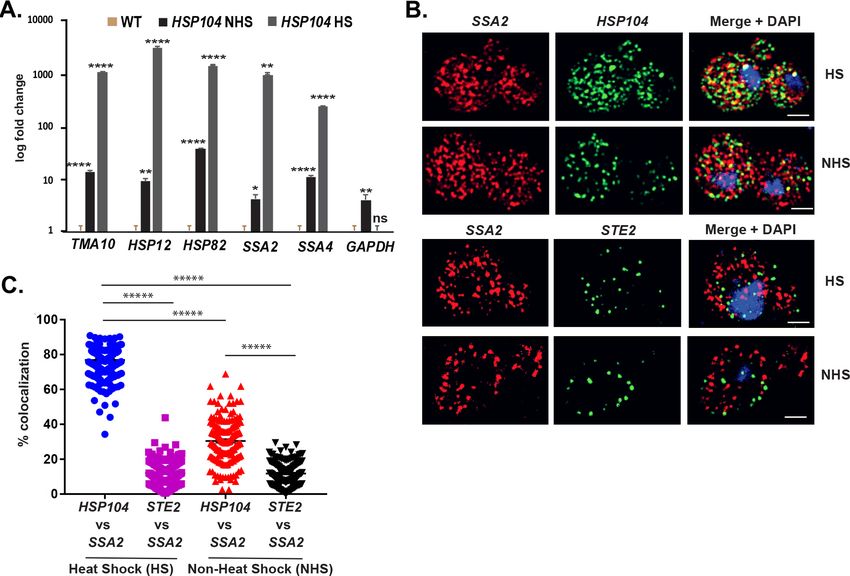
Research article                                                                                                               Cell Biology

Figure 1. mRNAs encoding secreted and membrane proteins involved in yeast mating undergo multiplexing. (A) RaPID RNA pulldown and RNA-seq
reveal transcript multiplexing in yeast. Different MS2 aptamer-tagged mRNAs (listed on the x axis) expressed from their genomic loci were precipitated
from MATa yeast (BY4742) by MS2-CP-GFP-SBP after formaldehyde crosslinking in vivo and cell lysis procedures (RaPID; see ’Materials and methods’).
RNA-seq was performed and the reads were plotted to yield a heat map for the relative enrichment of the tagged as well as non-tagged mRNAs (see
Figure 1 continued on next page

Nair, Zabezhinsky, et al. eLife 2021;10:e66050. DOI: https://doi.org/10.7554/eLife.66050                                                       4 of 32
Research article Cell Biology
Figure 1 continued
color key for approximate values). All non-coding RNAs (e.g., Ty elements, tRNAs, snoRNAs, ribosomal RNAs) were removed but are included in the
heat map of Figure 1—figure supplement 1. A list of the log2 transformed and control-subtracted expression values, as plotted here, is given in
Supplementary file 1. (B) mRNAs encoding proteins involved in mating in MATa cells are enriched in the pulldowns of SRO7, EXO70, and OM45
mRNAs. A selective heat map comprising the mRNAs encoding secreted and membrane proteins involved in yeast cell mating is shown. Positive
enrichment of cell type-specific MATa mRNAs (STE3, MFa1, MFa2, SAG1, AFB1) is shown in purple, while negative enrichment of the non-expressed
MATa mRNAs (MFA1, MFA2, STE2, AGA2, BAR1, STE6, ASG7) is shown in yellow.
The online version of this article includes the following figure supplement(s) for figure 1:
Figure supplement 1. mRNAs encoding secreted and membrane proteins involved in yeast mating multiplex into a single ribonucleoprotein particle.
mRNAs encoding mating pathway components co-localize to ER in both
vegetative-growing and pheromone-treated cells
It was important to determine whether mRNAs of the mating pathway localize to the ER in the cell
body, like other mSMPs (Kraut-Cohen et al., 2013), or perhaps to the bud/shmoo tip, like asymmet-
rically localized mRNAs that encode polarity and secretion factors (Aronov et al., 2007; Gelin-
Licht et al., 2012). A previous study has suggested that aptamer-tagged mating pathway mRNAs,
like MFA2, localize to P-bodies located in (or near) the shmoo tip and that these granules are func-
tional sites for transmitting the mating signal (Aronov et al., 2015). However, a more recent work
by us (Haimovich et al., 2016) has shown that endogenously expressed MS2 aptamer-tagged MFA2
mRNA does not localize to P-bodies and that P-bodies only form under conditions of mRNA overex-
pression, as used in Aronov et al., 2015. We examined the localization of endogenously expressed
MS2-tagged STE2 and AGA1 mRNAs, along an ER marker, either mCherry-Scs2 (Figure 2C) or -
Sec63 (Figure 2D), in both non-treated and a-factor (1 mM)-treated cells. We found that both
mRNAs co-localized either with nER or with cER, both with or without a-factor (images for mCherry-
Scs2 are shown in Figure 2C and quantified for mCherry-Sec63 in Figure 2D), and are not present in
the bud or shmoo tips (Figure 2C). In addition, we examined the localization of endogenously
expressed MS2 aptamer-tagged MFA1 and MFA2 mRNA in wild-type (WT) cells or cells lacking
SHE2, an ER-localized RBP involved in the asymmetric localization of mRNAs to the bud (but not
shmoo) tip (Genz et al., 2013). Both MFA1 and MFA2 mRNAs localized to the cell bodies in either
pheromone-treated and non-treated cells, and no difference in localization was observed upon the
deletion of SHE2 (Figure 2—figure supplement 1A). Thus, like other mSMPs, these mating pathway
mRNAs are not trafficked to the polarized extensions of yeast cells (Kraut-Cohen et al., 2013).
As RNA co-localization is predicted by RNA multiplexing, we tagged endogenous AGA2 with the
PP7 aptamer (Larson et al., 2011) in the cells expressing MS2-tagged STE2 and examined for co-
localization upon expression of their respective fluorescent protein-tagged aptamer-binding proteins
(e.g., PP7-PS-tomato and MS2-CP-GFP(x3)). Overall, we found that 65.5 ± 7.7% (average ± standard
deviation; n = 3 experiments) of PP7-tagged AGA2 mRNA co-localized with MS2-tagged STE2
mRNA (Figure 2D), of which 39.5 ± 3.5% co-localized in the cytoplasm and 60.5 ± 3.5% co-localized
in the nucleus. We validated the live imaging results using single-molecule fluorescence in situ
hybridization (smFISH), which is typically more sensitive than imaging using MS2-CP-GFP(x3) and
thus reveals a greater number of mRNAs (Figure 2E and F). Specific probes against native STE2 and
PP7 aptamer-tagged AGA2 were used to score localization of the STE2 and AGA2 mRNAs, respec-
tively (we note that native AGA2 mRNA is insufficiently long for smFISH). The majority (73.1 ± 1.0%)
of STE2 mRNA spots co-localized with PP7-tagged AGA2 mRNA spots in untreated cells, while 65.4
± 0.1% co-localized in cells treated with a-factor (Figure 2E and F), and co-localization was observed
both in the nucleus and cytoplasm. Thus, mating transperon mRNAs shown to multiplex using bio-
chemical means appear to co-localize before and after nuclear export.
As insertion of the PP7 aptamer sequence before the 3’UTR might potentially alter mRNA fate,
we examined the co-localization of endogenously expressed untagged mating mRNAs using smFISH
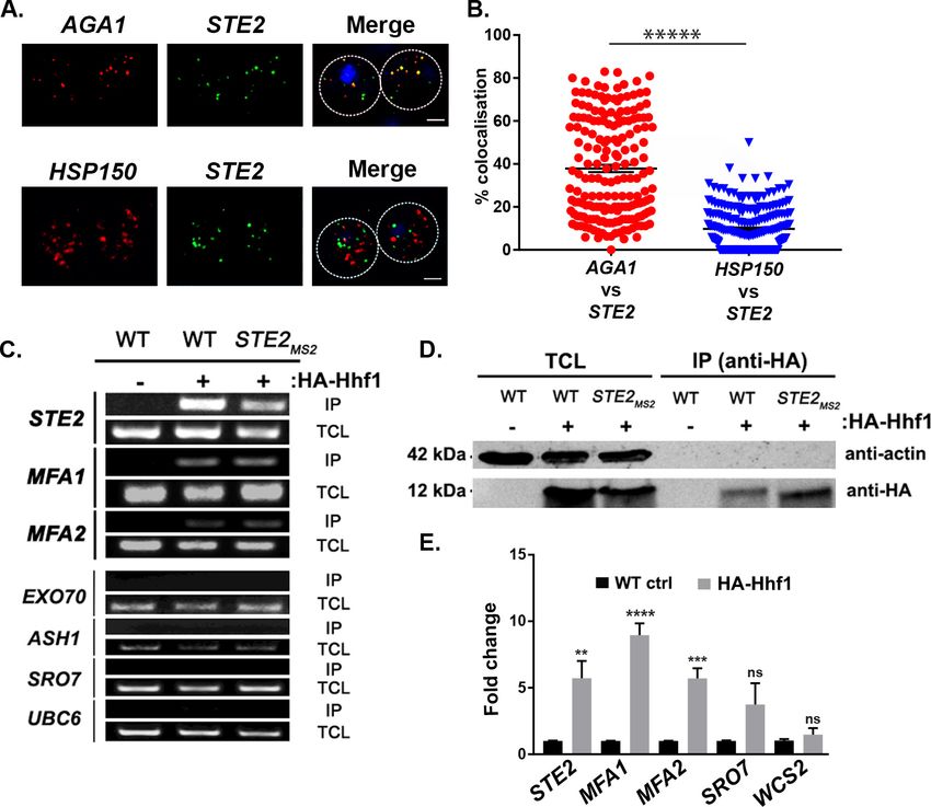
(Pizzinga et al., 2019; Tsanov et al., 2016). We examined co-localization of the AGA1 and STE2
mRNAs and, in parallel, that of the STE2 and HSP150 mRNAs in order to show specificity. Overall,
we found that 37.9 ± 1.7% (average ± standard error of the mean; SEM) of AGA1 mRNA co-localized
with STE2 mRNA, while only 9.8 ± 0.6% of HSP150 mRNA co-localized with STE2 mRNA (Figure 3A
and B; representative images are shown in Figure 3a and quantified in Figure 3B). Together, the
Nair, Zabezhinsky, et al. eLife 2021;10:e66050. DOI: https://doi.org/10.7554/eLife.66050 5 of 32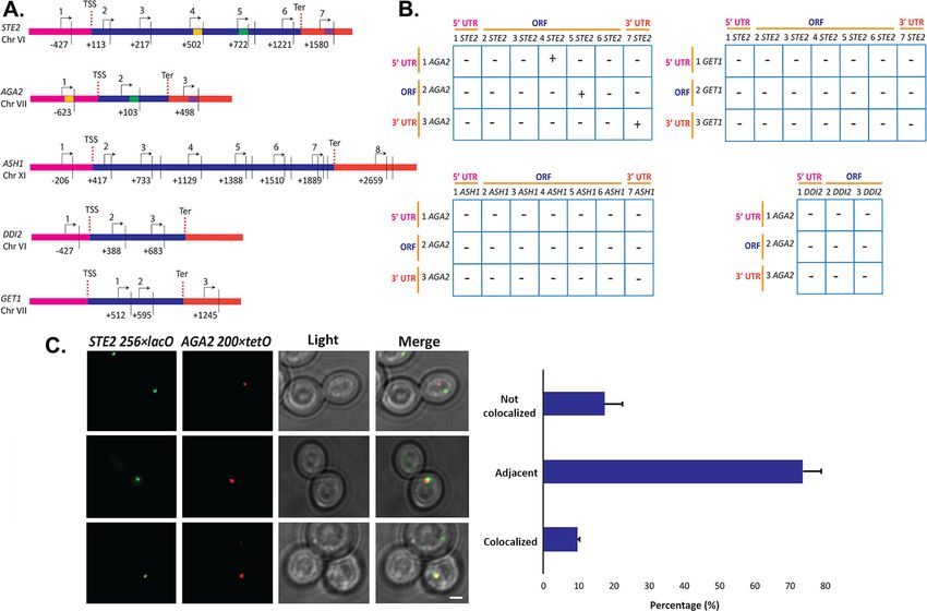
Research article                                                                                                               Cell Biology

                                    Figure 2. mRNAs encoding secreted and membrane proteins involved in mating multiplex in both MATa and
                                    MATa cells and localize to the ER. (A) A mating messenger ribonucleoprotein (mRNP) complex is also formed in
                                    MATa cells. MATa yeast (BY4741) expressing MS2 aptamer-tagged SAG1 from its genomic locus were grown to
                                    mid-log phase (O.D.600 = 0.5) and subjected to RaPID followed by qRT-PCR (RaPID-PCR; see ’Materials and
                                    methods’). RNA derived from the total cell extract (Input) or biotin-eluated fraction (RaPID) was analyzed by qRT-
                                    PCR using primer pairs corresponding to mRNAs expected to possibly multiplex (based on the results shown in
                                    Figure 1). Three biological repeats were performed and an unpaired t-test was used to compare the SAG1
                                    pulldown to the WT control pulldown; ****p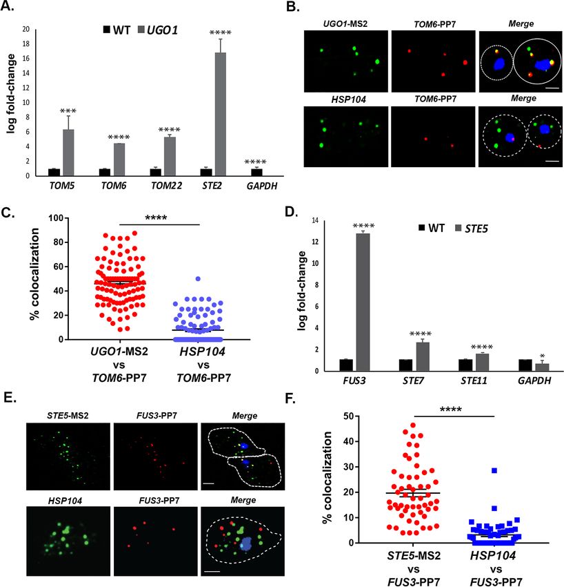
Research article                                                                                                                Cell Biology

                                    Figure 2 continued
                                    ER in pheromone-treated or -non-treated cells. Wild-type cells expressing MS2 aptamer-tagged STE2 or AGA1
                                    mRNAs (as listed) were transformed with single-copy plasmids expressing MS2-CP-GFP(x3) and either Scs2-
                                    mCherry (left panel) or Sec63-RFP (right panel). Cells were grown to mid-log phase and either a-factor (5 mM; +a-
                                    factor) or dimethyl sulfoxide (DMSO) (-a-factor) was added for 90 min, followed by fixation with 4%
                                    paraformaldehyde in a medium containing 3.5% sucrose and prior to imaging by confocal microscopy. Left panel:
                                    fluorescent images: mRNA – MS2-CP-GFP(x3) labeling; ER – Scs2-mCherry labeling; merge – merger of mRNA and
                                    ER windows; light – transmitted light. Size bar = 2 mm. Right panel: quantitative analysis of RNA co-localization:
                                    nER – nuclear ER; cER – cortical ER. (D) Mating mRNAs STE2 and AGA2 co-localize both inside and outside of the
                                    nucleus. Yeast expressing MS2 aptamer-tagged STE2 mRNA and PP7 aptamer-tagged AGA2 mRNA from its
                                    genomic locus were transformed with plasmids expressing MS2-CP-GFP(x3) and PP7-PS-tomato, while the nucleus
                                    was stained with 4,6-diamidino2-phenylindole (DAPI). STE2 mRNA – MS2-CP-GFP(x3) labeling; AGA2 mRNA –
                                    PP7-PS-tomato labeling; nucleus – DAPI labeling; merge – merger of mRNA and nucleus windows; light –
                                    transmitted light. Size bar = 2 mm. (E) Single-molecule fluorescence in situ hybridization (smFISH) validation of
                                    STE2 and AGA2 mRNA co-localization. Non-transformed cells from (D) were either treated with a-factor (10 mM) or
                                    left untreated and processed for smFISH labeling using non-overlapping FISH probes complementary to the STE2
                                    mRNA and PP7 aptamers, prior to labeling with DAPI. The representative image shown is from a single focal
                                    plane. AGA2 – Cy3 labeling; STE2 – Alexa488 labeling; merge – merged Cy3/Alexa488 windows with DAPI
                                    staining. Size bar = 2 mm. (F) Scatter plot of co-localization data from (E). Co-localization was assessed using the
                                    FISHquant algorithm (see ’Materials and methods’). Black lines indicate the avg. ± SEM for each sample. Each data
                                    point represents a single cell.
                                    The online version of this article includes the following figure supplement(s) for figure 2:
                                    Figure supplement 1. MFA1 and MFA2 mRNAs localize to the cell body.

                                    multiplexing and imaging results suggest that mating pathway mRNAs form particulate mRNP
                                    complexes.

                                    A histone H4 paralog binds to mating mRNP RNAs
                                    To determine how the mating mRNP particle assembles, we performed RaPID-MS using the MFA1,
                                    MFA2, and STE2 mRNAs either bearing or lacking their 3’UTRs, along with an unrelated control
                                    RNA, ASH1, as target RNAs. We identified a band of ~150 kDa that was present in all lanes upon sil-
                                    ver staining, except for the lanes corresponding to STE2 mRNA lacking its 3’UTR and ASH1 mRNA
                                    (Figure 3—figure supplement 1). Sequencing revealed that this band contained a core histone H4
                                    paralog, Hhf1. Although the predicted molecular mass of Hhf1 is ~11 kDa, the likelihood exists that
                                    it was crosslinked with another protein. Indeed, other crosslinked products were found in this band
                                    although none were shown to be connected to mRNAs of the mating mRNP (data not shown).
                                        To verify whether Hhf1 alone can precipitate the mating mRNP particle, we performed
                                    immunoprecipitation (IP) using HA-tagged Hhf1 and examined the precipitates for the presence of
                                    mating mRNAs. First, we found that HA-Hhf1 could precipitate either native or MS2 aptamer-tagged
                                    STE2, along with native MFA1 or MFA2 mRNA, but not EXO70, ASH1, SRO7, or UBC6 mRNA by
                                    RT-PCR (Figure 3C). Thus, we could verify a specific interaction between Hhf1 and RNAs of the mat-
                                    ing particle and, in addition, show that HA-Hhf1 migrates at around 12 kDa in SDS-PAGE gels
                                    (Figure 3D), as predicted. Next, we validated these results in HA-Hhf1 pulldowns using qRT-PCR,
                                    whereby we could detect native STE2, MFA1, and MFA2, but not SRO7 or WSC2, in the precipitates
                                    (Figure 3E). A similar experiment was performed with MATa cells expressing HA-Hhf1 that identi-
                                    fied both MFa1 and MFa2, but not SRO7 or WSC2 (n = 2 experiments; data not shown).

                                    Histone H4 depletion affects mating and co-localization of mRNA
                                    Although histones are principally known for their DNA-binding functions in nucleosome assembly
                                    (Wu and Grunstein, 2000), we determined whether Hhf1 or its paralog, Hhf2, plays a role in mating
                                    RNP assembly and mating. First, we examined whether deletions in H4 or other histone genes affect
                                    mating. We performed quantitative mating assays by crossing WT yeast against single gene muta-
                                    tions in the H2A, H2B, H3, and H4 paralog pairs. We found that only deletions in H4 (hhf1D or
                                    hhf2D) led to a significant decrease (~50–60%) in mating (Figure 4A). Since histone paralog pairs are
                                    essential, we created a conditional allele of HHF1 at its genomic locus by adding an auxin-induced
                                    degron sequence at the 5’ end (HA-AID-HHF1) along with an HA epitope, in hhf2D cells. This allele

Nair, Zabezhinsky, et al. eLife 2021;10:e66050. DOI: https://doi.org/10.7554/eLife.66050                                                        7 of 32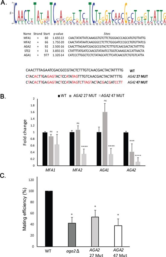
Research article                                                                                                              Cell Biology

                                    Figure 3. Mating pathway mRNAs co-localize and are co-precipitated by HA-Hhf1. (A) Endogenous STE2 and
                                    AGA1 mRNAs co-localize. Wild-type (WT) BY4741 cells were processed for single-molecule fluorescence in
                                    situ hybridization (smFISH) labeling using designated FISH probes complementary to the STE2 and AGA1 mRNAs,
                                    and STE2 and HSP150 mRNAs, prior to labeling with 4,6-diamidino2-phenylindole (DAPI). Dotted lines are
                                    representative of the cell outline observed in brightfield micrographs. AGA1 – Cy3 labeling; STE2 – Alexa488
                                    labeling; HSP150 – Cy5 labeling; merge – merged Cy3/Cy5 and Alexa-488 windows with DAPI staining. Size
                                    bar = 2 mm. The representative image shown is from a single focal plane. (B) Scatter plot of co-localization data
                                    from (A). Co-localization was assessed using the FISHquant algorithm (see ’Materials and methods’). Black lines
                                    indicate the average + SEM for each sample. Each data point represents a single cell. ***** indicates
                                    p-value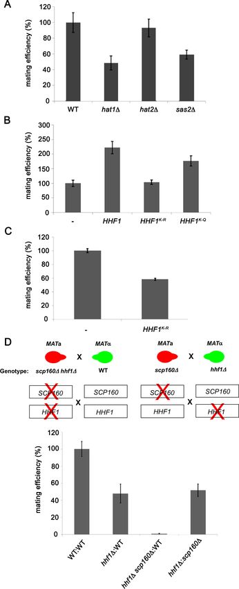
Research article                                                                                                            Cell Biology

                                    Figure 4. Histone H4 function is required for mating mRNP assembly and mating. (A) Histone H4 alone is required
                                    for mating. MATa yeast (BY4741) bearing mutations in H2A (hta1D, hta2D), H2B (htb1D, htb2D), H3 (hht1D, hht2D),
                                    and H4 (hhf1D, hhf2D) were grown to mid-log phase, crossed against MATa wild-type (WT) cells, and mating was
                                    scored using the quantitative mating assay. n = 3 experiments; *p-value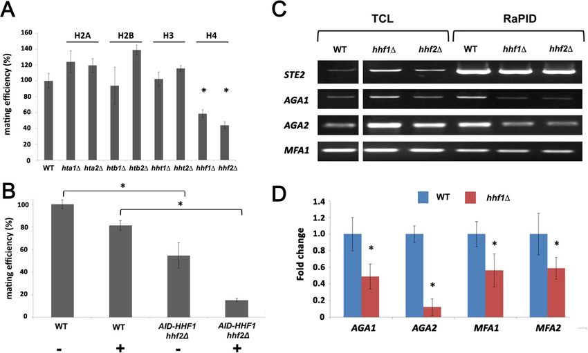
Research article                                                                                                     Cell Biology

                                       We analyzed the co-localization of AGA1 and STE2 mRNAs in WT cells and AID-HHF1 hhf2D cells
                                    either with or without the addition of 3-IAA (Figure 4—figure supplement 1E). While WT cells gave
                                    37.9 ± 1.7% co-localization, this was reduced to 15.9 ± 1.0 and 14.0 ± 0.9% (avg. ± SEM) either with-
                                    out or with 3-IAA addition, respectively. We note that the addition of auxin resulted in a greater
                                    number of AID-HHF1 hhf2D cells that showed no co-localization.

                                    Deletion of histone H4 alleles affects mating transperon assembly
                                    As the depletion of H4 greatly lessens the mating propensity of yeast, we examined whether this is a
                                    consequence of altered mRNP particle assembly. We first performed RaPID-PCR on STE2 mRNA
                                    derived from hhf1D and hhf2D cells, and measured the co-precipitation of other mating pathway
                                    mRNAs. We found decreased levels of AGA1, AGA2, and, to a lesser degree, MFA1 mRNA in the
                                    hhfD deletion strains (Figure 4C). This result was verified using RaPID-qPCR where the levels of
                                    bound AGA1, MFA1, and MFA2 mRNAs declined by ~50%, while those of AGA2 declined even
                                    more (Figure 4D). The results indicate that the reduction in H4 amounts results in defects in mating
                                    mRNP complex formation, which may account for the loss in mating efficiency (Figure 4B).

                                    Histone acetyl transferases and histone H4 acetylation are required for
                                    mating
                                    The N-terminus of histones is extensively modified by methylation, acetylation, and phosphorylation
                                    (Allis and Jenuwein, 2016). We examined the mating efficiency of yeast upon the deletion of three
                                    histone acetyl transferase genes (HATs; e.g., HAT1, HAT2, and SAS2) (Kurdistani and Grunstein,
                                    2003) that were found to be physically and genetically linked with the HHF1 gene and its product,
                                    according to the Saccharomyces Genome Database (SGD). We found that the deletion of HAT1 or
                                    SAS2 had the same reduced efficiency as observed with hhf1D cells (Figure 5A). This implies that
                                    the acetylated state of H4 may be necessary for function.
                                        To verify this, we mutated all N-terminal Hhf1 lysine residues to arginines (e.g., K6R, K9R, K13R,
                                    K17R, K32R; K-to-R mutant) to abolish acetylation by HATs, or to glutamines (e.g., K6Q, K9Q, K13Q,
                                    K17Q, K32Q; K-to-Q mutant), to mimic the acetylated state. We mated MATa cells expressing these
                                    mutants with WT MATa cells and assessed the mating efficiency. Overexpression of WT HHF1
                                    caused a >2-fold increase in mating efficiency (Figure 5B), while overexpression of the K-to-R
                                    mutant abolished it. Correspondingly, overexpression of the K-to-Q mutant yielded the same two-
                                    fold increase in mating efficiency as observed upon WT HHF1 overexpression (Figure 5B). Since the
                                    K-to-R mutation appeared to be non-functional, we expressed this mutant from the genome and
                                    examined mating efficiency. We found that the expression of this sole copy of HHF1 led to a 50%
                                    decrease in mating (Figure 5C), but without lowering HHF1 mRNA expression (e.g., level of HHF1K-
                                    to-R=1.7 ± 0.6-fold over HHF1 in WT cells [avg. ± SEM, n = 3]). While we cannot rule out the possibil-
                                    ity that the K-to-R mutation results in protein instability leading to reduced mating efficiency, never-
                                    theless, it appears that histone H4 acetylation is important for the mating process.

                                    Histone H4 and Scp160 work cooperatively to confer mating
                                    A previous work from the lab identified Scp160 as an ER-localized RBP involved in the delivery of
                                    certain mPOLs (e.g., SRO7) and mRNAs that confer the internal MAP kinase (MAPK) mating signal
                                    (e.g., FUS3, KAR3, STE7) to the shmoo tip upon pheromone treatment (Gelin-Licht et al., 2012).
                                    Deletion of SCP160 was shown to prevent the polarized trafficking of these mRNAs on cER, resulting
                                    in a loss in chemotropism, a 60–70% decrease in mating in heterozygous crosses, and a >98%
                                    decrease in homozygous crosses. As mating transperon RNAs also localize to ER (Figure 2C),
                                    although not asymmetrically localized to the bud or shmoo tips, we examined whether Hhf1 and
                                    Scp160 work together or separately on distinct signaling paths. To do this, we examined whether
                                    deletions of either HHF1 or SCP160 in the separate mating partners or together, as combined muta-
                                    tions in one of the mating partners, had additive/synergistic deleterious effects upon mating. We
                                    crossed MATa hhf1D cells against MATa scp160D cells and observed a block in mating similar to
                                    that of hhf1D x WT crosses (Figure 5D), indicating that there was no additive defect when single
                                    gene mutations are present in the separate mating partners. However, when hhf1D scp160D double
                                    mutants are crossed against WT cells, we observed a complete block in mating (i.e., sterility), remi-
                                    niscent of a scp160D homozygous cross (Gelin-Licht et al., 2012). This indicates that HHF1 and

Nair, Zabezhinsky, et al. eLife 2021;10:e66050. DOI: https://doi.org/10.7554/eLife.66050                                            10 of 32
Research article Cell Biology
Figure 5. Histone H4 acetylation is required for mating. (A) Histone acetyltransferases are required for mating.
Wild-type (WT; BY4741) and yeast lacking known acetyltransferase genes (hat1D, hat2D, and sas2D) were grown to
mid-log phase (O.D.600 = 0.5) and examined for mating with WT MATa cells (BY4742). Three quantitative mating
experiments were performed and gave similar results; p-valuesResearch article Cell Biology
Figure 5 continued
that either mimic or block N-terminal histone H4 acetylation increase or decrease mating efficiency, respectively.
MATa WT (BY4741) cells alone (-) or overexpressing HHF1 (HHF1), along with cells overexpressing HHF1 bearing
K-to-R (HHF1K-R) or K-to-Q (HHF1K-Q) mutations from a 2 um plasmid, were grown to mid-log phase and examined
for mating with MATa cells. Three experiments were performed; p-valuesResearch article Cell Biology
Figure 6. A motif in the mating mRNA coding region facilitates mRNP assembly and mating. (A) A conserved
motif is common to all five MATa mRNAs. MEME-ChIP analysis of the sequences of all five MATa mRNAs was
performed and revealed a 47-nucleotide consensus motif in the coding regions, shown schematically as a
sequence logo based on nucleotide representation. Also shown are two forms (AGA2 27-MUT and AGA2 47-MUT)
in which the motif was altered within the coding region to remove conserved nucleotides without altering the
amino acid sequence. (B) Mutation of the consensus motif in AGA2 leads to an inhibition in messenger
ribonucleoprotein (mRNP) particle assembly. MS2 aptamer-tagged STE2 cells bearing the aga2D deletion were
transformed with a single-copy plasmid expressing AGA2 or either of the two AGA2mut forms. Next, WT, AGA2
27-MUT, and AGA2 47-MUT cells expressing MS2-tagged STE2 were subjected to RaPID-qPCR with primers
against AGA1, AGA2 (note: not within mutated region), MFA1, and MFA2. The histogram indicates the levels of
precipitated mating mRNAs from wild-type (WT) (black) or AGA2 27-MUT and AGA2 47-MUT (dark and light grey,
Figure 6 continued on next page
Nair, Zabezhinsky, et al. eLife 2021;10:e66050. DOI: https://doi.org/10.7554/eLife.66050 13 of 32Research article Cell Biology
Figure 6 continued
respectively) cells after normalization for the level of STE2 mRNA pulldown. Three biological repeats were
performed and an unpaired t-test was performed to compare AGA2 27-MUT and AGA2 47-MUT with WT;
****pResearch article Cell Biology
Figure 7. Intergenic association of STE2 and AGA2 genes. (A) Schematic of the STE2, AGA2, ASH1, GET1, and DDI2 genes and the oligonucleotides
used for their amplification from 3C DNA samples. Coordinates correspond to TaqI sites (shown as vertical black bars); site numbering is relative to
ATG (+1). Forward primers used for 3C analysis were sense-strand identical (arrows) and positioned proximal to TaqI sites as indicated. Primers are
numbered to distinguish the pairs used in the PCR reactions shown in (B). 5’UTRs, ORFs, and 3’UTRs are color-coded, as indicated. Transcription start
sites (TSS) and termination sites (Ter) are indicated. (B) Matrix summarizing the intergenic association of represented genes as determined by 3C-PCR.
Primer pairs corresponding to the different genes listed in (A) were used in PCR reactions. ’+’ indicates PCR amplification and the interaction between
genes. ’–’ indicates no amplification. See Figure 7—figure supplement 1A for examples of both. 5’UTRs, ORFs, and 3’UTRs are indicated by broken
orange lines. (C) Live cell fluorescence microscopy of AGA2 224 tetR and STE2 256 lacO yeast grown to mid-log phase prior to imaging. Left
panel: STE2 256 lacO gene is labeled with GFP-lacI; AGA2 224 tetR is labeled with tetR-tdtomato; merge – merger of STE2 and AGA2 windows;
light – transmitted light. Size bar = 2 mm. Right panel: histogram of the data from three biological replicas (avg. ± std.dev.); co-localized – fully
overlapping signals; adjacent – partially overlapping signals; not co-localized – no overlap between signals. The representative image shown is from a
single focal plane.
The online version of this article includes the following figure supplement(s) for figure 7:
Figure supplement 1. Intergenic association of STE2 and AGA2 genes.
between STE2 and GET1, or AGA2 and DDI2, or between ASH1 and AGA2, for example (see Fig-
ure 7—figure supplement 1A, for example of latter).
To confirm the interaction between the STE2 and AGA2 genes, we created yeast bearing AGA2-
tagged upstream of the 5’UTR with 224 tetracycline repressor repeats (224 tetR) and STE2-tagged
upstream of its 5’UTR with 256 lac operator repeats (256 lacO) and performed live fluorescence
imaging using co-expressed tetR-tdTomato and GFP-tagged lacI, respectively. We observed 9.5%
fully co-localized signals (i.e., 100% overlapping) and 73.6% partially co-localized signals (10–25%
overlapping), where both loci appear adjacent to each other (Figure 7C). In contrast, only 17.3% of
the loci were not closely associated. As a control, we performed live imaging of AGA2 224 TetR
and the ASH1 gene tagged with 256 LacO repeats and the abovementioned fluorescent reporters
(Figure 7—figure supplement 1C and D), and observed far less locus co-localization or adjacent
loci (e.g., 2.3 and 27.2%, respectively), as compared to AGA2 224 TetR and STE2 256 LacO
(Figure 7C). These results suggest that the AGA2 and STE2 loci exist in close association within the
nucleus. Next, to determine if the allele interaction is transcription-mediated, we treated AGA2
224 TetR- and STE2 256 LacO-tagged cells with 1,10 phenanthroline, which inhibits general
transcription. We observed a modest increase (40–50%) in non-co-localized AGA2 and STE2 alleles
Nair, Zabezhinsky, et al. eLife 2021;10:e66050. DOI: https://doi.org/10.7554/eLife.66050 15 of 32Research article Cell Biology
(Figure 7—figure supplement 1E), which suggests that allele apposition may be mediated by tran-
scription. Likewise, the deletion of HHF1 alone reduced the level of allelic coupling in AGA2
224 TetR and STE2 256 LacO cells (Figure 7—figure supplement 1F), which could indicate a
structural role for H4 in leading to RNA multiplexing.
mRNAs encoding heat shock proteins multiplex to form a heat shock
RNP particle
Chowdhary et al., 2019 reported an intergenic association of HSP genes in yeast undergoing heat
shock. Based on this, we predicted that HSP mRNAs might also undergo multiplexing due to HSP
gene allelic coupling. Thus, we tagged HSP104 with the MS2 aptamer and performed RaPID to iden-
tify cohort HSP mRNAs in the pulldowns. Importantly, the mRNAs of HSP genes previously shown to
undergo intergenic interaction (e.g., TMA10, HSP12, HSP82, SSA2, and SSA4) (Chowdhary et al.,
2019) were found to co-precipitate both before, but much more after heat shock (Figure 8A). We
also examined the co-localization of HSP104 and SSA2 mRNAs under both non-heat shock and heat
Figure 8. mRNAs encoding heat shock proteins undergo enhanced multiplexing upon heat shock. MATa yeast strains (BY4741) expressing MS2
aptamer-tagged HSP104, UGO1, or STE5 from their genomic loci or untagged control (WT) cells were grown to mid-log phase (O.D.600 = 0.5) and
subjected to RaPID followed by qRT-PCR (RaPID-qRTPCR; see ’Materials and methods’). RNA derived from the total cell extracts or biotin-eluated
fractions was analyzed by qRT-PCR using primer pairs corresponding to mRNAs expected to multiplex (see listed genes) or not (e.g., GAPDH mRNA).
(A) mRNAs encoding heat shock proteins (HSPs) multiplex upon heat shock. Cells expressing MS2 aptamer-tagged HSP104 were either exposed to
heat shock (10 min; 40˚C; HS) or maintained at 30˚C (NHS) prior to fixation and RaPID-qRT-PCR. Three biological replicates were performed and an
unpaired t-test was used to compare each under non-heat and heat shock conditions with WT; ****pResearch article Cell Biology
shock conditions by smFISH (Figure 8B and C). Under non-heat shock conditions, we found that
30.4 ± 0.9% of HSP104 mRNA co-localized with SSA2 mRNA, whereas this increased to 76.8 ± 0.5%
(avg. ± SEM) upon heat shock (Figure 8B and C; representative images shown in Figure 8B and
quantified in Figure 8C). As a control, we visualized STE2 mRNA in co-localization experiments with
SSA2 mRNA. We found similar low levels of STE2 co-localization with SSA2 mRNA either with or
without heat shock (11.9 ± 0.5% and 11.7 ± 0.4%, respectively) (Figure 8B and C). Thus, like mating
pathway mRNAs, HSP mRNAs also multiplex into specific transperons, especially during heat shock.
We examined the HSP gene sequences for recognizable motifs and identified three potential
motifs (Figure 8—figure supplement 1A) present in nearly all HSP cohort genes (Figure 8—figure
supplement 1B). To determine whether histone H4 is involved in the assembly of the HSP trans-
peron, we performed a growth test for the different histone deletion mutants upon heat shock.
While single gene deletions did not affect doubling time after heat shock (Figure 8—figure supple-
ment 1C), AID-HHF1 hhf2D cells treated with 3-IAA showed a longer doubling time after heat shock
(at all times examined), as compared to WT cells or untreated AID-HHF1 hhf2D cells (Figure 8—fig-
ure supplement 1D). Moreover, we found that HSP mRNA (e.g., TMA10, HSP12, HSP82, SSA2, and
SSA4) multiplexing with HSP104 mRNA was reduced significantly in these cells after heat shock in
the presence of 3-IAA (Figure 8—figure supplement 2A and B).
We next analyzed HSP104 and SSA2 mRNA co-localization in both WT and AID-HHF1 hhf2D cells,
either with or without the addition of 3-IAA (Figure 8—figure supplement 2C). Upon heat shock,
we observed reduced mRNA co-localization in AID-HHF1 hhf2D cells either with (51.5 ± 0.9%;
avg. ± SEM) or without (55.8 ± 0.9%; avg. ± SEM) the addition of 3-IAA, as compared to WT cells
(76.8 ± 0.5%). We note that cells with no co-localized HSP104 and SSA2 mRNA were observed only
in 3-IAA-treated AID-HHF1 hhf2D cells. Since changes in RNA expression might affect co-localiza-
tion, we analyzed HSP104 and SSA2 expression using qRT-PCR. Importantly, we did not observe
changes in expression without the addition of 3-IAA under either heat shock or non-heat shock con-
ditions. An increase in HSP104 and SSA2 gene expression was observed under non-heat shock con-
ditions after the addition of 3-IAA; however, no overall differences were observed between auxin-
treated and -non-treated AID-HHF1 hhf2D cells, as compared to WT cells, under heat shock condi-
tions (Figure 8—figure supplement 2D). Together, these results suggest that histone H4 plays a
role in both HSP mRNA multiplexing and the subsequent cell recovery after heat shock.
mRNAs encoding mitochondrial outer membrane proteins or MAPK
proteins also multiplex
To further identify examples of mRNA multiplexing, we employed existing MS2-tagged genes
encoding proteins that undergo (or are likely to undergo) complex formation in yeast cells. We first
examined whether mRNAs encoding MOMPs multiplex using tagged UGO1 mRNA. Since the basal
expression of UGO1 mRNA was low (i.e., few puncta observed), we induced expression by growing
cells on a non-fermentable carbon source (2% glycerol) before performing RaPID or subsequent fluo-
rescence microscopy experiments. The pulldown of UGO1 mRNA led to co-precipitation of cohort
MOMP mRNAs (e.g., TOM5, TOM6, TOM22; Figure 9A), but not GAPDH mRNA, suggesting that a
specific mRNA multiplex can be detected. Since we initially observed co-precipitation of the mating
mRNAs along with OM45 (Figure 1A and B), which codes for another MOMP, we examined for the
precipitation of STE2 mRNA with UGO1 mRNA. Indeed, STE2 mRNA could co-precipitate with
UGO1 mRNA, suggesting a possible interaction between MOMP and mating pathway mRNA
complexes.
To verify the interaction between MOMP mRNAs, we tagged TOM6 with the PP7 aptamer in cells
expressing MS2-tagged UGO1 and measured mRNA co-localization using smFISH. We found that
46.3 ± 1.7% (avg. ± SEM) of PP7-tagged TOM6 mRNA co-localized with MS2-tagged UGO1 mRNA
(Figure 9B and C). As control, we visualized native HSP104 mRNA in co-localization experiments
with PP7-tagged TOM6 mRNA using smFISH. We found 7.7 ± 1.1% of HSP104 mRNA co-localized
with PP7-tagged TOM6 mRNA (Figure 9B and C). We note that less HSP104 RNA spots were
observed on a non-fermentable carbon source (2% glycerol; 6.8 ± 0.4 spots of HSP104 mRNA;
avg. ± SEM), as compared to experiments performed on a glucose-containing medium (e.g.,
50.1 ± 0.4 spots of HSP104 mRNA; avg. ± SEM) (Figure 8B), indicating that gene expression was
affected. Overall, however, we show that MOMP mRNAs are able to form multiplexes and to co-
localize.
Nair, Zabezhinsky, et al. eLife 2021;10:e66050. DOI: https://doi.org/10.7554/eLife.66050 17 of 32Research article Cell Biology
Figure 9. mRNAs encoding mitochondrial outer membrane and MAPK proteins multiplex to form different
transperons. (A) Mitochondrial outer membrane protein (MOMP) mRNAs multiplex. Cells expressing MS2
aptamer-tagged UGO1 were grown in a medium containing glycerol as a carbon source, prior to fixation and
RaPID-qRT-PCR. Three biological replicates were performed and an unpaired t-test was used to compare the
UGO1 pulldown to the wild-type (WT) control pulldown. ****pResearch article Cell Biology
Figure 9 continued
brightfield micrographs. STE5 – Cy3 labeling; FUS3 – Cy5 labeling; HSP104 – Alexa488 labeling; merge – merged
Cy3/Alexa-488 and Cy5 windows with DAPI staining. Size bar = 2 mm. The representative image shown is from a
single focal plane. (F) Scatter plot of data from (E) showing the proportion of co-localized smFISH foci. RNA
localization was scored using the FISHquant algorithm (see ’Materials and methods’). Each dot represents a single
cell. Black lines indicate avg. ± SEM distribution.
Finally, we examined whether mRNAs encoding non-secreted cytoplasmic components of the
yeast mating pathway (e.g., MAP kinase pathway genes: STE5, FUS3, STE7, and STE11) undergo
multiplexing like mRNAs encoding their secreted counterparts (Figures 1 and 2A, B and
6C, and Figure 6—figure supplement 1B). We tagged STE5 mRNA with the MS2 aptamer and
treated the cells for 1 hr with a-factor (0.5 mM) to induce gene expression. RaPID pulldown of STE5
mRNA after pheromone induction led to a significant co-precipitation of FUS3 mRNA, although only
slight, but significant, increases in STE7 or STE11 mRNA pulldown were detected (Figure 9D). We
verified the association of MS2-tagged FUS3 and PP7-tagged STE5 mRNA in cells using smFISH and
observed 19.6 ± 1.4% (avg. ± SEM) co-localization in the presence of a-factor (Figure 9E and F). As
a control, we visualized HSP104 mRNA co-localization with PP7-tagged FUS3 mRNA, but found only
3.2 ± 0.6% co-localization (Figure 9E and F). Thus, mRNAs encoding components of the phero-
mone-activated MAPK cascade also appear to form specific multiplexes.
Discussion
Prokaryotes coordinate the expression of genes encoding proteins involved in the same biological
process/context by sequential placement in chromosomes in order to generate polycistronic mes-
sages (operons) that can be translationally controlled. Perhaps due to the greater complexity
observed both at the gene and cellular levels, eukaryotes have preferentially relied on the discontig-
uous distribution of genes over different chromosomes. In this case, the co-regulation of gene
expression presumably becomes dependent on shared transcriptional control elements and specific
RBPs that interact selectively with certain transcripts and confer trafficking to sites of co-translation.
However, given that the number of RBPs is limited and that they can interact with a wide number of
potential transcripts, it is unclear if and how defined subsets of mRNAs are packaged together in
order to facilitate co-translation.
We hypothesized that mRNAs encoding proteins involved in the same process might assemble
into macromolecular complexes composed of multiplexed transcripts and specific RBPs to form dis-
crete RNP complexes. By performing mRNA pulldown and RNA-seq experiments, we first identified
a subset of mRNAs that encode secreted components involved in the mating of a-haploid cells. This
mRNP complex included mRNAs encoding the a-pheromone receptor (STE3), a-mating factor
(MFa1, MFa2), a-agglutinin (SAG1), and a-pheromone blocker (AFB1) (Figures 1A, B and 2A, and
Figure 1—figure supplement 1). Correspondingly, we identified a mating mRNP complex from
MATa cells that included mRNAs encoding the a-mating factor receptor (STE2), a-mating phero-
mone (MFA1, MFA2), and a-agglutinins (AGA1, AGA2) (Figure 2B). Thus, we demonstrated that
mRNAs coding for proteins of the same cellular process multiplex into mRNP complexes, presum-
ably to allow for their delivery to the same intracellular site for local translation. Additional experi-
ments in which we identified the existence of other mRNA subsets that multiplex, like those
encoding HSPs, MOMPs, and MAPK pathway proteins (Figures 8 and 9), clearly support this idea.
Thus, mRNA multiplexing in trans appears to form functional transperons (i.e., RNA operons
[Keene, 2007; Keene and Tenenbaum, 2002]) relevant to cell physiology (see below). While we do
not know the full extent of RNA multiplexing, we might predict that analogous transperons (i.e., con-
taining other transcripts) form within cells to modulate other physical processes. For example, recent
works by Ashe and colleagues have shown that mRNAs encoding either translation factors or glyco-
lytic enzymes co-localize and undergo co-translation in yeast (Morales-Polanco et al., 2021;
Pizzinga et al., 2019). In addition, mRNAs encoding proteins in hetero-oligomeric/multisubunit
complexes (proteasome, exocyst, COPI, FAS, ribosome, etc.) might form multiplexes to facilitate
polypeptide complex assembly upon co-translation, as shown at the protein level (Shiber et al.,
2018), even if dedicated assembly chaperones are needed. In our study, we may have missed
Nair, Zabezhinsky, et al. eLife 2021;10:e66050. DOI: https://doi.org/10.7554/eLife.66050 19 of 32Research article Cell Biology
multiplexes in the initial RaPID-seq experiment (Figure 1 and Figure 1—figure supplement 1) for
several reasons. First, RNA multiplexing may require elevated gene expression, as in the case of the
HSP or MOMP complexes (Figures 8 and 9A–C), which require induction conditions to visualize the
RNA granules, as opposed to the normal growth conditions employed initially. Second, the RaPID-
seq pulldowns employed only 0.01% formaldehyde in the crosslinking procedure, as initially pub-
lished (Slobodin and Gerst, 2010), and not the 0.1–0.5% formaldehyde we have employed in
the later work (Zabezhinsky et al., 2016). Third, the precipitated complexes were stringently
washed and, thus, these different factors may have disrupted weaker transperons or missed them
entirely.
Importantly, by using a variety of molecular and single-cell imaging techniques (e.g., Figure 3C;
live fluorescence imaging) we show specific and robust physical interactions between genes located
on different chromosomes that encode mRNAs that form transperons (Figure 7 and Figure 7—fig-
ure supplement 1A and B). This observation of allelic coupling is strengthened by the work of
Chowdhary et al., 2019, where the intergenic association of yeast HSP genes was shown. Other
chromatin conformation capture studies have revealed intergenic associations that lead to the so-
called transcription factories associated with different transcriptional regulators (e.g., promoters,
transcription factors). For example, Schoenfelder et al., 2010 showed interchromosomal interac-
tions between Klf4-regulated globin genes in fetal liver cells that allowed for co-regulated gene tran-
scription. Papantonis et al., 2012 revealed that TNF-a responsive coding and miRNA genes in
human endothelial cells undergo intrachromosomal interactions regulated by NFkB upon cytokine
induction. Thus, specialized transcriptional factories created through allelic coupling may be a com-
mon mechanism for co-regulated gene expression in eukaryotes.
How allelic coupling comes about to form these factories and resulting transperons is not yet
clear. However, we identified a role for histone H4 in the assembly of the mating transperon
(Figures 3C, E and 4C, D, and Figure 4—figure supplement 1E) and its physiological consequences
(i.e., pheromone responsiveness of both mating partners and mating; Figure 4A and B, and Fig-
ure 6—figure supplement 2A). RaPID-MS pulldowns of mating mRNAs encoding secreted proteins
identified the H4 paralog, Hhf1, as binding to the STE2, MFA1, and MFA2 mRNAs, but not to ASH1
mRNA or STE2 mRNA lacking its 3’UTR (Figure 3—figure supplement 1). Thus, specific H4-RNA
interactions are likely to be involved in transperon assembly. Hhf1 pulldowns directly precipitated
the mating transperon (Figure 3C and E) and the deletion of either paralog (HHF1 or HHF2) led to
defects in RNA multiplexing (Figure 4C and D) and mating (Figure 4A and B). Likewise, a combina-
tion of HHF2 deletion and an auxin-induced degradative form of Hhf1 resulted in a complete block
in mating and loss of mRNA co-localization under conditions in which cell viability was not affected
(Figure 4B and Figure 4—figure supplement 1C and E). Similar results were obtained using the
HSP mRNAs, which also form physiologically functional transperons and co-localize in a manner
dependent upon histone H4 (Figure 8, Figure 8—figure supplement 1D, and Figure 8—figure
supplement 2). This suggests that both Hhf1 and Hhf2 are important for the formation of the mRNA
complexes identified here.
Mutations in the amino terminal histone H4 acetylation sites had similar effects as gene deletions,
whereby inactivating K-to-R mutations inhibited mating (Figure 5B and C). In contrast, K-to-Q muta-
tions (or H4 overexpression) improved mating (Figure 5B). Thus, histone H4 paralogs, but no other
histones (Figure 4A and Figure 8—figure supplement 1C), appear to affect the transmission of
physiological signals relevant to RNA multiplexing. More work is required to demonstrate the role
histone H4 plays in transperon formation and whether H4 is present in these multiplexes post-assem-
bly and export from the nucleus. Although we found that fluorescent protein-tagged Hhf1 labeled
the nucleus, we were unable to observe H4 in the cytoplasm or show co-localization there with STE2
mRNA (data not shown). Thus, we do not know whether this non-canonical interaction of H4 with
mRNA is exclusive to the nucleus or not. Canonical histone functions relate mainly to DNA-protein
interactions that facilitate nucleosome formation. However, the requirement of acetylation for H4
function in mating (Figure 5A–C) suggests that histone H4 might participate in the formation of a
DNA-RNA intermediate upon gene transcription. This mechanism potentially allows for RNA multi-
plexing, provided that the loci encoding mRNAs intended for assembly in trans also are in close
apposition, as evidenced here (Figure 7B and C, and Figure 7—figure supplement 1A,B and F).
Results showing that a transcription inhibitor affects allelic coupling imply that gene apposition and
transcription may be connected (Figure 7—figure supplement 1E). Moreover, the fact that H4
Nair, Zabezhinsky, et al. eLife 2021;10:e66050. DOI: https://doi.org/10.7554/eLife.66050 20 of 32Research article Cell Biology
deletion also affects allelic coupling may implicate an involvement of chromatin architecture in trans-
peron formation (Figure 7—figure supplement 1F).
Our lab is interested in how mRNA localization affects cell physiology, and a previous work has
demonstrated that yeast mSMPs (including MFA2) localize to ER in the cell body (Kraut-
Cohen et al., 2013). Here, we verified that endogenously expressed STE2 and AGA1 mRNAs also
localize to ER in the cell body, both before and after treatment with pheromone (Figure 2C). More-
over, these mRNAs co-localize both prior to and post nuclear export (Figure 2D–F). This finding
demonstrates once again that mSMPs are not asymmetrically localized to the polarized extensions
of yeast (e.g., bud and shmoo tips), unlike ASH1 and polarity-establishing mRNAs (e.g., SRO7,
SEC4, CDC42) in budding cells, which is mediated by She2 (Aronov et al., 2007), or SRO7 and
FUS3 mRNAs in shmooing cells, which is mediated by Scp160 (Gelin-Licht et al., 2012). It also dem-
onstrates that RNA multiplexing and transperon formation occur within the nucleus (Figures 2D–F
and 8B). Since this work and previous studies have revealed that mRNAs encoding secreted mating
pathway components, MOMPs, and pheromone-activated MAPK pathway components localize to
the ER, mitochondria, and ER of the shmoo tip, respectively (Gadir et al., 2011; Gelin-Licht et al.,
2012; Zabezhinsky et al., 2016), we predict that these mRNAs are targeted as intact transperons.
Moreover, as Scp160 binds MAPK pathway mRNAs (e.g., FUS3, STE7, KAR3) and delivers them to
the shmoo tip upon pheromone treatment (Gelin-Licht et al., 2012), we predict that Scp160 might
traffick this MAPK transperon.
An earlier work has (Aronov et al., 2015) suggested that mRNAs encoding mating components,
like MFA2, localize preferentially to P-bodies in the shmoos and shmoo tips of pheromone-treated
yeast, and that P-bodies are necessary for transmission of the mating signal and subsequent mating.
However, these results were obtained under conditions of mRNA overexpression. In contrast, we
found that neither endogenously expressed MS2-tagged MFA2 (Haimovich et al., 2016) nor STE2
mRNA (Figure 6—figure supplement 2C) localize to P-bodies, nor do P-bodies form under normal
growth conditions conducive to the expression of these mRNAs and to mating (Figure 6—figure
supplement 2C; top panel). In addition, we show here that MFA2 and other mating mRNAs (e.g.
MFA1, STE2, and AGA1) do not localize to the shmoos or shmoo tips, but rather to ER present in
the cell body (Figure 2C and Figure 2—figure supplement 1). Moreover, the deletion of genes nec-
essary for P-body formation (e.g., PAT1, DHH1) had no significant effect upon mating (Figure 6—
figure supplement 2B). As mating pathway mRNAs localize to P-bodies only upon stress conditions
(e.g., gene overexpression, glucose starvation) (Haimovich et al., 2016 and Figure 6—figure sup-
plement 2C), it would appear that P-bodies are not sites for mating mRNA localization or transmis-
sion of the mating signal under normal growth conditions. Our findings re-emphasize the
importance of (i) measuring mRNA localization under native conditions of gene expression; (ii) con-
sidering the possibility that RNA overexpression and/or the insertion of RNA aptamers may induce
artifacts (e.g., mRNA mislocalization, P-body formation, or both); and (iii) examining mRNA integrity/
localization using additional techniques (e.g., Northern analysis, RNA-seq, smFISH) (Garcia and
Parker, 2015; Haimovich et al., 2016; Heinrich et al., 2017) for secondary validation.
As cis-acting RNA elements may work in conjunction with specific RNA-binding proteins to confer
multiplexing, we analyzed the coding regions and 3’UTRs of the mating mRNAs from the two yeast
haplotypes. Sequence analysis revealed the presence of conserved motifs present in the mating
mRNAs from either MATa or MATa cells (Figure 6A). We mutated the MATa sequence element in
the AGA2 gene and found that it affected transperon assembly similar to the deletion of AGA2
(aga2D) (Figure 6B). Importantly, this correlated with an inhibition in cellular responsiveness (i.e.,
shmooing) toward its mating partner, as well as the responsiveness of its mating partner (Figure 6—
figure supplement 2A), resulting in significant defects in mating (Figure 6C). Thus, defects in trans-
peron formation result in significant changes in cell physiology, and we predict that a yet unidenti-
fied RBP interacts with this element to facilitate RNA assembly and, possibly, trafficking. The MATa
element is unlikely to be the H4-interacting motif itself, since it was not identified among the mating
mRNAs from MATa cells, which have other shared motifs (Figure 6—figure supplement 1C). Like-
wise, removal of the 3’UTR from STE2 appeared to reduce H4 binding (Figure 3—figure supple-
ment 1), possibly indicating its role in histone association. Additional RaPID-MS experiments should
help reveal the identity of this protein, as well as those interacting with the motifs in mating mRNAs
from MATa cells (Figure 6—figure supplement 1C), and genetic analyses should reveal their contri-
bution to the mating process.
Nair, Zabezhinsky, et al. eLife 2021;10:e66050. DOI: https://doi.org/10.7554/eLife.66050 21 of 32You can also read