The Initiation of Eukaryotic DNA Replication - UConn MCB
←
→
Page content transcription
If your browser does not render page correctly, please read the page content below
Annual Review of Biochemistry
                                                                                                                                     The Initiation of Eukaryotic
                                                                                                                                     DNA Replication
                                                                                                                                     Alessandro Costa1 and John F.X. Diffley2
Annu. Rev. Biochem. 2022.91:107-131. Downloaded from www.annualreviews.org
Access provided by University of Connecticut on 09/08/22. For personal use only.

                                                                                                                                     1
                                                                                                                                       Macromolecular Machines Laboratory, The Francis Crick Institute, London, UK;
                                                                                                                                     email: alessandro.costa@crick.ac.uk
                                                                                                                                     2
                                                                                                                                       Chromosome Replication Laboratory, The Francis Crick Institute, London, UK;
                                                                                                                                     email: john.diffley@crick.ac.uk

                                                                                   Annu. Rev. Biochem. 2022. 91:107–31               Keywords
                                                                                   First published as a Review in Advance on
                                                                                                                                     DNA replication, cell cycle, cryo–electron microscopy
                                                                                   March 23, 2022

                                                                                   The Annual Review of Biochemistry is online at    Abstract
                                                                                   biochem.annualreviews.org
                                                                                                                                     DNA replication in eukaryotic cells initiates from large numbers of sites
                                                                                   https://doi.org/10.1146/annurev-biochem-072321-
                                                                                                                                     called replication origins. Initiation of replication from these origins must
                                                                                   110228
                                                                                                                                     be tightly controlled to ensure the entire genome is precisely duplicated in
                                                                                   Copyright © 2022 by Annual Reviews.
                                                                                                                                     each cell cycle. This is accomplished through the regulation of the first two
                                                                                   All rights reserved
                                                                                                                                     steps in replication: loading and activation of the replicative DNA helicase.
                                                                                                                                     Here we describe what is known about the mechanism and regulation of
                                                                                                                                     these two reactions from a genetic, biochemical, and structural perspective,
                                                                                                                                     focusing on recent progress using proteins from budding yeast.

                                                                                                                                                                                                      107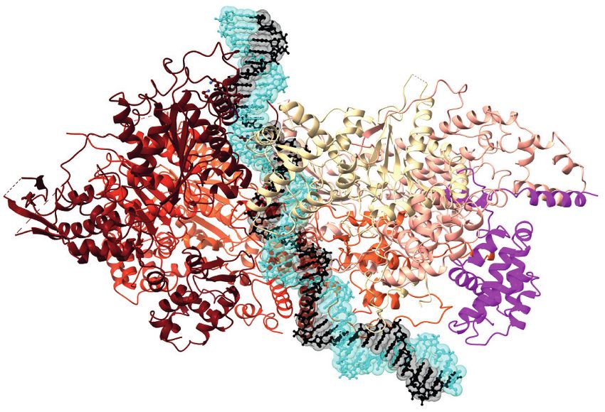
Contents
                                                                                         1. INTRODUCTION . . . . . . . . . . . . . . . . . . . . . . . . . . . . . . . . . . . . . . . . . . . . . . . . . . . . . . . . . . .      108
                                                                                         2. REPLICATION ORIGINS AND THE INITIATOR . . . . . . . . . . . . . . . . . . . . . . . . . . .                                             110
                                                                                            2.1. Eukaryotic DNA Replication Origins . . . . . . . . . . . . . . . . . . . . . . . . . . . . . . . . . . . . . .                     110
                                                                                            2.2. Mechanism of Origin Recognition by the Origin Recognition Complex . . . . . .                                                      112
                                                                                         3. HELICASE LOADING . . . . . . . . . . . . . . . . . . . . . . . . . . . . . . . . . . . . . . . . . . . . . . . . . . . . . . .          113
                                                                                            3.1. Structural Mechanism of First MCM Ring Loading . . . . . . . . . . . . . . . . . . . . . . . . .                                   113
                                                                                            3.2. Structural Mechanism of Second MCM Hexamer Loading . . . . . . . . . . . . . . . . . .                                             115
                                                                                            3.3. Regulation of Helicase Loading . . . . . . . . . . . . . . . . . . . . . . . . . . . . . . . . . . . . . . . . . . . .             116
                                                                                         4. HELICASE ACTIVATION . . . . . . . . . . . . . . . . . . . . . . . . . . . . . . . . . . . . . . . . . . . . . . . . . . . .             117
                                                                                            4.1. Overview of Helicase Activation . . . . . . . . . . . . . . . . . . . . . . . . . . . . . . . . . . . . . . . . . . .              117
                                                                                            4.2. Structural Mechanism of Helicase Activation and Replisome Assembly . . . . . . .                                                   119
Annu. Rev. Biochem. 2022.91:107-131. Downloaded from www.annualreviews.org
Access provided by University of Connecticut on 09/08/22. For personal use only.

                                                                                         5. CONCLUSIONS . . . . . . . . . . . . . . . . . . . . . . . . . . . . . . . . . . . . . . . . . . . . . . . . . . . . . . . . . . . . .   123

                                                                                         1. INTRODUCTION
                                                                                         The emergence of complex, multicellular life on Earth was enabled by the evolution of molecular
                                                                                         machines and regulatory pathways that bring about the accurate and rapid duplication of large
                                                                                         genomes. In bacteria, like Escherichia coli, the entire genome is contained on a single circular chro-
                                                                                         mosome and is replicated by two DNA replication machines (replisomes) traveling in opposite
                                                                                         directions from a single initiation site (oriC) known as a replication origin (1). As a consequence,
                                                                                         the time required to replicate the entire genome (the synthetic or S phase) in bacteria is propor-
                                                                                         tional to the genome size: Addition of new genes or genetic pathways would come at the cost of
                                                                                         a longer S phase. Eukaryotic cells initiate replication from many replication origins distributed
                                                                                         across the genome. Because of this replication strategy, the length of S phase in eukaryotes is
                                                                                         dependent upon the frequency of origins in the genome and is independent of overall genome
                                                                                         size. Disconnecting genome size from S phase length was crucial for supporting the evolution of
                                                                                         the large genomes—up to 150 billion base pairs (bp)—seen in modern eukaryotes. For such a
                                                                                         strategy to be effective, however, the firing of individual origins must be tightly regulated and
                                                                                         coordinated so that the genome is copied exactly once in each cell cycle—nothing can remain
                                                                                         unreplicated, and nothing can be replicated more than once.
                                                                                             Eukaryotes arose from an organism related to extant archaeal prokaryotes, most likely from
                                                                                         an organism closely related to current Asgardarchaeota (2). Orthologs of most of the key proteins
                                                                                         that are involved in eukaryotic DNA replication initiation can be found in archaeal genomes (3).
                                                                                         Moreover, one can see the beginnings of coordinated, multi-origin replication in Archaea. For ex-
                                                                                         ample, archaeal species in the Sulfolobus genus contain three replication origins on a single circular
                                                                                         chromosome. Each origin in Sulfolobus is flanked by a gene encoding its own dedicated initiator—a
                                                                                         protein that binds specifically to that origin and promotes initiation. How Archaea regulate these
                                                                                         origins to ensure they fire once per cell cycle is currently largely unknown.
                                                                                             At the heart of all replisomes is a DNA helicase, and the loading of the helicase by initiator
                                                                                         proteins is a key step in initiating replication. Replicative DNA helicases are hexameric rings that
                                                                                         encircle single-stranded DNA (ssDNA) at replication forks, using ATP hydrolysis to translocate,
                                                                                         displacing the complementary, nontranslocation strand as they proceed (4). Consequently, to es-
                                                                                         tablish bidirectional replication forks at origins in double-stranded DNA, the two DNA strands in
                                                                                         the origin must first be separated in order for two DNA helicases to be loaded. In E. coli, the DnaA

                                                                                   108   Costa   •   Diffley
1                                     2

                                                                                                      Helicase loading                    Helicase activation

                                                                                                                              CDK               DDK               Rad53

                                                                                   Figure 1
                                                                                   ●1 Loading of the eukaryotic replicative helicase onto DNA replication origins leads to the formation of a

                                                                                   head-to-head DH of MCM that encircles duplex DNA. ●      2 Activation involves the assembly of two CMG

                                                                                   holo-helicases that unwind the DNA, providing the single-stranded DNA template for the replicative
                                                                                   polymerases. Upon cell-cycle transition into S phase, the activities of CDK and DDK promote CMG
                                                                                   formation. At the same time, CDK blocks further DH loading to prevent rereplication. When DNA damage
                                                                                   is detected during S phase, the Rad53 checkpoint kinase prevents late-origin firing. Abbreviations: CDK,
Annu. Rev. Biochem. 2022.91:107-131. Downloaded from www.annualreviews.org
Access provided by University of Connecticut on 09/08/22. For personal use only.

                                                                                   cyclin dependent kinase; CMG, Cdc45–MCM–GINS; DDK, Dbf4 dependent kinase; DH, double hexamer;
                                                                                   GINS, go-ichi-nii-san; MCM, minichromosome maintenance; S phase, synthetic phase.

                                                                                   initiator accomplishes this by binding to oriC and generating ssDNA in an adjacent DNA unwind-
                                                                                   ing element; two DnaB helicase hexamers are then recruited via interactions with DnaA, with one
                                                                                   hexamer encircling each of the two single strands of DNA. In eukaryotes, the origin recognition
                                                                                   complex (ORC) initiator loads the minichromosome maintenance (MCM) helicase—comprising
                                                                                   six related subunits, Mcm2–7—around duplex DNA as an inactive head-to-head double hexamer
                                                                                   (DH) in a process we refer to as helicase loading (Figure 1). The DH then undergoes a series
                                                                                   of structural changes leading to helicase activation. In this step, MCM generates untwisted DNA
                                                                                   within its central channel and then is reconfigured so that each MCM ring encircles one strand
                                                                                   of DNA. During helicase activation, two additional proteins—Cdc45 (cell division cycle 45) and
                                                                                   the GINS heterotetramer (go-ichi-nii-san, which means 5-1-2-3 in Japanese), consisting of Sld5
                                                                                   (synthetic lethal with Dpb11), Psf1 (partner of Sld5 1), Psf2, and Psf3—become stably associated
                                                                                   with each MCM ring to form the active replicative helicase called CMG (Cdc45–MCM–GINS).
                                                                                       The control of the helicase loading and activation steps by global cell cycle regulators ensures
                                                                                   that genome duplication is appropriately coupled to cell growth, environmental cues, chromo-
                                                                                   some segregation, and cell division and also ensures that no origin fires more than once in any
                                                                                   cell division cycle. The eukaryotic cell cycle is controlled by cyclin dependent kinase (CDK) and
                                                                                   its opposing ubiquitin ligase, the anaphase promoting complex or cyclosome (APC/C). Bistable
                                                                                   switches, driven by system-level feedback mechanisms, divide the cell cycle into two states: One
                                                                                   state, which is characterized by low CDK and high APC/C activities, extends from the end of
                                                                                   mitosis through to the end of G1 phase; the alternate state, which is characterized by high CDK
                                                                                   and low APC/C activities, extends from S phase through mitosis (5). Helicase loading can oc-
                                                                                   cur only during the low CDK period, while helicase activation can occur only during the high
                                                                                   CDK period. Large numbers of origins can have helicase loaded before entry into S phase; dur-
                                                                                   ing S phase, as loaded helicases become activated, new helicases cannot be loaded, thus ensur-
                                                                                   ing origins cannot fire more than once in a cell cycle. As a consequence, there is no urgency for
                                                                                   all origins to fire immediately, and indeed, most cells have a temporal program of origin firing,
                                                                                   with origins being activated throughout S phase in a predictable pattern (6).
                                                                                       In addition to being a fascinating and fundamental biological process, DNA replication also
                                                                                   has significance for human health: Defects in replication initiation and its regulation are responsi-
                                                                                   ble for a series of congenital human diseases (7); moreover, oncogene expression can misregulate
                                                                                   origin firing, contributing to genomic instability in cancer (8). Here, we review progress in un-
                                                                                   derstanding the mechanisms and regulation of helicase loading and activation in eukaryotes. We

                                                                                                                                www.annualreviews.org   •   The Initiation of Eukaryotic DNA Replication   109
focus on progress in understanding this in the budding yeast Saccharomyces cerevisiae, which has
                                                                                         been enabled by incisive genetics and driven more recently by advances in single-particle cryo–
                                                                                         electron microscopy (cryo-EM) and the reconstitution of helicase loading and activation with
                                                                                         purified proteins (9–11).

                                                                                         2. REPLICATION ORIGINS AND THE INITIATOR
                                                                                         2.1. Eukaryotic DNA Replication Origins
                                                                                         The first eukaryotic DNA replication origins isolated and characterized were from the S. cere-
                                                                                         visiae genome. These autonomously replicating sequences (ARSs) are both the genetic elements
                                                                                         required to initiate replication and the biochemical sites where replication begins (12–18). All
                                                                                         ARSs contain a T-rich, nonpalindromic ARS consensus sequence (ACS) in an A domain (19),
                                                                                         which is essential but not sufficient for origin function (20–22). In a biochemical tour de force,
Annu. Rev. Biochem. 2022.91:107-131. Downloaded from www.annualreviews.org

                                                                                         Bell and Stillman (23) identified and purified ORC as a factor that specifically recognizes the ACS.
Access provided by University of Connecticut on 09/08/22. For personal use only.

                                                                                         ORC is a six-subunit complex that requires ATP binding but not ATP hydrolysis to recognize and
                                                                                         stably bind the ACS. ORC protects the ACS from cleavage by DNase1 and generates DNase1 hy-
                                                                                         persensitive sites in adjacent sequences. This diagnostic cleavage signature was used to show ORC
                                                                                         is bound at replication origins in vivo (24). The genes encoding all six ORC subunits are essen-
                                                                                         tial for viability in budding yeast, and hypomorphic orc mutants exhibit defects in initiating DNA
                                                                                         replication (25–31). Homologs of the yeast ORC subunits are found in all of the main eukaryotic
                                                                                         lineages (32), and protein complexes containing human, Xenopus, and Drosophila homologs of ORC
                                                                                         subunits have been characterized (33–36). ORC is required for replication in Xenopus egg extracts
                                                                                         (36), and ORC subunits are essential for replication in human cells (37), though some cancer cell
                                                                                         lines appear to be able to proliferate without Orc1 and Orc2 (38). Taken together, these results
                                                                                         show that ORC is the replication initiator protein in budding yeast, a role likely retained across
                                                                                         Eukarya.
                                                                                             In addition to the ACS, a flanking sequence called the B domain is required for origin function.
                                                                                         B domains vary somewhat in length, but in general, yeast origins are contained within 100–200-
                                                                                         bp nucleosome-free regions (39). An early analysis of the ARS1 origin identified three sequence
                                                                                         elements, B1, B2, and B3 (Figure 2a), within the B domain that are important, but not essential,
                                                                                         for origin function (40). The B3 element is the binding site for the general transcription factor
                                                                                         Abf1, which functions to position a nucleosome adjacent to the origin (41). The B1 element, which
                                                                                         lies next to the ACS, contributes to ORC binding affinity (42, 43) and can thus be considered part
                                                                                         of the extended ORC-binding site.
                                                                                             Understanding the function of the B2 element has been important in elucidating the mech-
                                                                                         anism of helicase loading. The B2 element of ARS1 contains a near match to the ACS in the
                                                                                         opposite orientation to the A-domain ACS. ORC can bind to the B2 element, but this binding
                                                                                         is greatly enhanced when the A element is mutated, indicating that two ORC molecules cannot
                                                                                         bind simultaneously to ARS1 (23). Unbiased approaches have shown that the optimal B2 sequence
                                                                                         corresponds to the ACS (44), suggesting that the function of B2 is to bind ORC. DNase1 foot-
                                                                                         printing with purified ORC revealed additional protections and hypersensitive sites beyond the
                                                                                         B2 element that are not accounted for by the A/B1 or B2 elements, indicating that there are likely
                                                                                         to be additional, weaker ORC-binding sites in the B domain (23, 42). Indeed, the occurrence of
                                                                                         multiple near matches to the ACS downstream and in the opposite orientation of the main ACS
                                                                                         was a feature of origins noted as early as 1988 (45). More recently, two ORC-binding sites (A/B1)
                                                                                         separated by random DNA sequences were shown to be functional as replication origins but only
                                                                                         when placed in opposing orientations; functional ARSs were generated with sites anywhere be-
                                                                                         tween 25 and 400 bp apart (22), indicating that the distance between sites is less important than

                                                                                   110   Costa   •   Diffley
a                ORC                                                     Abf1

                                                                                                           ACS          B1                 B2                            B3

                                                                                                                                         ORC

                                                                                                                                                                         180°      20°
                                                                                                                                                                                           K371
                                                                                                                                                                                           R369
                                                                                                b                                                           Orc1
Annu. Rev. Biochem. 2022.91:107-131. Downloaded from www.annualreviews.org

                                                                                                                                                       basic patch
Access provided by University of Connecticut on 09/08/22. For personal use only.

                                                                                                                                                                                       Y372
                                                                                                                                                                                 R367

                                                                                                                                                                                   K362
                                                                                                                                                            Orc2                F360

                                                                                                                                                              W396

                                                                                                                                                                  F485          Y486

                                                                                                                                                                     Orc4
                                                                                                Orc1    Orc4     Orc2   Orc5     Orc3    Orc6

                                                                                   Figure 2
                                                                                   Linear structure of the ARS1 origin of replication and its recognition mechanism. (a) The ACS and B1
                                                                                   elements constitute one binding site for ORC. The B2 element is an inverted ORC-binding site. The B3
                                                                                   element is recognized by the general transcription factor, Abf1. (b) ORC wraps around and bends duplex
                                                                                   DNA at the ACS and B1 sites. Base recognition is performed by residues clustered in a basic patch N
                                                                                   terminal of the AAA+ domain in Orc1, the ISM element of Orc3, and an insertion in the WHD beta hairpin
                                                                                   of Orc4. Abbreviations: AAA+, ATPase associated with various cellular activities; ACS, ARS consensus
                                                                                   sequence; ARS, autonomously replicating sequence; ISM, initiator-specific motif; ORC, origin recognition
                                                                                   complex; WHD, winged helix domain.

                                                                                   the orientation of the sites. Taken together, these results suggest that one function, perhaps the
                                                                                   primary function, of B domains is to provide sites for ORC binding in the opposite orientation to
                                                                                   the A/B1 ORC-binding site; the B2 element at ARS1 is an example of such a site.
                                                                                      B domains have additional features that likely contribute to origin function. Because of the
                                                                                   asymmetric distribution of adenine and thymine residues in the Watson and Crick strands, they
                                                                                   have an inherent tendency to exclude nucleosome occupancy (39). This base composition feature
                                                                                   also means that B domains have lower helical stability; that is, they are inherently easier to unwind
                                                                                   (46).
                                                                                      At approximately the same time that the first yeast replication origins were being identified,
                                                                                   Harland and Laskey (47) showed that a variety of DNA molecules from bacteria, bacteriophages,
                                                                                   and animal viruses could replicate efficiently when microinjected into Xenopus laevis eggs, indi-
                                                                                   cating that a requirement for sequence-defined replication origins is not a universal feature of

                                                                                                                               www.annualreviews.org    •   The Initiation of Eukaryotic DNA Replication   111
eukaryotic replication. They further showed that these microinjected DNAs replicated just once
                                                                                         in a cell cycle (47), demonstrating that specific DNA sequences were also not required for restrict-
                                                                                         ing replication to once per cell cycle. As described in Section 3, the system ensuring once per cell
                                                                                         cycle replication in eukaryotes, including yeast and metazoans, does not operate at the level of
                                                                                         individual origin sequences.
                                                                                             Nonetheless, replication in many metazoan cells begins at defined, specific sites. The method-
                                                                                         ology for identifying origins and the current view of what defines a metazoan replication origin
                                                                                         have been reviewed extensively (48, 49). The only DNA element found thus far at many origins is
                                                                                         a G-rich element that does not have a clear consensus sequence but rather shares a propensity for
                                                                                         forming a noncanonical four-stranded helical structure called the G quadruplex (G4). In contrast
                                                                                         to the budding yeast ORC, human ORC appears to bind DNA and promote initiation in a largely
                                                                                         sequence-independent manner (50, 51); however, consistent with the origin mapping data, human
                                                                                         ORC has been shown to exhibit preferential binding to G4 ssDNA (52). Metazoan replication
Annu. Rev. Biochem. 2022.91:107-131. Downloaded from www.annualreviews.org

                                                                                         origins tend to have decreased nucleosome densities, and a number of local histone modifications
Access provided by University of Connecticut on 09/08/22. For personal use only.

                                                                                         have been found to correlate with origin positions and/or ORC binding. Of these, H4K20me2
                                                                                         interacts directly with the bromo-adjacent homology (BAH) domain of Orc1 (53). ORC may also
                                                                                         be recruited to specifically modified histones via interactions with the HBO1 acetyltransferase
                                                                                         (54) and the LRWD1/ORCA protein (55, 56).

                                                                                         2.2. Mechanism of Origin Recognition by the Origin Recognition Complex
                                                                                         ORC is a hetero-hexameric assembly composed of five homologous proteins (Orc1–5), which
                                                                                         form a crescent moon shape, and an Orc6 subunit bound in the periphery. Orc1–5 contain an
                                                                                         N-terminal AAA+ (ATPase associated with various cellular activities) module and a C-terminal
                                                                                         DNA-binding domain. Several ATPase sites of ORC have become inactivated during evolution,
                                                                                         with only Orc1, Orc4, and Orc5 retaining the ability to bind ATP, while hydrolysis occurs only at
                                                                                         one bipartite catalytic center (formed between the arginine finger in Orc4 and the ATP-binding
                                                                                         site in Orc1, Orc4>Orc1) (57). Orc6 is structurally related to the transcription factor TFIIB,
                                                                                         which is composed of a tandem repeat of cyclin-box folds (58, 59) with the C-terminal end docked
                                                                                         onto Orc3 (60). Crystallographic and cryo-EM studies on yeast, Drosophila, and human complexes
                                                                                         showed that DNA binds the central cavity of the Orc1–5 crescent (61–64). Structures of the meta-
                                                                                         zoan complexes show that DNA binding can be impaired by ATPase autoinhibition. In the autoin-
                                                                                         hibited state, the AAA+ module of Orc1 is positioned away from the Orc4 subunit, which disrupts
                                                                                         the Orc4>Orc1 active site and at the same time occludes the DNA-binding cavity of ORC (60,
                                                                                         63). Autoinhibition is released upon ATP binding and the interaction with additional initiators
                                                                                         (61).
                                                                                             The structure of S. cerevisiae ORC bound to ACS DNA reveals how specific origin sequences
                                                                                         are recognized at the origin of replication (62). DNA contacts are mediated by distinct ORC
                                                                                         elements, whose structure was previously described for archaeal Orc1 orthologs in complex with
                                                                                         origin DNA (65, 66). These elements include an insertion in the AAA+ domain known as the
                                                                                         initiator-specific motif (ISM) (with Orc1, 4, 5, and 3 in the eukaryotic complex forming a spiral
                                                                                         around DNA), as well as the winged helix domain (WHD). Additionally, in the S. cerevisiae ORC–
                                                                                         DNA structure, a helical insertion in the Orc4 WHD beta hairpin engages in the recognition of
                                                                                         thymine and adenine bases in the ACS (Figure 2b), and its mutation alters origin discrimination
                                                                                         and firing patterns in cells (67). Other base-recognition elements are seen only in the S. cerevisiae
                                                                                         ORC structure and engage a thymine-rich stretch in the ACS minor groove, all through hydrogen
                                                                                         bonding with thymine O2. One is a tryptophan in the Orc2 ISM, and the second is a patch of
                                                                                         basic residues that map onto a loop between an N-terminal BAH appendix and the AAA+ domain

                                                                                   112   Costa   •   Diffley
of Orc1 (62). The Orc2 ISM tryptophan and the Orc4 WHD helical insertion exist only in a
subgroup of Saccharomyces-related budding yeasts and evolved together with origin sequence
specificity (67). Conversely, the Orc1 basic patch is universally conserved in eukaryotes.
Nucleosome interaction may also have a role in recruiting ORC onto origins. The crystal
structure of the N-terminal domain of Orc1 bound to a nucleosome core particle explains how
this engagement occurs, showing a BAH domain that binds H2A and H2B via a canonical acidic
patch interaction (68). As described in Section 2.1, nucleosome recognition in higher eukaryotes
may involve Orc1 BAH engagement of the H4K20me2 mark (53).
Duplex DNA threaded through the ORC central channel becomes bent and is bound by Orc6
(Figure 2b) as it emerges on the C-terminal side of the crescent (61, 62, 64). During the helicase-
recruitment process, the Cdc6 protein becomes transiently associated with ORC through a mech-
anism controlled by ATP binding of Cdc6. To create the space required for Cdc6 recruitment,
the WHD of Orc2 disengages from DNA, which in turns leads to the reconstitution of a new
Annu. Rev. Biochem. 2022.91:107-131. Downloaded from www.annualreviews.org
Orc1>Cdc6 active site and heptameric complex formation (61, 64). Cdc6 closes the ORC ring,
Access provided by University of Connecticut on 09/08/22. For personal use only.
topologically entrapping duplex DNA, while also maintaining a bent DNA conformation, as in
the isolated ORC.
3. HELICASE LOADING
To explain how replication from nonspecific start sites could nonetheless be precisely regulated
to once per cell cycle in metazoan cells, Harland and Laskey (47, 69) proposed that chromatin
is licensed prior to S phase with a factor that binds frequently and nonspecifically to chromatin
and is required for replication; this license is removed during DNA replication, so the replicated
chromatin is unlicensed and therefore unable to rereplicate. Using Xenopus egg extracts, Blow and
Laskey (70) showed that permeabilization of nuclei after replication, for example, by detergent
treatment, allowed chromatin to be relicensed in egg extracts for another round of replication.
This led them to propose that the licensing factor cannot cross the nuclear envelope and can
access chromatin only when the nuclear envelope breaks down in mitosis. Exploiting the egg ex-
tract replication system, MCM and ORC were identified as essential components of the licensing
reaction (36, 71–73).
Genomic footprinting and chromatin immunoprecipitation experiments using synchronized
budding yeast cells showed that ORC remains bound to origins throughout the cell cycle (74–
76). Genomic footprinting identified an extended protection across the B domain (74). This pre-
replicative complex (pre-RC) is present at origins from the end of mitosis until S phase (74) and
requires the Cdc6 protein (77). Subsequently, in both Xenopus and budding yeast, Cdc6 was found
to be required for MCM loading onto chromatin (78, 79), suggesting that licensing in Xenopus
and pre-RC assembly in yeast are likely to be the same thing. ORC and Cdc6 can be removed
from chromatin by salt extraction without removing MCM (79), indicating that these proteins
load MCM onto chromatin but, once MCM is loaded, are no longer required to retain MCM
on chromatin. The reconstitution of this reaction with purified proteins (9, 10) has provided an
approach to understand this loading reaction in detail, discussed in the following three sections.
3.1. Structural Mechanism of First MCM Ring Loading
The six distinct subunits in the MCM ring form two tiers, an N-terminal oligomerization do-
main and a AAA+ ATPase tier, embellished by C-terminal WHD extensions (80). In the loading-
competent state, MCM exists as an open ring (81, 82) containing a discontinuity between Mcm2
and Mcm5, which is used as an entry gate for DNA threading (83). The structure and mechanism
of the loading factor Cdt1 appear different in yeast and metazoans. In yeast, Cdt1 is stably bound
www.annualreviews.org • The Initiation of Eukaryotic DNA Replication 113to MCM (10) and contains an inactive dioxygenase fold important for loading (81, 82). In higher
eukaryotes, Cdt1 does not form a stable complex with MCM and, in place of the dioxygenase
fold, it contains an N-terminal intrinsically disordered domain with a role in liquid–liquid phase
separation (61, 84).
The middle (M) and C-terminal (C) WHD modules of Cdt1 are universally conserved and
their essential role in helicase loading can be explained by the cryo-EM structure of yeast MCM–
Cdt1. They bind to the side of MCM (wrapping around Mcm2, 6, and 4) and contribute to keeping
the Mcm2–5 gate open, which is required for DNA access to the helicase central channel (83). Bio-
chemical data indicate the Mcm3 WHD plays a key role in the recruitment and loading of MCM
onto origin DNA and also triggers ATP hydrolysis by the ORC–Cdc6 complex (85). This is in line
with recent cryo-EM studies where the MCM recruitment reaction was slowed by truncating the
Mcm6 WHD domain (86). Here, the Mcm3 WHD, together with the Mcm7 WHD, was found
engaged with the C-terminal face of the ORC–Cdc6 ring, contacting Orc1, Orc2, and Cdc6, in a
Annu. Rev. Biochem. 2022.91:107-131. Downloaded from www.annualreviews.org
so-called engagement complex. Upon binding of Mcm4 by the Orc1 WHD, a preinsertion state
Access provided by University of Connecticut on 09/08/22. For personal use only.
is stabilized in which the bent DNA emerging from the ORC–Cdc6 ring becomes aligned with
the Mcm2–5 gate (Figure 3). In this configuration, the DNA appears ideally poised for threading
into the MCM central channel (86). Robust ORC–Cdc6–Cdt1–MCM (OCCM) formation can
be achieved with wild-type proteins by using the slowly hydrolyzable ATP analog ATPγS (85,
86). Both the preinsertion complex visualized with the Mcm6 truncation mutant and the ATPγS-
stabilized OCCM complex closely resemble structural intermediates observed only in early time
points of the ATP-dependent DH loading reaction from time-resolved two-dimensional cryo-EM
1 2 3 4 5 6
PDB ID:5ZR1 PDB ID:6WGG PDB ID:5V8F PDB ID:6RQC Modeled PDB ID:6F0L
1st Cdc6 and DNA threaded 1st MCM loaded 2nd MCM
ORC MCM–Cdt1 into MCM 2nd ORC MCM–Cdt1 double hexamer
recruited recruited by ORC channel recruited recruited loaded
Orc6
Cdc6 Cdt1
N
N N N N
N B2
N
Orc1–5
ACS ACS MCM OCCM
C MO MOC–MC M–M
Figure 3
Structural mechanism of MCM DH loading onto origin DNA. Diagrams are based on experimentally determined or modelled atomic
structures. Six steps are required for the sequential, quasi-symmetric loading of a DH. ●
1 ORC binds and bends the ACS/B1 site.
●2 Cdc6 binds to ORC and recruits a first MCM–Cdt1 complex. In this preinsertion complex, bent DNA is aligned with the Mcm2–5
gate. ●
3 DNA is threaded through the Mcm2–5 gate to form the OCCM complex, in which ORC binds the C-terminal side of the
MCM ring. ● 4 OCCM is disassembled and ORC binds to the N-terminal side of a first loaded MCM ring, forming the MO complex.
●5 Cdc6 engages ORC and recruits MCM–Cdt1, forming a second preinsertion complex. This structural intermediate is modelled by
superposing the MO and preinsertion complex, and it recapitulates two-dimensional images obtained by time-resolved cryo-EM
imaging of the MCM loading reaction. ● 6 A DH of MCM encircles duplex DNA. Abbreviations: ACS, ARS consensus sequence; ARS,
autonomously replicating sequence; cryo-EM, cryo–electron microscopy; DH, double hexamer; MCM, minichromosome maintenance;
MO, MCM–ORC; OCCM, ORC–Cdc6–Cdt1–MCM; ORC, origin recognition complex; PDB ID, Protein Data Bank identifier.
114 Costa • Diffleyanalysis (59). This observation supports the notion that OCCM is a bona fide loading intermediate
(Figure 3).
In the OCCM complex, the C-terminal WHD tier of ORC/Cdc6 sits on top of the MCM
WHDs, so that the two rings are aligned and both harbor duplex DNA in their central chan-
nels. As part of the global reconfiguration from MCM–Cdt1 to OCCM, the C-terminal WHD
of the Mcm6 becomes restrained to the side of the MCM through engagement with a linker
region of Cdt1 that connects the M and C domains. The Mcm5 WHD also moves away from
the pore center facilitating DNA access into the MCM channel (87). The MCM ring transitions
from a lock-washer shape to a planar structure as it engages the ORC–Cdc6 complex. At the same
time, several ORC–DNA contacts are lost, allowing bent DNA to straighten, pass through the
Mcm2–5 gate, and enter into the helicase central channel (86). In the OCCM complex, the MCM
ring clamps around duplex DNA; however, the 2–5 gate remains ajar, raising the question of how
topological closure of the first MCM ring around duplex DNA is achieved (87). Single-molecule
Annu. Rev. Biochem. 2022.91:107-131. Downloaded from www.annualreviews.org
analyses provide important insights into OCCM disassembly, which is required to complete MCM
Access provided by University of Connecticut on 09/08/22. For personal use only.
loading. First, Cdc6 is released upon ATP hydrolysis in the Orc1>Cdc6 active site. Then Cdt1
is ejected, and the Mcm2–5 gate is shut around DNA (88, 89). Analysis of ATPase-defective mu-
tants has indicated that ATP hydrolysis by the Orc1>Cdc6 and Orc4>Orc1 sites is not absolutely
required for MCM loading; however, ATP hydrolysis by MCM subunits is essential for loading
(90, 91). It remains to be established which MCM ATPase sites need to fire and in what order to
lock a first loaded MCM ring around duplex DNA. Release of ORC from the C-terminal face of
MCM is understood to be promoted by hydrolysis at the Orc4>Orc1 ATPase site (57), and single-
molecule data indicate that this is important for the completion of DH loading onto origin DNA
(88). ATP hydrolysis by Orc1>Cdc6 is also important for disassembling aberrant or incomplete
intermediates and has been proposed to act as a licensing proofreader (85, 90, 91).
3.2. Structural Mechanism of Second MCM Hexamer Loading
In vitro reconstitution using mutant MCM variants established that the loading of the first and
second MCM rings employ the same Mcm3 WHD element, indicating that both helicases are
loaded via the OCCM mechanism (92). Two ORC binding events, to inverted sites on origin
DNA, were found to be required for efficient DH formation to occur (22). This suggested that
the orientation of ORC binding to DNA is important for the engagement of the N-terminal do-
mains of two MCM rings that leads to DH formation (22). Single-molecule spectroscopy studies
indicated that helicase loading is sequential, suggesting that the first loaded MCM ring plays a
role in the recruitment of the second ring (88, 89). Recent time-resolved cryo-EM experiments
explained how sequential helicase loading occurs. When imaging the MCM loading reaction re-
constituted on the ARS1 origin of replication, a new loading intermediate was visualized, con-
taining MCM bound to ORC (MO) on DNA, after the release of Cdt1 and Cdc6 that comes
with OCCM disassembly (Figure 3). Single-particle reconstruction showed that the N-terminal
domain of Orc6 bridges across the closed Mcm2–5 gate, which can occur only when ATP is hy-
drolyzed (88). Therefore, MO contains an ATP-hydrolysis-dependent, fully loaded single MCM
ring encircling duplex DNA (59). Single-loaded rings were frequently seen in single-molecule ex-
periments (93), suggesting they may not be uncommon. The interaction between MCM and ORC
in MO is completely different from that of OCCM, as ORC no longer engages the C-terminal
WHD elements in MCM. Rather, it contacts the N-terminal tier and binds DNA in an inverted
configuration, which supports the formation of a symmetric DH (22).
While fluorescence spectroscopy experiments established that two Cdt1 and two Cdc6
molecules are involved in loading a DH onto origins of replication, the data support a mechanism
whereby one ORC molecule could load both the first and the second MCM rings (89). Cryo-EM
www.annualreviews.org • The Initiation of Eukaryotic DNA Replication 115experiments describe two distinct and inverted binding sites for ORC on MCM before loading
of the first MCM ring (C-terminal MCM-interacting in OCCM) and after loading (N-terminal
MCM-interacting in MO) (Figure 3). As ORC employs distinct functional elements for OCCM
and MO formation, one could envisage that ORC swaps its position on the same MCM ring, while
never losing contacts with the single-loaded helicase particle. However, single-particle analysis
identified a molecular species in which one ORC particle engages N-terminal MCM while a sec-
ond ORC particle engages the C-terminal end, indicating that helicase loading can occur using two
distinct ORC molecules (59). This scenario is consistent with ORC titration experiments showing
that the increase in MCM loading efficiency at increasing ORC concentrations is not linear (22). It
is thus likely that DH formation can be accomplished by either one or two ORC molecules (22, 89).
In the fully loaded MCM DH, the two helicases become interlocked via six B domains (five of
which are zinc fingers) in the N-terminal module, as well as a helical extension in Mcm7 that en-
gages Mcm5 in trans (80) (Figure 3). All MCM subunits contact DNA, via the zinc finger elements
Annu. Rev. Biochem. 2022.91:107-131. Downloaded from www.annualreviews.org
and a set of beta hairpins that project inside the MCM central channel in the N-terminal mod-
Access provided by University of Connecticut on 09/08/22. For personal use only.
ule, as well as a helix-2 insertion and a presensor 1 beta hairpin that embellish the AAA+ module
(94, 95). The two rings are slightly offset in this complex, resulting in a cockeyed configuration
that causes a constriction in the central channel, at the interface between the two MCM rings.
Duplex DNA can snake through this constriction, becoming bent but not melted (94, 95). Both
DNA strands in the double helix are touched by MCM subunits within each helicase ring. These
observations provide a rationale for why only passive sliding and not unidirectional translocation
occurs in the loaded MCM DH (9, 10). Helicase activation that occurs upon CMG formation in
S phase is required to achieve DNA melting, translocation strand selection, and replication-fork
establishment.
3.3. Regulation of Helicase Loading
The timing of pre-RC appearance (74) correlates closely with the destruction of the mitotic cyclin
Clb2 and expression of the CDK inhibitor Sic1, which suggested CDK may be an inhibitor of pre-
RC assembly. This was demonstrated by showing that inactivation of CDK in G2 could drive the
inappropriate assembly of pre-RCs, even in the absence of functional APC/C or other mitotic
regulators (96, 97).
CDK directly inhibits helicase loading by phosphorylating ORC, Cdc6, and MCM. CDK
phosphorylation of degron sequences in Cdc6 targets it for degradation via the Skp1–cullin–F
box protein (SCF)Cdc4 E3 ubiquitin ligase (98–102). CDK phosphorylation of a nuclear import-
export cassette shared between Mcm2 and 3 causes relocation of the MCM complex, along with
associated Cdt1, from the nucleus to the cytoplasm (103–107). CDK phosphorylates the Orc2
and 6 subunits of ORC and directly inhibits ORC’s ability to load MCM in vitro (85, 108)
(Figure 1). Moreover, Clb5–CDK can bind to a short sequence motif [Arg-X-Leu (RXL)] in
Orc6, sterically interfering with MCM loading (109). Cdc6 degradation and MCM nuclear exclu-
sion can be triggered by the late G1 cyclins (Cln1–3) (99, 103) as well as the S phase and mitotic
cyclins (Clb1–6). Cln1–3, however, cannot directly trigger helicase activation; that is done by the
downstream S phase cyclins, Clb5 and 6. This generates a mechanism analogous to an airlock,
in which the cell shuts down helicase loading before, not concomitant with, helicase activation,
ensuring that relicensing of fired origins cannot accidentally occur during the transition between
cell states.
Only when all three inhibitory pathways (Cdc6, MCM, and ORC phosphorylation) were
bypassed in G2 arrested cells was substantial rereplication detected by flow cytometry (110),
indicating that these three inhibitory pathways are at least partially overlapping. The mechanisms
116 Costa • Diffleyinvolved in preventing MCM loading outside of G1 phase evolve rapidly (111). This is partly
because the loss of any single pathway is not lethal and partly because regulatory mechanisms can
be swapped between pre-RC components without compromising the block to rereplication (111).
As a consequence of this rapid evolution, the block to MCM loading is quite different in meta-
zoans. Firstly, the APC/C plays a direct role by targeting the Cdt1 inhibitor Geminin for degra-
dation (112), and secondly, Cdt1 is targeted for degradation during S phase by interaction with
DNA-bound proliferating cell nuclear antigen (PCNA) (113, 114). CDK also directly inhibits pre-
RC components; for example, CDK targets ORC1 for degradation during S phase (115). Recently
it has been shown that Drosophila ORC, Cdc6, and as previously mentioned, Cdt1 have intrinsi-
cally disordered domains that drive liquid–liquid phase separation, and CDK phosphorylation of
Orc1 and Cdt1 prevents this (84).
4. HELICASE ACTIVATION
Annu. Rev. Biochem. 2022.91:107-131. Downloaded from www.annualreviews.org
Access provided by University of Connecticut on 09/08/22. For personal use only.
4.1. Overview of Helicase Activation
Many of the factors required for helicase activation (firing factors) were identified by Araki (116)
and colleagues, beginning with the gene encoding the largest subunit of DNA polymerase ε (Pol ε),
POL2. Pol ε is the leading strand DNA polymerase (117) but is also essential for CMG formation
after MCM loading (11). The elegant genetic screens used to discover the remaining firing factors,
Sld3–7, Sld2, DNA polymerase B 11 (Dpb11), GINS, and Mcm10, have been described previously
(116). In addition to these components, two protein kinases—CDK and Dbf4 dependent kinase
(DDK)—play direct, essential roles in helicase activation (118).
Helicase activation begins with the stepwise assembly of a preinitiation complex (pre-IC) (119)
that includes the firing factors described in the previous paragraph (Figure 4). CDK and DDK
are inactive during G1 phase because they each have a regulatory subunit (cyclin and Dbf4, re-
spectively) that is targeted for degradation by APC/C. When APC/C is inactivated at the end
of G1 phase, these regulatory subunits can accumulate and activate their respective kinases. The
substrates for DDK are the Mcm6 and Mcm4 subunits of the MCM DH. Mutation of DDK phos-
phorylation sites to alanine in these subunits greatly reduces DNA replication (120, 121), and there
are numerous mutants in MCM subunits, including phosphomimicking mutants, that can bypass
the requirement for DDK in vivo (120–123). DDK can efficiently phosphorylate MCM, but only
when it is loaded into DHs on DNA (124), and phosphorylation of MCM alone is necessary to
activate the helicase with purified proteins (11). MCM phosphorylation by DDK generates bind-
ing sites for the Sld3 protein (120); Sld3, in complex with Sld7, then recruits Cdc45 to the MCM
DH (125). The substrates for CDK are Sld2 and Sld3 (126–128). Sld2 contains multiple CDK
phosphorylation sites; most of these sites are required to promote CDK phosphorylation of a sin-
gle essential site, Thr84 (127, 129). Two CDK sites in Sld3, Thr600, and Ser622, are essential for
replication in yeast (126, 128) and are conserved in the human ortholog of Sld3, Treslin/TICRR
(130–134). Phosphorylation of Sld2 and Sld3 is necessary and sufficient to activate the MCM he-
licase with purified proteins (11). The phosphorylation of Sld2 and Sld3 generates binding sites
for pairs of BRCA1 C-terminal (BRCT) repeats in Dpb11 (TopBP1 in humans); BRCT repeats 1
and 2 of Dpb11 bind phospho-Sld3, and repeats 3 and 4 bind phospho-Sld2. These interactions
can be rendered constitutive by phosphomimicking mutants (Sld2) (126, 128) and direct protein
fusion (Sld3–Dpb11) (128), and when these bypass mutants are combined, DNA replication can
be induced in the absence of CDK activity (126, 128). Thus, phosphorylation of MCM by DDK
recruits Sld3/7 and Cdc45; CDK phosphorylation of Sld2 and 3 then recruits Sld2 to the MCM
DH via Dpb11. Sld2 in turn recruits Pol ε and GINS (135).
www.annualreviews.org • The Initiation of Eukaryotic DNA Replication 117MCM
ATP DDK
P P
P P
P P
P P
GINS
Sld3/7 Dbp11
Annu. Rev. Biochem. 2022.91:107-131. Downloaded from www.annualreviews.org
Cdc45
Access provided by University of Connecticut on 09/08/22. For personal use only.
Sld2
CDK
Pol ε
ATP
P P
P P
Preinitiation complex
ATP
DNA untwisting
?
Origin melting
Mcm10
ADP + Pi Fork establishment
Figure 4
Helicase activation. DNA-loaded DHs are phosphorylated by DDK. Phospho-Mcm4 and -Mcm6
are recognized by Sld3, which exists in complex with homo-dimeric Sld7 and recruits Cdc45 onto MCM.
CDK targets Sld3 as well as Sld2. Phospho-Sld2 and phospho-Sld3 bind to Dpb11, which also binds Pol ε
and GINS. Sld2, Dpb11, GINS, Pol ε, Sld3/7, and Cdc45 binding to a DH forms the preinitiation complex.
Release of ADP and binding of ATP leads to stable CMG formation, concomitant with DH interface
disruption and origin DNA untwisting. Addition of Mcm10 switches on ATPase-powered DNA unwinding
by MCM, causing two CMG particles to cross their paths, which establishes bidirectional replication forks.
Abbreviations: CDK, cyclin dependent kinase; CMG, Cdc45–MCM–GINS; DDK, Dbf4 dependent kinase;
DH, double hexamer; GINS, go-ichi-nii-san; Mcm, minichromosome maintenance; Pol ε, DNA polymerase ε.
118 Costa • DiffleySeveral of the firing factors have been shown to be present in cells at levels well below the num-
ber of origins used in S phase, and overexpression of subsets of these firing factors can accelerate
initiation from late-firing origins (125, 136). This suggests a model for replication timing in which
sequential action of limiting firing factors, driven by some underlying difference in origin context,
results in an ordered firing of origins during S phase (137). Late origin firing is inhibited in re-
sponse to DNA replication stress or DNA damage by the DNA damage checkpoint kinase Rad53
(138, 139), and this occurs via phosphorylation and inactivation of Sld3 and Dbf4 (140, 141).
Generation of this pre-IC has an important effect on the MCM DH: It triggers release of the
ADP that was generated during MCM loading and remained stably bound to MCM until this
point (142). This allows MCM to bind ATP; it is ATP binding, without hydrolysis, that drives the
next transformation. In this step, a number of things appear to happen concomitantly: The DH
separates into individual hexamers, Cdc45 and GINS become stably bound in a CMG complex,
and the first DNA untwisting of 6–7 bp per CMG is seen (142). Because this is a relatively short
Annu. Rev. Biochem. 2022.91:107-131. Downloaded from www.annualreviews.org
region of untwisting, it is likely that the lagging strand template has not yet been evicted from the
Access provided by University of Connecticut on 09/08/22. For personal use only.
MCM central channel (142) (Figure 4). In the final step, an additional protein, Mcm10, together
with ATP hydrolysis, converts this inactive CMG into an active helicase, translocating in a 3 -to-
5 direction on the leading-strand template with the N-terminal domain of MCM at the front
of the helicase, implying that the two CMGs must pass each other by translocating on opposing
strands in the origin (142, 143). DNA sequence appears to play little or no role in the helicase
activation step since DHs pushed away from their position of loading by RNA polymerase are
still competent for firing (144).
MCM subunits, along with Cdc45, are required for both the initiation and elongation phases
of DNA replication (145–147) and MCM, along with Cdc45, moves with replication forks during
elongation (75). MCM exhibits weak DNA helicase activity (148, 149), but the CMG has robust
helicase activity (150–152) and forms the core of the replisome at replication forks (153). Cdc45 is
one of the firing factors limiting for origin firing (125), so its recycling after replication termination
may play some role in coordinating the completion of replication from early-firing origins with
the activation of late-firing origins.
4.2. Structural Mechanism of Helicase Activation and Replisome Assembly
A structural understanding of the origin firing process remains to be achieved. Several firing fac-
tors transiently bind to the DNA-loaded MCM DH in the pre-IC on the path to CMG assembly.
It is unclear whether these factors engage individual MCM rings in the DH or rather bind across
the MCM DH structure, recognizing the symmetry of loaded helicases poised to support bidirec-
tional replication. As discussed in Section 4.1, in the cascade of molecular events leading to helicase
activation, the first step involves DDK phosphorylation of DNA-loaded MCM (124, 154). DDK
docks onto Mcm2 and phosphorylates Mcm4 and Mcm6 (155). Whether docking and phosphory-
lation occur on the same ring or across the DH remains to be established. It is also unclear whether
or not conversion of one DH into two CMGs is cooperative, with the binding of firing factors to
one MCM ring promoting the recruitment of firing factors on the second ring of the DH. Crys-
tallographic characterization of the Sld3/7 complex, which recruits Cdc45 to the DNA-loaded
DH, provides tantalizing evidence in support of a cooperative mechanism. In particular, the struc-
ture of the Sld3/7 oligomerization domain reveals that Sld7 forms a homodimer, which would
place two copies of Sld3 in a position suitable to read Mcm4/6 phosphorylation symmetrically in
both helicase rings across the MCM DH (156). Single-molecule work shows that Sld3-mediated
recruitment of Cdc45 is sequential (157), arguing against a purely symmetric mechanism for the
simultaneous recruitment of two sets of helicase activators by symmetric firing factors. Likewise,
www.annualreviews.org • The Initiation of Eukaryotic DNA Replication 119the structural mechanism for the recruitment of GINS to loaded MCMs remains to be explored.
An endogenous complex containing phospho-Sld2, Dpb11, Pol ε, and GINS has been purified
from yeast cells (135). It is unknown whether this complex contains any homo-dimerization do-
main like Sld3/7, which would support a symmetric mechanism for the recruitment of two GINS
molecules.
Mutations that destabilize the MCM DHs still support CMG formation and initial DNA un-
twisting but block DNA unwinding and origin firing (158). These data invite a model whereby
CMG formation occurs independently on two loaded MCM rings. On the path to CMG conver-
sion, the firing factors that recruit Cdc45 and GINS to the MCM might still serve a bridging role
between the two DNA-loaded helicases, even when the DH interface is disrupted. Despite their
differences, one unifying theme of all proposed helicase activation mechanisms is that building
two CMGs from one symmetric DH is required to establish bidirectional replication.
The structural analysis of the in vitro conversion of MCM DHs to CMGs at origins of repli-
Annu. Rev. Biochem. 2022.91:107-131. Downloaded from www.annualreviews.org
cation is limited to low-resolution negative stain electron microscopy (142). This is due to the
Access provided by University of Connecticut on 09/08/22. For personal use only.
complexity and relatively low efficiency of CMG formation in vitro, which poses exceptional
challenges to high-resolution imaging. However, overexpression approaches led to describing the
three-dimensional structure of the CMG using electron microscopy at increasingly higher resolu-
tion. A first inspection of the CMG structure immediately suggests a mechanism whereby GINS
and Cdc45 recruitment would cause disruption of the MCM DH interface. GINS and Cdc45
latch across the side of the helicase ring, engaging the Mcm3, 5, and 2 subunits and connecting
the N-terminal and ATPase tiers (150, 159, 160). The GINS subunit Psf2, in particular, engages
the A domain of Mcm5. This contact is incompatible with an interaction that stabilizes MCM
dimerization in the DH, involving a helical extension of Mcm7 that engages the A domain of
Mcm5 in trans (80). This Mcm7 element is shorter in Metazoa and might not play the same DH-
stabilization function, suggesting that some differences in the structural mechanism of helicase
activation might exist in yeast compared to those in higher eukaryotes.
Co-overexpression of the 11 members of the CMG complex has also allowed a structural and
functional understanding of the replicative helicase at the replication fork. Cryo-EM structures of
yeast, Drosophila, and human assemblies all indicate that the CMG engages a DNA fork substrate
by encircling ssDNA (143, 159, 161–164), and biochemical data show that unwinding involves
translocation along the leading-strand template via a steric exclusion mechanism (151, 165–168).
Here, the lagging-strand template is kept outside of the helicase central channel, most efficiently
in the presence of the ssDNA-binding protein replication protein A (RPA), which stimulates un-
winding (167). Several findings support the notion that a replisome reconstituted by combining
the overexpressed CMG and Pol ε complexes on an artificial DNA fork has the same structure
and function as a replisome built in vitro by recapitulating the origin activation cellular path-
way (169–171). First, the same Pol ε elements that are essential for (a) cell viability (172, 173),
(b) GINS recruitment to the MCM (174), and (c) DNA replication in vitro have been shown to
bridge MCM and GINS in the cryo-EM structure (164). These findings provide insights into
the noncatalytic role of Pol ε as a firing factor that is essential for helicase activation. Second, in
both systems, the directionality of DNA translocation positions the N-terminal MCM domain in
the CMG at the leading edge of the advancing helicase. As introduced before, this implies that
two separated CMG particles originating from the same DH must cross their paths to achieve
replication-fork establishment (142, 143, 161). During this process, the ejected strand of one ring
becomes the translocation strand of the opposite ring, ensuring that origin firing occurs only
if both MCM helicases transition from a duplex-DNA to a ssDNA interaction. Third, Mcm10
stimulates DNA unwinding in both systems (142, 165, 175). Mcm10 facilitates the transition be-
tween a duplex DNA–interacting to a single-strand-interacting form of the CMG, which may
120 Costa • Diffleyitself stimulate ATP hydrolysis (142, 175, 176). Origin firing by Mcm10 therefore requires the
ejection of the lagging strand template from the MCM central channel; however, it is unknown
what subunit interface in the MCM ring might function as a DNA gate for ssDNA eviction during
replication-fork establishment. Two observations suggest that the DNA-ejection gate might not
be the same gate used for helicase loading (Mcm2–5). First, in the cryo-EM structure of CMG–Pol
ε, which is formed before lagging-strand exclusion, the Mcm2–5 gate appears securely shut, with
GINS engaging Mcm5 and Cdc45 plugging the Mcm2/Mcm5 opening in the N-terminal half of
the helicase ring (150). On the C-terminal half, the Pol ε subunits Pol2 and Dpb2 straddle the
Mcm2 and Mcm5 ATPase domains, respectively. Collectively, GINS, Cdc45, and Pol ε binding to
MCM seals off both ends of the Mcm2–5 DNA-entry gate, making it a less likely candidate for
the lagging-strand-ejection gate (164). Second, a CMG variant containing a rapamycin-inducible
linkage between Mcm2 and Mcm5 efficiently transitions from a duplex-interacting to a ssDNA-
interacting state, indicating that one or more exit routes for lagging strand ejection may exist that
Annu. Rev. Biochem. 2022.91:107-131. Downloaded from www.annualreviews.org
differ from the Mcm2–5 gate (176). How Mcm10 acts at the molecular level to promote lagging
Access provided by University of Connecticut on 09/08/22. For personal use only.
strand ejection and stimulate DNA unwinding still needs to be established. It is, for example,
unknown whether a transition from duplex- to ssDNA-interacting helicases occurs before two
helicases meet, allowing both CMGs to cross their paths and establishing no new physical inter-
action. Alternatively, lagging strand ejection could occur in the moment when the two helicases
translocate past one another.
The structural mechanism of DNA unwinding after replication-fork establishment is start-
ing to be elucidated by cryo-EM imaging of the actively translocating CMG, or a supercomplex
that also includes chromosome transmission fidelity 4 (Ctf4) and the fork-stabilization complex,
Csm3–Tof1–Mrc1 (163) that stimulates DNA unwinding and replication-fork progression (177,
178). Duplex DNA is split by a set of beta hairpin loops that project into the helicase central chan-
nel from the N-terminal oligonucleotide/oligosaccharide-binding fold oligomerization domain of
MCM. Here, a conserved Mcm7 Phe363 element engages in π–π interactions with a paired base
at the fork nexus, and the beta hairpins of Mcm3 divert the lagging strand toward a gap between
the Mcm3 and Mcm5 B domains (Figure 5) (161–163). Different CMG conformers observed in
both the yeast and Drosophila helicases provide insights into how ATP binding in different AAA+
active sites around the MCM ring stabilizes different modes of DNA engagement in the ATPase
tier in a manner that suggests N terminus–to–C terminus, 3 -to-5 movement (161, 163). This
translocation would involve substrate rotation within the inner perimeter of the MCM ring. The
same rotary cycling mechanism has been proposed for AAA+ protein translocases (179), as well as
viral bacterial and archaeal helicases that have the opposite polarity of DNA unwinding, suggest-
ing a conserved mechanism of movement along peptide and nucleic acid polymers for hexameric
ATPases (180–183).
The ssDNA exposed through the unwinding action of the CMG serves as a template for prim-
ing by Pol α and lagging and leading DNA synthesis by Pol δ and ε (184). An RNA primer can
be synthesized by Pol α on the lagging-, but not the leading-, strand template, meaning that the
orientation of Pol α relative to the CMG helicase is critical in this process. Pol α can be linked to
the CMG via the replisome-organizing factor Ctf4, a homo-trimeric complex that is capable of
concomitantly binding core replisome components, as well as other client proteins, via conserved
recognition motifs (Figure 5) (185–188). When engaged with Ctf4, Pol α is positioned at the front
of the advancing replisome (188) and in proximity to the Mcm3–5 interface, through which the
lagging-strand tail is channeled at the DNA–fork nexus (162, 163). This architecture suggests a
simple structural mechanism for lagging-strand priming. Ctf4 links the CMG to up to two copies
of Pol α (185, 188), which recapitulates stoichiometries observed in single-molecule studies on the
replisome during DNA synthesis (189). Alternatively, Ctf4 has been observed to bridge two CMG
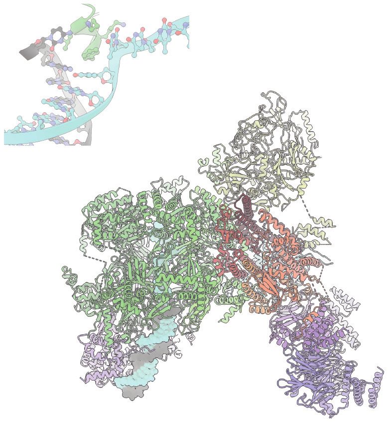
www.annualreviews.org • The Initiation of Eukaryotic DNA Replication 121F367 K364
L366 3’
5’ Flexible
F363 polymerase
domain
Mcm7
Pol ε
Cdc45
AAA+
MCM GINS
Pol α
NTD
Annu. Rev. Biochem. 2022.91:107-131. Downloaded from www.annualreviews.org
Access provided by University of Connecticut on 09/08/22. For personal use only.
Tof2
Csm3
Tof1 Ctf4 DNA2
Chl1
Parental DNA
2nd CMG
Figure 5
Composite atomic model that combines the CMG complex bound to DNA, Csm3–Tof1, and Ctf4 (PDB ID:
6SKL) and a structure of CMG bound to DNA and Pol ε (PDB ID: 6HV9). GINS and Cdc45 bind to the
side of an MCM ring, here viewed from the side. Parental duplex DNA enters the N-terminal face of the
MCM ring, where Csm3–Tof1 and Ctf4 are located. Ctf4 forms a homo-trimeric disk and functions as a
molecular hub onto which multiple factors can dock. Pol ε engages the AAA+ face of the MCM ring and is
poised to capture the leading-strand template as it exits from the helicase central channel. Abbreviations:
AAA+, ATPase associated with various cellular activities; CMG, Cdc45–MCM–GINS; Ctf, chromosome
transmission fidelity; GINS, go-ichi-nii-san; MCM, minichromosome maintenance; NTD, N-terminal
domain; PDB ID, Protein Data Bank identifier; Pol, DNA polymerase.
helicases, which led to the proposal that diverging helicases might remain linked during bidirec-
tional replication (188). These tantalizing observations predict a key role for Ctf4 in replisome
function; however, DNA replication establishment and fork progression rates in vitro are unaf-
fected by the presence or absence of Ctf4 (169), and single-molecule experiments have shown that
interactions between divergent replisomes are not required for replication in Xenopus egg extracts
(190). Thus, the main role of Ctf4 must be linking the replisome with factors that play genome
maintenance functions distinct from DNA replication (Figure 5). In fact, deletion of Ctf4 shows a
strong sister chromatid cohesion defect (191), and cellular studies implicated Ctf4, together with
noncatalytic elements in Mcm2 and Pol α, in recycling of parental histones onto the duplicated
lagging strand (192). These are frontier topics in chromosome biology that fall outside the scope
of this article.
The crystal structure of the catalytic domain of Pol ε trapped on a DNA substrate (193) and
the cryo-EM structure of the ternary complex of Pol δ bound to DNA and PCNA (194) explain
the structural mechanism of processive DNA synthesis by the leading- and lagging-strand poly-
merases. However, how these functions are coordinated at the fork remains largely unknown.
Biochemical reconstitution indicates that Pol δ plays an important role during the establishment
of leading strand synthesis, suggesting that substrate handoff between Pol δ and Pol ε must occur
before processive DNA replication is established (178). Structural work suggests that, while Pol ε
is anchored to the CMG, the catalytic domain of Pol2 is free to move with respect to the replisome
core (195), visiting configurations that appear distinct from the rigid structure recently described
122 Costa • DiffleyYou can also read