FANCD2 directly inhibits DNA2 nuclease at stalled replication forks and acts as a RAD51 mediator in strand exchange
←
→
Page content transcription
If your browser does not render page correctly, please read the page content below
bioRxiv preprint doi: https://doi.org/10.1101/2021.07.08.450798; this version posted July 9, 2021. The copyright holder for this preprint
       (which was not certified by peer review) is the author/funder. All rights reserved. No reuse allowed without permission.

   FANCD2 directly inhibits DNA2 nuclease at stalled replication forks and acts as a
   RAD51 mediator in strand exchange

   Wenpeng Liu1,5,7, Ivan Roubal1, Piotr Polaczek1, Yuan Meng2,5, Won-chae Choe1, Marie-
   Christine Caron3, Carl A. Sedgeman1, Yu Xi5, Changwei Liu2,5, Qiong Wu2, Li Zheng2, Jean-
   Yves Masson3,4, Binghui Shen2, and Judith L. Campbell1*
   1
       Braun Laboratories, California Institute of Technology, Pasadena, CA 91125, USA
   2
       Department of Cancer Genetics and Epigenetics, Beckman Research Institute, City of Hope,
   1500 East Duarte Road, Duarte, CA 91010-3000, USA
   3
       Genome Stability Laboratory, CHU de Québec Research Center, HDQ Pavilion, Oncology
   Division, 9 McMahon, Québec City, QC, G1R 3S3, Canada
   4
       Department of Molecular Biology, Medical Biochemistry and Pathology; Laval University
   Cancer Research Center, Québec City, QC, G1V 0A6, Canada
   5
       Colleges of Life Sciences, Zhejiang University, Hangzhou, Zhejiang 310027, China
   7
       Current address: Department of Biochemistry, Vanderbilt University School of Medicine, 613
   Light Hall, 2215 Garland Avenue, Nashville, Tennessee 37232, USA

   Correspondence: jcampbel@caltech.edu

                                                                    1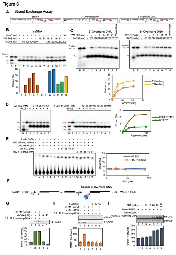
bioRxiv preprint doi: https://doi.org/10.1101/2021.07.08.450798; this version posted July 9, 2021. The copyright holder for this preprint
       (which was not certified by peer review) is the author/funder. All rights reserved. No reuse allowed without permission.

   Summary
   FANCD2 protein, a key coordinator and effector of the interstrand crosslink repair pathway, is
   also required to prevent excessive nascent strand degradation at hydroxyurea induced stalled
   forks. The mechanisms of the fork protection are not well studied. Here, we purified FANCD2
   to study how FANCD2 regulates DNA resection at stalled forks. In vitro, we showed that
   FANCD2 inhibits fork degradation in two ways: 1) it inhibits DNA2 nuclease activity by directly
   binding to DNA2. 2) independent of dimerization with FANCI, FANCD2 itself stabilizes RAD51
   filaments to inhibit various nucleases, including DNA2. More unexpectedly, FANCD2 acts as
   a RAD51 mediator to stimulate the strand exchange activity of RAD51, and does so by
   enhancing ssDNA binding of RAD51. Our work biochemically explains mechanisms by which
   FANCD2 protects stalled forks and further provides a simple molecular explanation for genetic
   interactions between FANCD2 and the BRCA2 mediator.

   Introduction
   Successful completion of DNA replication requires the integration of many proteins and
   pathways that protect, repair and/or restart replication forks. The principles underlying how
   these pathways interact and are regulated to maximize genome stability have yet to be
   determined. Fanconi anemia is a rare disease of bone marrow failure, developmental
   abnormalities, and cancer predisposition. At the cellular level it is diagnosed by sensitivity to
   DNA ICLs (interstrand crosslinking agents) and genome instability. Fanconi anemia is a
   multigenic disease defined by at least 22 complementation groups, including many regulatory
   components, nucleolytic activities, and HDR (homology directed repair) genes. The
   component genes suggest a coherent pathway for maintaining genome stability during DNA
   replication that goes beyond ICL repair and includes response to many additional types of
   replication stress(Boisvert and Howlett, 2014; Federico et al., 2018). The multigenic character
   of the FA pathway lends itself to comprehensive genetic and biochemical dissection(Carr and
   Lambert, 2013; Chaudhury et al., 2013; Chaudhury et al., 2014; Hashimoto et al., 2010;
   Kottemann and Smogorzewska, 2013; Lossaint et al., 2013; Petermann et al., 2010;
   Raghunandan et al., 2015; Schlacher et al., 2011; Schlacher et al., 2012; Sobeck et al., 2006;
   Yeo et al., 2014).
         FANCD2 is a key regulator of the FA pathway and the focus of our current
   studies(Kottemann and Smogorzewska, 2013; Timmers et al., 2001). During canonical
   replication-coupled repair of ICLs, after a replication fork encounters an ICL, FANCD2 and a
   related protein FANCI, are phosphorylated by activated ATR kinase. A FANCD2/FANCI
   heterodimer is also formed, and FANCD2 in this heterodimer, but not free FANCD2, is mono-
   ubiquitylated by the FA core complex containing nine FA proteins, including the FANCL E3
   ligase complex and several associated proteins. FANCD2-ubi is involved in both activation of
   repair events and also is directly required in the later enzymatic repair steps at strand breaks

                                                                    2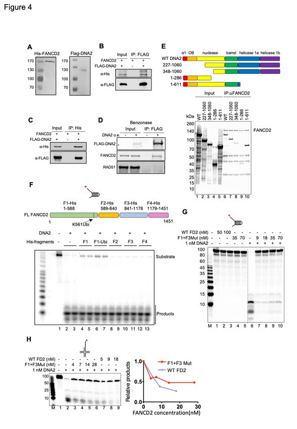
bioRxiv preprint doi: https://doi.org/10.1101/2021.07.08.450798; this version posted July 9, 2021. The copyright holder for this preprint
       (which was not certified by peer review) is the author/funder. All rights reserved. No reuse allowed without permission.

   (Knipscheer et al., 2009; Kottemann and Smogorzewska, 2013; Long et al., 2014; Long et al.,
   2011; Long and Walter, 2012; Raschle et al., 2008). The role of ubiquitin is to enforce stable
   binding of FANCD2/FANCI to DNA, specifically by clamping FANCD2-ubi/FANCI heterodimers
   onto DNA for DNA repair(Alcon et al., 2020; Rennie et al., 2020; Tan et al., 2020; Wang et al.,
   2020).
         FANCD2 also appears to have both ubiquitin-independent and FANCI-independent
   functions. In addition to its role in ICL repair, FANCD2 is required in the BRCA2-regulated,
   RAD51-mediated replication fork protection pathway, irrespective of the source of DNA
   damage(Chaudhury et al., 2013; Raghunandan et al., 2015; Schlacher et al., 2011; Schlacher
   et al., 2012). While several studies of fork protection implied that non-ubiquitylatable FANCD2
   (FANCD2-K561R) could not restore fork protection to patient-derived FANCD2-defective
   cells(Kais et al., 2016; Schlacher et al., 2012), other results support that FANCD2 is likely to
   have constitutive functions, at least for low levels of replication stress, such as endogenous
   stress(Boisvert and Howlett, 2014; Federico et al., 2018). With respect to ubiquitylation, study
   of FANCD2 knockout and knock-in cell lines showed that cells expressing only non-
   ubiquitylatable FANCD2-K561R had much less severe phenotypes than cells with a FANCD2
   knockout (Tian et al., 2017). Complementary studies showed that mutants defective in the
   trans-acting FA core complex components responsible for ubiquitylation of FANCD2 are less
   sensitive to replication fork stalling agents than FANCD2 knockdowns or knockouts
   (Thompson et al., 2017). Importantly, one of us reported that FANCD2 can protect forks by
   different mechanisms than FANCA/C/G, members of the core complex(Liu et al., 2020).
   FANCD2 has been shown to interact with RAD51, a key player/regulator in fork protection,
   and to do so in a ubiquitylation-independent, but HU-stimulated manner(Chen et al., 2017).
   FANCD2 also has FANCI independent functions(Chaudhury et al., 2013; Dubois et al., 2019;
   Thompson et al., 2017). FANCD2 deficient cells are HU and aphidicolin (a DNA polymerase
   inhibitor) sensitive, while FANCI cells are not (Thompson et al., 2017). These results warrant
   studies of ubiquitin- and FANCI- independent roles of FANCD2.
         Fork protection implies protection from nucleases. Several nucleases have been
   implicated in nascent DNA degradation at stalled forks, in both fork-protection proficient and
   fork-protection deficient cells (Schlacher et al., 2011; Schlacher et al., 2012; Thangavel et al.,
   2015). Prominent among these is the DNA2 helicase/nuclease, which seems to play a role in
   several different fork protection sub-pathways (Liu et al., 2020; Rickman et al., 2020). DNA2
   is essential for replication in normal yeast cells(Zheng et al., 2020). Multi-tasking DNA2
   removes long 5’ ssDNA flaps in the presence of Pif1 helicase during non-canonical Okazaki
   fragment processing(Budd et al., 2006; Diffley, 2020; Zheng et al., 2020); performs long-range
   resection of DSBs to provide 3’ ends for BRCA2-mediated, RAD51 filament formation and
   strand invasion during HDR; and prevents deleterious fork reversal in yeast(Hu et al., 2012).
   At stalled forks, human DNA2 is required for repair of stalled replication forks, where it

                                                                    3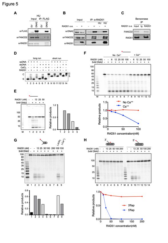
bioRxiv preprint doi: https://doi.org/10.1101/2021.07.08.450798; this version posted July 9, 2021. The copyright holder for this preprint
       (which was not certified by peer review) is the author/funder. All rights reserved. No reuse allowed without permission.

   mediates limited resection, putatively on a reversed fork, to promote fork restart, in replication
   fork protection proficient cells(Thangavel et al., 2015). We discovered that DNA2 deficient
   cells are sensitive to inter- or intra-strand crosslinks induced by cisplatin or formaldehyde.
   Paradoxically, depletion of DNA2 in cells deficient in FANCD2 rescued ICL sensitivity in
   FANCD2 mutants, suggesting that DNA2 became toxic in the absence of fork
   protection(Karanja et al., 2014; Karanja et al., 2012). We and others provided direct evidence
   that in cells defective in fork protection, DNA2-mediated over-resection occurs at replication
   forks stalled by exogenous DNA damaging agents or replication inhibition and that FANCD2
   is likely to regulate this resection(Higgs et al., 2015; Higgs et al., 2018; Jiang et al., 2015; Liu
   et al., 2016; Thangavel et al., 2015; Tian et al., 2017; Wang et al., 2015). The question remains
   as to how FANCD2 regulates DNA2, however. It is not clear if this is direct or indirect because,
   for instance, RAD51 is also inhibitory to over-resection, apparently through DNA2 inhibition,
   in vivo(Wang et al., 2015) and because FANCD2 and RAD51 are epistatically linked in fork
   protection(Schlacher et al., 2012).
               Recently, several studies have suggested that FANCD2 and BRCA2 perform parallel
   or compensatory functions in fork protection and fork recovery after collapse(Kais et al., 2016;
   Lachaud and Rouse, 2016; Michl et al., 2016). Since BRCA2 is thought to stabilize RAD51
   filaments, we hypothesized that FANCD2 may provide a backup source of this BRCA2 function
   in response to replication stress. This mechanism is supported by the fact FANCD2 alone and
   FANCD2/FANCI heterodimers interact physically with RAD5(Berti et al., 2013; Chen et al.,
   2017; Sato et al., 2016; Thangavel et al., 2015; Thangavel et al., 2010). FANCD2/FANCI
   complexes have been shown to increase RAD51 levels on DNA, but the specific contribution
   of FANCD2 itself and the relationship of this observation to suppression of BRCA2-/- defects
   has not been established.
           In this work we delved deeper into the mechanisms of FANCD2 fork protection by
   comparing the effect of FANCD2 deficiency on replication forks stalled due to acute or chronic
   replication stress induced by different damaging agents. Our in vivo results confirm that
   FANCD2 is required to protect stalled replication forks from DNA2-dependent over-resection
   after acute stress. They also revealed, however, that FANCD2 has an additional and opposing
   function that is needed for promoting resection after prolonged stress. We identified at least
   two potential mechanisms by which FANCD2 protects nascent DNA from nucleolytic resection
   in vitro: 1) FANCD2 inhibits DNA2 nuclease activity and binds directly to the DNA2 nuclease
   domain; and 2) FANCD2 stabilizes RAD51 ssDNA filaments which prevent nucleolytic
   digestion by multiple nucleases. Surprisingly, we also find that FANCD2, like BRCA2, acts as
   a mediator in RAD51-dependent strand exchange. FANCD2 promotes RAD51 mediated
   strand exchange activity by stabilizing RAD51 on ssDNA, but not, like BRCA2 by inhibiting the
   RAD51 ATPase or RAD51 dsDNA binding activities, and not requiring FANCD2 DNA binding
   motifs. All the in vitro activities of FANCD2 are FANCI independent and interaction with DNA2

                                                                    4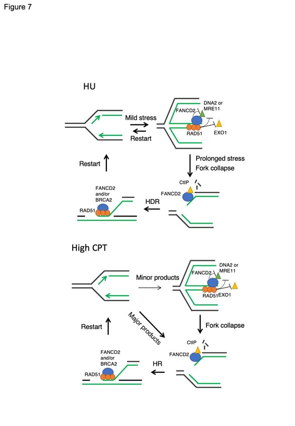
bioRxiv preprint doi: https://doi.org/10.1101/2021.07.08.450798; this version posted July 9, 2021. The copyright holder for this preprint
       (which was not certified by peer review) is the author/funder. All rights reserved. No reuse allowed without permission.

   is ubiquitin independent in vitro. The ability to stimulate strand exchange adds a novel
   mechanistic explanation for the well-documented dependency of BRCA2-/- tumors on FANCD2,
   the suppression of BRCA1/2-/- phenotypes by elevated levels of FANCD2(Kais et al., 2016;
   Lachaud and Rouse, 2016; Michl et al., 2016), and adds a new dimension to how FANCD2
   deficiency leads to inhibition of fork protection and genome instability(Schlacher et al., 2012).

   Results

   FANCD2 Plays Different Roles in Repair at Different Extents/Stages of Fork Stalling

   FANCD2 has been demonstrated by iPOND to associate with replication forks, and to increase
   four to five fold in the presence of HU(Dungrawala et al., 2015). To further verify the
   association of FANCD2 with replication forks stalled by HU, we labeled nascent DNA strands
   with the thymidine analog BrdU in the presence of HU under conditions that specifically mark
   nascent ssDNA (single-stranded DNA) and used immunofluorescence to monitor localization
   of FANCD2 and gH2AX(Couch et al., 2013). We find that gH2AX foci and FANCD2 foci co-
   localize with BrdU-marked nascent ssDNA foci (Figure 1A). We then compared the level of
   association of FANCD2 with replication forks in the absence and presence of HU by
   immunofluorescence (see Method Details). We detected FANCD2/EdU associated foci only
   after HU treatment (Figure 1B). (Note that the images were magnified and the ratio of cells
   with >4 FANCD2/EdU colocalized foci to total cells was determined). The difference between
   IPOND and immunofluorescence (IF) in observed FANCD2 association with the nascent DNA
   in the absence of HU may be due to assay detection resolution. We conclude that FANCD2
   substantially recruited to stalled replication forks in the presence of HU.

         Multiple pathways are involved in stalled replication fork repair, and forks undergo
   sequential changes in architecture during chronic stalling(Helleday et al., 2008; Lemacon et
   al., 2017; Petermann et al., 2010; Quinet et al., 2017). A major question in FANCD2 mediated
   repair is what structure is being acted upon and whether this substrate changes during the
   course of fork pausing and processing(Petermann et al., 2010; Piberger et al., 2019). To
   investigate this question, we monitored resection in FANCD2 deficient cells at various extents
   of stalling with either HU, CPT (camptothecin), or cisplatin, each of which induces a different
   type of replication stress. Initially we used RPA2 phosphorylation as a surrogate for measuring
   ssDNA arising during resection in the presence of HU. Cells were treated with HU for 0-8 h
   and nuclear extracts prepared. At all-time points, we observe drastically increased RPA-p
   levels (resection) in nuclear extracts of HU-treated PD20 FANCD2-/- deficient cells compared
   to PD20:FANCD2 (FANCD2-complemented) cells, where there was very little resection
   (Figure 1C). This is consistent with other published results showing that FANCD2 protects
   stalled forks from nascent DNA degradation. Unexpectedly, however, we see a change in the
   pattern over the time course. In PD20 cells, we observe an initial increase in resection at early

                                                                    5
bioRxiv preprint doi: https://doi.org/10.1101/2021.07.08.450798; this version posted July 9, 2021. The copyright holder for this preprint
(which was not certified by peer review) is the author/funder. All rights reserved. No reuse allowed without permission.
times, 2-4 h of treatment, but then a decline in resection with treatment up to 8 h, though there
is still substantial over-resection. We interpret the decline of RPA-p at later times in the
continued presence of HU as due to conversion of the initial, RPA-p marked damage that is
protected by FANCD2, into other lesions, such as DSBs or gaps (collapse) and that FANCD2
is required for resection of the latter structures. A change in structure is supported by neutral
COMET assay (Figure S1A). The proposed requirement for FANCD2 on fork collapse is
consistent with the fact that FANCD2 tethers CtIP to collapsed forks, and CtIP stimulates
resection needed for repair after extensive ICL-induced stalling(Murina et al., 2014; Unno et
al., 2014; Yeo et al., 2014), either by recruitment and stimulation of BLM and DNA2 nuclease
to resect the end of the break(Ceppi et al., 2020; Hoa et al., 2015; Yeo et al., 2014), or by
recruitment of pol theta and stimulation of alternative non-homologous end joining (alt-
NHEJ)(Badie et al., 2015; Zhang and Jasin, 2011).
The proposal that FANCD2 can have two opposing effects on resection is also supported
by the fact that PD20 cells treated with CPT (camptothecin) behave differently from those
treated with HU (Figure 1D). While CPT can also induce fork slowing or stalling(Ray Chaudhuri
et al., 2012), it rapidly induces DSBs when the replication fork encounters sites of the CPT-
induced Top1-DNA cleavage complexes(Berti et al., 2020; Whelan and Rothenberg, 2021).
We do not see an overall increase in resection after CPT treatment in the absence of FANCD2
in the PD20 cells compared to the complemented cells (Figure 1D and Figure S1C), which we
attribute to the dominance of DSB lesions that require FANCD2 for resection. We do still see
that in the absence of FANCD2, there is over-resection at early times compared to later times
(Figure 1D, lanes 3 and 4 compared to lanes 5 and 6) in PD20 cells, similar to HU, suggesting
there is some fork stalling at earlier times with fork collapse after chronic exposure, as with
HU(Berti et al., 2020; Whelan and Rothenberg, 2021). We obtained supporting data for the
difference in replication fork structure caused by HU and CPT using neutral COMET assays.
In HU, after mild treatment, there is a low level of DSBs that increase as the extent of HU
treatment is increased (Figure S1A). With CPT, there is a greater level of DSBs at early times
and they increase rapidly and extensively with prolonged treatment (Figure S1B).
Cisplatin treatment phenocopies CPT since RPA2-p in FANCD2-deficient cells prepared
by knockdown of FANCD2, is significantly decreased after prolonged treatment with either
compound (Figure S1D). Comparison of the results with HU to those with CPT suggests that
transiently stalled, possibly reversed replication forks, rather than DSBs, are the primary sites
of FANCD2 negative regulation of resection and that different repair mechanisms may function
on CPT and cisplatin damage, as recently proposed by others(Couch et al., 2013; Rickman et
al., 2020).
In summary, our results suggest that FANCD2 is required to protect from over-resection
at early times after damage on forks transiently stalled by HU. FANCD2, however, may play
a different role during chronic stalling that leads to fork collapse to DSBs or to other types of
6bioRxiv preprint doi: https://doi.org/10.1101/2021.07.08.450798; this version posted July 9, 2021. The copyright holder for this preprint
(which was not certified by peer review) is the author/funder. All rights reserved. No reuse allowed without permission.
damage, when it may actually be required for resection and repair.
FANCD2-mediated Regulation of End Resection Occurs on Nascent DNA upon
Replication Stress
To more directly investigate replication fork-coupled resection in FANCD2-deficient cells, we
next used single molecule tracking of nascent DNA before and after brief (4h) HU treatment.
As reported previously, we see over-resection in the FANCD2 depleted cells, just as we saw
using RPA-p as the readout for resection (Figure 1C). The resection we observe might occur
either on single-stranded “flaps” on uncoupled forks, on forks with gaps due to re-priming, or
on the regressed arm of reversed forks(Piberger et al., 2019; Quinet et al., 2019; Zellweger et
al., 2015). Regressed forks arise when forks stall, template strands rewind, and nascent
strands anneal, forming a four-way junction similar to a Holliday junction(Higgins et al., 1976).
Zellweger et al. demonstrated that both uncoupled forks with gaps and reversed forks
accumulate in cells treated with low levels of HU(Zellweger et al., 2015). To test the model
that over-resection in FANCD2-deficient cells occurred on reversed forks, we monitored
resection in FANCD2-depleted cells and in cells co-depleted for FANCD2 and RAD51, the
SMARCAL1 translocase or ZRANB3 translocases, all of which have been reported to mediate
HU-induced fork reversal and to be required for fork degradation in the absence of the
FA/BRCA fork protection pathway(Betous et al., 2013; Bhat et al., 2015; Couch et al., 2013;
Kolinjivadi et al., 2017b; Lemacon et al., 2017; Mijic et al., 2017; Taglialatela et al., 2017;
Vujanovic et al., 2017; Zellweger et al., 2015). We find that co-depletion of either RAD51 or
SMARCAL1 or ZRANB3 with FANCD2 resulted in significant rescue of over-resection (Figure
2A and 2b). Similar results were obtained with BRCA2 knockdowns (Figure 2B),as reported
by numerous studies. The results suggest that the over-resection upon limited HU treatment
in FANCD2 deficient cells occurs primarily on reversed fork substrates. Furthermore, both
SMARCAL1 and ZRANB3 are likely required to produce the substrate since knockdown of
either one completely protected from observable nascent DNA degradation.
We also monitored the effect of SMARCAL1 and ZRANB3 on resection after CPT
treatment using the RPA-p western blot assay. Knockdown of SMARCAL1 slightly reduced
resection but ZRANB3 siRNA did not after CPT treatment (Figure S2A). Co-depletion of
FANCD2 and FANCM, another factor capable of reversing forks in vitro, also reduced
resection (Figure S2A). However, we see more residual resection after ZRANB3 depletion in
the assays for RPA-p after CPT treatment (Figure S2A) than we see in the DNA fiber
experiment after HU treatment (Figure 2B), supporting our proposal that reversed fork
structures may not be the sole substrates for resection after CPT treatment. Thus, the RPA-p
assay may also be detecting resection at forks collapsed to DSBs when the fork encounters
the single-strand break induced by CPT treatment (or at non-specific DSBs) rather than after
7bioRxiv preprint doi: https://doi.org/10.1101/2021.07.08.450798; this version posted July 9, 2021. The copyright holder for this preprint
(which was not certified by peer review) is the author/funder. All rights reserved. No reuse allowed without permission.
decay of a reversed fork substrate that failed to be repaired or at gaps behind the
fork(Hashimoto et al., 2010). CPT damage may have different requirements for restart,
consistent with the difference in the level of resection after CPT treatment compared to results
with HU (Figure 1C and 1D).
MUS81 and SLX1/4 are structure specific endonucleases that can cleave unrepaired,
long-lived reversed forks such as arise at an ICL or after prolonged fork stalling by other agents,
to produce DSBs. To directly test if such DSBs were substrates for over-resection, we co-
depleted FANCD2 and MUS81 or SLX4 to see if knockdown could reduce HU-induced fork
degradation (Figure 2C). Depletion of MUS81 and SLX4 did not affect the HU-induced nascent
degradation observed in the absence of FANCD2, supporting that the fork degradation
detected in the absence of FANCD2 by fiber tracking is not due to previous endonucleolytic
digestion (by these nucleases) but rather by resection of reversed forks.
DNA2 or MRE11 Depletion Reverses the Over-resection of Nascent DNA in the Absence
of FANCD2 upon Replication Stress
Following up on our observation that knockdown of DNA2 can rescue the cisplatin and
formaldehyde sensitivity of the FANCD2-/- PD20 patient cell line(Karanja et al., 2014), we
confirm that degradation of nascent DNA in FANCD2-deficient cells upon HU treatment can
be rescued by knockdown of DNA2 but also by knockdown of MRE11 nuclease (Figure 2D
and Figure S2B), just as knockdown of DNA2 significantly decreases this RPA-p, i. e. resection,
in cisplatin treated FANCD2-/- cells with brief cisplatin treatment (Figure S2C, lane 4). MRE11
and DNA2 may function as alternative nucleases or function sequentially, as they do at DSBs
(Symington and Gautier, 2011). We also tested the role of DNA2 after brief CPT treatment
using the RPA-p assay; the results show that DNA2 but not CtIP knockdown reverses the
over-resection in the absence of FANCD2 upon short CPT treatment (Figure S2D and S2E).
In the presence of FANCD2, knockdown of CtIP reduces resection, consistent with an
important role for FANCD2 mentioned above in CtIP recruitment and the ability of CtIP to
stimulate both MRE11 and WRN/BLM-DNA2 mediated resection in vitro(Ceppi et al., 2020).
The difference between MRE11 and CtIP in the absence of FANCD2 is consistent with 2-
D gel studies in yeast that show that Sae2, the CtIP ortholog, only participates in resection at
late collapsed forks and not at early reversed-fork or uncoupled-fork intermediates(Colosio et
al., 2016). They are also consistent with our proposal that the requirement for FANCD2 after
chronic exposure to HU occurs at collapsed rather than reversed forks.
DNA2 Depletion Can Rescue Replication Restart Defects Observed in the Absence of
FANCD2
8bioRxiv preprint doi: https://doi.org/10.1101/2021.07.08.450798; this version posted July 9, 2021. The copyright holder for this preprint
(which was not certified by peer review) is the author/funder. All rights reserved. No reuse allowed without permission.
We next tested if the over-resection leads to replication restart defects such as are found in
aphidicolin treated FANCD2 deficient cells(Yeo et al., 2014). We performed DNA combing
assays to determine stalled fork restart efficiency after HU or cisplatin treatment (Figure S2F).
We found that restart efficiency is moderately decreased in FANCD2 or DNA2 knockdown in
U2OS cells, consistent with previous studies(Yeo et al., 2014). However, combining DNA2
and FANCD2 knockdown in U2OS cells rescues the restart defects in both HU and cisplatin
treatment. These results reveal that nascent strand degradation in FANCD2 deficient cells can
reduce restart efficiency, and that rescue of nascent DNA degradation by depleting DNA2
restores restart efficiency.
FANCD2 Inhibits DNA2 Nuclease Activity in vitro Providing a Mechanism for FANCD2's
in vivo Role at Reversed Forks
Since over-resection in PD20 cells treated with HU is massively increased over the FANCD2-
complemented cells at all extents of treatments (Figure 1C), and since FANCD2 and DNA2
have been shown to interact in vivo in a DNA-independent fashion, suggesting a
protein/protein interaction(Karanja et al., 2012), we tested whether FANCD2 directly regulates
degradation by DNA2 nuclease. FANCD2-His was purified from SF9 insect cells (Figure
S3A)(Roques et al., 2009) and was shown bind to dsDNA (Figure S3B) and to be free of
nuclease activities under the conditions used here (Figure 3A). When FANCD2 was added to
a DNA2 nuclease reaction, significant inhibition of DNA2 nuclease was observed, even in the
presence of high levels (5 nM) of DNA2 (Figure 3B and Figure S3C). Inhibition is likely due to
FANCD2 protein since all reactions contained the same amount of FANCD2 diluent. In these
experiments, the substrate partially mimics a stalled replication fork with single-stranded DNA
arms at the dsDNA junction. DNA2 processes substrates with several different configurations,
such as unligated 5’ flaps on Okazaki fragments or on base excision repair intermediates, or
5’ overhangs on regressed replication forks during replication fork stress/stalling or during DSB
resection during homologous recombination. As shown in Figures 3C and 3D and Figure S3C,
FANCD2 inhibits DNA2 nuclease on each of these structures. (Sequences of all
oligonucleotide substrates used are provided in Table S2. The substrates mimicking reversed
fork DNAs with or without a 5’ overhang (Figure 3D) were generated and validated as
described in Figure S3D).
FANCD2 forms stable complexes with FANCI in vivo and in vitro, although only 20%
of the FANCD2 in the cell co-IPs with FANCI(Alcon et al., 2020; Tan et al., 2020). We also
tested if FANCI inhibited DNA2 and showed that there was no inhibition of DNA2 nuclease,
except at the highest FANCI concentrations (Figure 3E). When FANCI was added along with
FANCD2, the small amount of nuclease activity remaining after inhibition of DNA2 by FANCD2
was not further inhibited by FANCI (Figure 3F).
9bioRxiv preprint doi: https://doi.org/10.1101/2021.07.08.450798; this version posted July 9, 2021. The copyright holder for this preprint
(which was not certified by peer review) is the author/funder. All rights reserved. No reuse allowed without permission.
We next addressed the specificity of FANCD2 inhibition for DNA2. Since depletion of
MRE11 suppressed the nascent DNA degradation in FANCD2 knockdowns (Figure 2D), we
tested the ability of FANCD2 to inhibit MRE11. MRE11 activity on duplex DNA was inhibited
by FANCD2, also consistent with a protein/protein interaction(Roques et al., 2009) (Figure 3G).
We also tested the effect of FANCD2 on EXO1 (Figure 3H). EXO1 was not inhibited by
FANCD2, so there is specificity to the inhibition of MRE11 and DNA2 by FANCD2.
How Does FANCD2 Inhibit DNA2 Nuclease?
We next asked whether inhibition was mediated by a DNA2/FANCD2 protein/protein
interaction and/or by a FANCD2 protein/DNA interaction. We purified His-tagged hFANCD2
from E. coli and FLAG-tagged hDNA2 protein from human cells as described in Method
Details(Takahashi et al., 2014) (Figure 4A). Immunoprecipitation experiments show that
purified DNA2 and FANCD2 bind directly and strongly to each other (Figure 4B and 4C) and
that the interaction is independent of DNA (Figure 4D), further supporting that in vivo
interaction may also be direct, consistent with our previous finding that DNA2 and FANCD2
also reciprocally coIP in extracts of CPT-treated cells and that the interaction is independent
of DNA(Karanja et al., 2012).
Using site-directed mutagenesis we identified a region on DNA2 in the N terminus
spanning a.a (amino acid) 227 to 348 that severely reduces coimmunoprecipitation with full-
length FANCD2 (Figure 4E and Methods). This region includes the canonical DEK nuclease
family active site motifs(Budd and Campbell, 2000; Yang, 2011), suggesting how the
interaction might interfere with nuclease function. We next used a complete set of fragments
of FANCD2 to determine the region that inhibits DNA2, a functional assay for “interaction”.
As shown in Figure 4F, fragment F1, a.a. 1-588 was the only sub-fragment that inhibited DNA2.
This region contains the FANCD2 ubiquitylation site (a.a. K561), and addition of a ubiquitin
coding sequence to F1 fragment, designated F1-ubi, increased the efficiency of inhibition.
These results imply that a protein/protein interaction is needed to inhibit DNA2 nuclease
activity.
We also investigated whether DNA binding by FANCD2 was involved in the inhibition of
DNA2. To do so, we used FANCD2-F1+F3Mut, which is defective, though not completely
blocked, in DNA binding (Figure S4A and S4B)(Niraj et al., 2017). Like WT FANCD2, FANCD2-
F1+F3Mut showed no nuclease activity itself (Figure 4G, controls, left) and strongly inhibited
DNA2 nuclease on the fork structure (Figure 4G, right) and with only marginally reduced
potency on the reversed fork structures (Figure 4H), although it bound to ssDNA 10-fold less
efficiently than WT FANCD2 (Figure S4B), consistent with previous characterization of the
FANCD2-F1+F3Mut protein(Niraj et al., 2017). This suggests that direct protein/protein
interaction contributes to DNA2 inhibition and that inhibition does not occur by simply blocking
the substrate or competing with DNA2 for the substrate.
10bioRxiv preprint doi: https://doi.org/10.1101/2021.07.08.450798; this version posted July 9, 2021. The copyright holder for this preprint
(which was not certified by peer review) is the author/funder. All rights reserved. No reuse allowed without permission.
Since the FANCD2-F1+F3Mut protein showed residual binding to DNA, however, to
further test whether DNA2 inhibition is through a protein/protein interaction, we investigated
whether inhibition by FANCD2 is species-specific. Yeast lacks a FANCD2 ortholog, and we
hypothesized that yeast DNA2 would only be inhibited by FANCD2 if inhibition was mediated
by occlusion/sequestration of DNA, thus preventing binding by DNA2. We observed no
inhibition of yeast DNA2 by FANCD2, even at great molar excess FANCD2, on either the
forked substrate or the reversed fork substrate (Figure S4C and S4D). Note that yDNA2 is
more active than hDNA2, as also reported by others(Kumar et al., 2017), accounting for the
concentrations used. The lack of inhibition of yeast DNA2 by FANCD2 supports, though it does
not prove, that inhibition of hDNA2 by FANCD2 involved a species-specific and therefore likely
a physiologically significant protein/protein interaction.
DNA2 is also Inhibited by RAD51 Filaments
In addition to rescue by DNA2 nuclease depletion or specific chemical inhibition of nucleases,
failure of BRCA2 or FANCD2 fork protection and resulting nascent DNA degradation has been
shown to be rescued by elevated RAD51 levels or by stabilization of RAD51 filaments(Bhat et
al., 2018; Schlacher et al., 2012; Wang et al., 2015). Furthermore, FANCD2 and RAD51 show
epistatic interaction in nascent DNA degradation assays, i.e. destabilization of RAD51
filaments does not lead to further degradation of nascent DNA in FANCD2 deficient
cells(Schlacher et al., 2012) and see also(Hashimoto et al., 2010; Hashimoto et al., 2012;
Higgs and Stewart, 2016; Kolinjivadi et al., 2017b; Schlacher et al., 2011; Schlacher et al.,
2012; Taglialatela et al., 2017; Thangavel et al., 2015; Wang et al., 2015; Zadorozhny et al.,
2017). To explore potential molecular interplay between FANCD2 and RAD51 in regulating
DNA2-mediated resection, we first looked at whether there is physical interaction between
DNA2, RAD51, and FANCD2 in HU-treated cells. We show that RAD51 co-IPs with FLAG-
DNA2 and with endogenous FANCD2 (Figure 5A). Reciprocally, we immunoprecipitated
RAD51 and showed that both FANCD2 and endogenous DNA2 coimmunoprecipitated (Figure
5B). The RAD51 immunoprecipitation in Figure 5B, which was analyzed under different gel
conditions from the a-FLAG-DNA2 IP in Figure 5A, reveals that both mono-ubiquitylated and
non-ubiquitylated FANCD2 are present in the co-immunoprecipitates. We then repeated these
experiments after treating extract with Benzonase (Figure 5C). The RAD51/FANCD2
interaction was still observed (Figure 5C) and is therefore not dependent on DNA, in keeping
with previous observations(Sato et al., 2016). However, since RAD51 was not found in a DNA2
IP after treatment of extracts with Benzonase (Figure 4D), we conclude the interaction of
DNA2 with RAD51 requires DNA and is not direct.
Although DNA2 and RAD51 don’t appear to interact directly, RAD51 filaments have been
implicated in regulating DNA2-mediated resection(Wang et al., 2015). We tested for inhibition
of FLAG-DNA2 nuclease by recombinant RAD51 protein. To distinguish between inhibition by
RAD51 protein alone and inhibition by RAD51 filament formation, we took advantage of the
11bioRxiv preprint doi: https://doi.org/10.1101/2021.07.08.450798; this version posted July 9, 2021. The copyright holder for this preprint
(which was not certified by peer review) is the author/funder. All rights reserved. No reuse allowed without permission.
demonstration that Ca2+ permits filament formation but modulates ATP hydrolysis and inhibits
dissolution of the filaments, resulting in stabilization(Bugreev and Mazin, 2004). As shown
previously and in Figure 5D, RAD51 filaments can be formed in the presence of ATP and are
more stable in the presence of Ca2+ than in its absence (compare lanes 2 and 4 and lanes 10
and 12, Figure 5D). We then determined the effect of increasing amounts of RAD51 on DNA2
nuclease activity. As shown in Figure 5E and 5F, RAD51 inhibits DNA2 degradation in the
presence of Ca2+ but inhibits much less in the absence of Ca2+, where filaments are less stable.
The Ca2+ effect suggests that inhibition is due to RAD51 filaments. RAD51 also inhibits DNA2
on forked and 5’-flap substrates (Figure 5G and 5H). RAD51 filament stabilization might block
many nucleases [ Figure S5A and (Kolinjivadi et al., 2017b)], bringing into question the
physiological significance of RAD51 inhibition of DNA2. Therefore, we turned our attention to
the study of additional functions for RAD51 and FANCD2 interaction in fork protection.
FANCD2 Stimulates Strand Exchange by High Concentrations of RAD51
We were struck by the fact that BRCA2-/- cells and FANCD2-/- show non-epistatic interactions
such as synthetic lethality and that over-expression of FANCD2 suppresses BRCA-/-
phenotypes(Kais et al., 2016; Michl et al., 2016). Furthermore, like BRCA2, FANCD2 interacts
physically and robustly with RAD51 [Figure 5 and (Chen et al., 2017; Sato et al., 2016)], and
RAD51 has been shown to localize to stalled forks in cells lacking BRCA2(Kolinjivadi et al.,
2017b). We hypothesized that FANCD2 might, similarly to BRCA2, stimulate RAD51-mediated
strand exchange(Jensen et al., 2010; Thorslund et al., 2010). While FANCD2 does not
enhance RAD51-mediated D-loop assays with resected plasmid substrates(Dubois et al.,
2019), complete strand exchange assays with oligonucleotides were never tested. Both
reactions are linked to DNA recombination, but mechanistically they are different. In D-loop
assays strand invasion into a supercoiled DNA recipient is measured and is thought to
represent a search for homology(Carreira et al., 2009; Carreira and Kowalczykowski, 2011).
Strand exchange assays, in contrast, measure a complete transfer of DNA strands (see
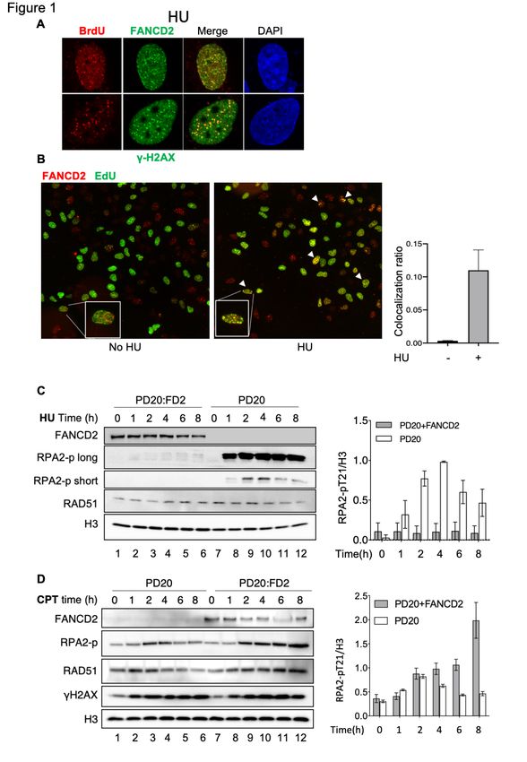
schematic in Figure 6A). As indicated, RAD51 catalyzes the exchange of the labeled strand in
the duplex to ssDNA to form the strand exchange product(Jensen et al., 2010; Thorslund et
al., 2010). High concentrations of RAD51, however, have been shown to be inhibitory in this
assay(Jensen et al., 2010; Thorslund et al., 2010). To measure strand exchange, RAD51 was
incubated, in the presence or absence of FANCD2, with unlabeled ssDNA (Figure 6B, pilot
experiment), with duplex DNA with a 3' ssDNA overhang (Figure 6C), or with duplex DNA with
a 5' ssDNA overhang (Figure 6C) to allow filament formation. Fully duplex DNA containing a
32
P labeled strand complementary to the ssDNA or respective overhang DNA was then added.
Stimulation of strand exchange by RAD51 is shown for ssDNA in Figure 6B, lanes 1-4.
Inhibition at high RAD51 levels is shown in Figure 6B, lane 5. Such inhibition is proposed to
arise once ssDNA is saturated with RAD51, allowing the excess RAD51 to bind to the labeled
12bioRxiv preprint doi: https://doi.org/10.1101/2021.07.08.450798; this version posted July 9, 2021. The copyright holder for this preprint
(which was not certified by peer review) is the author/funder. All rights reserved. No reuse allowed without permission.
dsDNA donor, which inhibits exchange(Jensen et al., 2010; Thorslund et al., 2010). Supporting
the hypothesis that the inhibition by high levels of RAD51 can be due to binding of excess
RAD51 to duplex DNA, we showed that addition of a dI-dC oligonucleotide relieves inhibition,
presumably by successfully competing with the labeled duplex donor for excess RAD51
binding in the assays (Figure 6B, lane 12). We then studied whether FANCD2 stimulated
RAD51 at high RAD51 concentrations, as has been shown for BRCA2(Jensen et al., 2010;
Thorslund et al., 2010). As shown in Figure 6B (lanes 6-11) and Figure 6C, although FANCD2
has no strand exchange activity on its own, FANCD2, indeed, reproducibly stimulates strand
exchange by high concentrations of RAD51 and does so in a concentration dependent manner.
Strand exchange involving duplex DNA with a 3’ or 5’ overhang, more closely resembling a
filament on resected DNA, was stimulated more efficiently than with ssDNA, suggesting that
stimulation may occur on DNA with ds/ss junctions and may occur at gaps as well as at ssDNA
tails (Figure 6B, C). Several controls that strand exchange was occurring were performed.
Reversing the order of addition of substrates, i.e., formation of RAD51 filaments on dsDNA
and then addition of ssDNA, did not lead to exchange (Figure S6); thus, we are not observing
inverse strand exchange(Mazina et al., 2017). Addition of cold oligonucleotide to the stop
reaction does not change the products, supporting that the strand exchange products are not
formed due to denaturation and renaturation in the stop mixture (Figure 6C, lanes labeled cold
oligo)(Jensen et al., 2010). We conclude that FANCD2 stimulates RAD51 mediated strand
exchange.
We next interrogated the mechanism of FANCD2 stimulation of RAD51. BRCA2 DNA
binding is required for stimulation of strand exchange, and BRCA2 is thought to stimulate
strand exchange in several ways: by stabilizing RAD51 filaments through inhibiting RAD51
DNA-dependent ATPase, by promoting the handoff of ssDNA from RPA to RAD51, and by
nucleating filament formation on ssDNA while inhibiting filament formation on duplex DNA. To
determine if FANCD2 DNA binding was required for stimulation of strand exchange, we tested
if the FANCD2 DNA binding mutant described above stimulated strand exchange(Niraj et al.,
2017). Although FANCD2-F1+F3Mut showed approximately ten-fold reduction in ssDNA
binding at 10 nM (Figure S4B), FANCD2-F1+F3Mut protein stimulates strand exchange even
more efficiently than FANCD2 WT (Figure 6D). We next determined if FANCD2 inhibits RAD51
DNA-dependent ATPase. Surprisingly, unlike BRCA2, FANCD2 does not inhibit RAD51 DNA-
dependent ATPase (Figure 6E), and thus may not be acting to stabilize RAD51/ssDNA
filaments by blocking the ATPase.
We finally tested if FANCD2 plays a role in targeting RAD51 preferentially to ssDNA by
inhibiting nucleation on dsDNA. We carried out DNA binding experiments using biotin-
streptavidin pull-downs (Figure 6F). We first demonstrated that dsDNA inhibits RAD51 binding
to a biotin-labeled 3’ overhang substrate (Figure 6G). We then added FANCD2 and found that
FANCD2, unlike what has been demonstrated for BRCA2, which has been shown to
13bioRxiv preprint doi: https://doi.org/10.1101/2021.07.08.450798; this version posted July 9, 2021. The copyright holder for this preprint
(which was not certified by peer review) is the author/funder. All rights reserved. No reuse allowed without permission.
specifically overcome dsDNA inhibition of RAD51 binding to overhang DNA(Jensen et al.,
2010; Thorslund et al., 2010), did not stimulate association of RAD51 with the 3’ overhang
substrate in the presence of excess dsDNA (Figure 6H). Thus, FANCD2 is not stimulating
RAD51 by reducing binding to dsDNA and is more likely stabilizing RAD51/ssDNA filaments.
Supporting this interpretation, FANCD2 does stimulate the accumulation of RAD51/ssDNA
complexes in the absence of dsDNA (Figure 6I), consistent with previous characterization of
RAD51/FANCD2/FANCI interaction with DNA(Sato et al., 2016). In summary, we suggest that
FANCD2 stimulates strand exchange by directly promoting, either through nucleation,
assembly, or filament stabilization, RAD51/ssDNA filament formation, rather than by
competing with dsDNA. This FANCD2-mediated stabilization does not involve inhibition of
RAD51 ATPase and does not require optimum FANCD2 DNA binding activity. Stimulation of
strand exchange with these characteristics suggests that the molecular role of FANCD2 in fork
protection and restart may involve stimulation of formation of or stabilization of RAD51
filaments in addition to the direct inhibition of DNA2 shown in Figure 4. The results further
suggest that interaction of FANCD2 with RAD51 protein [Figure 5 and(Sato et al., 2016)]
contributes to strand exchange, and therefore that this may contribute to the ability of elevated
levels of FANCD2 to suppress some BRCA2-deficiencies(Ceccaldi et al., 2015; Michl et al.,
2016).
Discussion
FANCD2 has been studied for decades and much has been learned about its cellular and
structural. What is unknown, however, despite extensive cellular and structural
characterization, are the biochemical activities it uses to accomplish and coordinate its diverse
in vivo roles. During our studies of cellular responses to DNA replication stress and
maintenance of genome stability, our work on the DNA2 helicase/nuclease, a protein involved
in resection necessary for replication fork protection, confirmed that FANCD2 regulates DNA2
resection. This led us to undertake a biochemical reconstitution of study of stalled fork
resection to identify the important biochemical functions of DNA2, i.e. resolution and
reconstitution. A model summarizing our results is shown in Figure 7.
Two Opposing Roles for FANCD2 at Stressed Replication Forks
In pursuing the discovery that FANCD2 and DNA2 show synthetic viability in the presence of
ICLs, we now demonstrated that over-resection of nascent DNA that occurs in FANCD2
deficient cells after brief replication stress induction by either HU, CPT or cisplatin, is
suppressed by depletion of DNA2, correlating with the cisplatin resistance of FANCD2/DNA2
doubly deficient cells, consistent with other recent reports(Higgs et al., 2018; Rickman et al.,
2020). We further demonstrate, for the first time, however, that after HU treatment this over-
resection occurs on stalled forks reversed by SMARCAL1 and ZRANB3 as well as RAD51
(Figure 2). DSBs do not seem to be a substrate for DNA2 hyper-resection in the absence of
14bioRxiv preprint doi: https://doi.org/10.1101/2021.07.08.450798; this version posted July 9, 2021. The copyright holder for this preprint
(which was not certified by peer review) is the author/funder. All rights reserved. No reuse allowed without permission.
FANCD2 since SLX4 and MUS81 are not required for nascent DNA degradation during short
HU treatment.
Unexpectedly, we found that this DNA2-inhibitory function of FANCD2 is only observed at
low levels of replication stress. In contrast, with chronic, or more extensive replication fork
stress, we find that FANCD2 is required for resection, rather than inhibiting it. To explain the
difference between short and extended HU, and between HU and CPT in FANCD2 deficient
cells, we propose that during extended HU treatment, the stalled replication forks
progressively accumulate structures such as gaps due to fork uncoupling/repriming or DSBs
due to fork collapse (Figure 7). These structures require FANCD2 for resection, which is, in
turn essential for their repair by post-replication mechanisms(Cong et al., 2019; Lemacon et
al., 2017; Panzarino et al., 2019; Petermann et al., 2010; Piberger et al., 2019; Quinet et al.,
2019; Ray Chaudhuri et al., 2015). Indeed, FANCD2 was previously shown to stimulate
resection at forks stalled by ICLs for 24 h by recruiting CtIP(Murina et al., 2014; Unno et al.,
2014; Yeo et al., 2014). CtIP may be involved in HR and/or to recruit pol theta to promote
resection for alt-NHEJ(Kais et al., 2016). We suggest that we are detecting a similar function
for FANCD2, that is stimulation of resection specifically at HU stalled forks that fail to be
repaired during fork reversal and that progress into collapse or restart (Figure 7). A similar
model applies to CPT-treated cells, but the requirement for FANCD2 for repair is much more
prominent, since DSBs are the primary fork damage and much more rapidly induced by the
Top1/DNA cleavage complexes when they are encountered by progressing replication forks
than they are by HU (Figure 7, CPT).
FANCD2 Inhibits DNA2 in vitro
To address this complexity, we undertook to reconstitute FANCD2-dependent reactions with
purified proteins. We then showed that FANCD2 protein mediates direct inhibition of the
resection activity of DNA2 nuclease, uncovering a molecular mechanism for how FANCD2
mediates fork protection at reversed forks at low level of stress. We find that mutant FANCD2
protein that is defective in DNA binding inhibits DNA2 as well as wild-type FANCD2.
Furthermore, since FANCD2 does not inhibit yeast DNA2, we propose that the inhibition
involves a protein/protein interaction. We have previously shown that the two proteins interact
in vivo and now verify by immunoprecipitation that highly purified DNA2 and FANCD2 interact
in the absence of DNA. Two recent reports found that FANCD2 is purified as a dimer(Tan et
al., 2020) and suggested that the dimer is not capable of DNA binding(Alcon et al., 2020; Alcon
et al., 2019). It was proposed that the DNA binding defect might be due to sequestration of
the DNA binding domain(Alcon et al., 2020; Alcon et al., 2019). Interestingly, FANCI did not
efficiently stimulate the FANCD2-mediated inhibition of DNA2, nor did FANCI inhibit DNA2
significantly on its own. Failure to stimulate FANCD2 inhibition might be explained if the
FANCD2 that inhibits DNA2 is in a dimeric form, which has also been reported to fail to interact
15bioRxiv preprint doi: https://doi.org/10.1101/2021.07.08.450798; this version posted July 9, 2021. The copyright holder for this preprint
(which was not certified by peer review) is the author/funder. All rights reserved. No reuse allowed without permission.
with FANCI(Alcon et al., 2020). The structural studies suggest that incubation of dimeric
FANCD2 with FANCI leads to formation of a heterodimer that can close around DNA. Our
finding suggests that this heterodimerization may not be necessary for inhibition of DNA2,
though heterodimers may participate when present. We propose that free FANCD2 may
directly bind to DNA2 and inhibit its nuclease activity. Otherwise, FANCD2 and/or
FANCD2/FANCI heterodimer stabilize RAD51 to protect DNA from DNA2 mediated resection.
FANCD2 also interacts with several additional nucleases, such as SLX4-associated
enzymes(Boisvert and Howlett, 2014) and is important for recruiting FAN1 nuclease to
chromatin. FANCD2/FANCI inhibits FAN1 in vitro(Sato et al., 2016).
Links between FANCD2 and RAD51 in Fork Protection: FANCD2 Stimulates Strand
Exchange by High Levels of RAD51.
We investigated and found that FANCD2, DNA2, and RAD51 coimmunoprecipitate in the
presence of DNA, suggesting that RAD51 might control DNA2. RAD51 overexpression
suppresses the fork protection defect of FANCD2 cells(Schlacher et al., 2012), and unstable
RAD51 filaments arising due to the dominant RAD51T131P mutation in FANCR/RAD51 cases
of Fanconi anemia lead to hyper-resection that can be reduced by DNA2 depletion, but not by
Mre11 depletion, in vivo(Wang et al., 2015). interaction results led us to test the ability of
RAD51 to inhibit DNA2. We found that RAD51 filaments indeed inhibit DNA2 biochemically.
There was no inhibition in the absence of ATP. RAD51 also inhibits MRE11 at gaps behind
forks, but not at fork junctions, the latter being where we propose DNA2 is primarily
acting(Hashimoto et al., 2010; Kolinjivadi et al., 2017a; Kolinjivadi et al., 2017b).
We then asked what function of FANCD2 influenced RAD51. FANCD2 and BRCA2 are
synthetically lethal and FANCD2 overexpression suppresses the replication fork protection
defect of BRCA-/- cells, suggesting parallel, rather than epistatic functions(Kais et al., 2016;
Michl et al., 2016). Furthermore, FANCD2/FANCI complexes stabilize RAD51 filaments (Sato).
Therefore, we tested whether FANCD2 had activities similar to BRCA2 protein, a RAD51
mediator. We found that FANCD2 stimulates RAD51 strand exchange, overcoming inhibition
of strand exchange by high levels of RAD51 and does so with similar stoichiometry to that
reported for BRCA2(Jensen et al., 2010; Thorslund et al., 2010). BRCA2 stimulates strand
exchange on one level by acting as a mediator in the exchange of RPA for RAD51 on resected
overhangs. Second, BRCA2 promotes RAD51 ssDNA filament formation through inhibition of
non-productive or inhibitory binding of RAD51 to dsDNA, presumably by competition between
BRCA2 and RAD51 for DNA binding(Jensen et al., 2010; Thorslund et al., 2010). BRCA2 uses
BRC repeats 1-4 to inhibit RAD51 ATPase and thus to stabilize filaments, but BRCA2 also
uses BRC repeat 6-8 to promote nucleation of RAD51 on ssDNA and thus stimulate strand
exchange(Carreira et al., 2009; Carreira and Kowalczykowski, 2011). We did not find that
FANCD2 inhibited RAD51 ATPase, nor did it overcome the inhibition of RAD51 binding to
16bioRxiv preprint doi: https://doi.org/10.1101/2021.07.08.450798; this version posted July 9, 2021. The copyright holder for this preprint
(which was not certified by peer review) is the author/funder. All rights reserved. No reuse allowed without permission.
ssDNA by dsDNA (biotin pull down assays). Thus, FANCD2 is acting differently from BRCA2
or MMS22L/TONSL(Piwko et al., 2016). Furthermore, the DNA-binding-defective FANCD2-
F1+F3Mut protein, was even more efficient than WT FANCD2 in enhancing strand exchange.
This result suggests that stimulation of strand exchange by FANCD2 involves a significant
FANCD2/RAD51 protein/protein interaction and that this in turn helps stabilize RAD51
filaments. Interestingly, the FANCD2/FANCI complex stabilizes RAD51 filaments, and FANCI
DNA binding motifs are necessary but the FANCD2 DNA binding motifs are not necessary
(Sato et al., 2016), in accordance with our observation that FANCD2 DNA binding mutants
stimulate strand exchange even in the absence of FANCI. Stimulation of strand exchange by
FANCD2 most likely involves stabilization in some way of the RAD51 filament, perhaps by
preventing end release, as suggested previously for the FANCD2/FANCI complex(Sato et al.,
2016) or by altering the filament structure in multiple ways, as demonstrated for RAD51
paralog(Belan et al., 2021; Berti et al., 2020; Cejka, 2021; Liu et al., 2011; Roy et al., 2021;
Sullivan and Bernstein, 2018; Taylor, 1958; Taylor et al., 2015).
Given that optimum FANCD2 DNA binding is not important for stimulating strand
exchange, FANCD2 might, alternatively perform a chaperone function for RAD51 filament
assembly. FANCD2 has been shown to have a histone chaperone/nucleosome assembly
function, and a FANCD2 mutation blocking this activity renders stalled replication forks prone
to degradation(Higgs et al., 2018). Furthermore, the histone chaperone function of FANCD2
is stimulated by histone H3K4 methylation, since in BOD1L or SETD1A depleted cells, which
are defective in H3K4 methylation, RAD51 filaments are destabilized and stalled forks
degraded by DNA2. Thus, mutations of the FANCD2 histone chaperone activity or
suppression of H3K4me correlate with destabilization of RAD51 filaments and degradation of
ICL-stalled forks, largely by DNA2, albeit by a SMARCAL1/ZRANB3 independent
intermediate(Higgs et al., 2018). The chaperone proposition is reasonable because many
proteins with histone chaperone function, defined as promotion of nucleosome assembly, have
been shown to promote other non-nucleosome protein assemblies, and FANCD2 might assist
complex formation between RAD51 and DNA as well as promoting histone association and
appropriate chromatin structure at stalled forks. Again, a similar function has been proposed
for the RAD51 paralogs(Belan et al., 2021; Cejka, 2021; Roy et al., 2021).
There are several steps in dealing with fork stress at which stimulation of RAD51-
mediated strand exchange could support fork protection and restart of stalled forks in
vivo(Bhat and Cortez, 2018). First, promotion of strand exchange could stimulate RAD51 in
replication fork reversal, which is BRCA2 independent(Berti et al., 2020; Mijic et al., 2017;
Zellweger et al., 2015), and the reversed forks might slow replication and facilitate repair. The
reversed forks might be substrates for FANCD2-regulated DNA2-mediated processing,
leading to replication fork restart(Thangavel et al., 2015). Two studies did show that cells
deficient in FANCD2 failed to restrain synthesis in the presence of HU or aphidicolin, possibly
17bioRxiv preprint doi: https://doi.org/10.1101/2021.07.08.450798; this version posted July 9, 2021. The copyright holder for this preprint
(which was not certified by peer review) is the author/funder. All rights reserved. No reuse allowed without permission.
by failing in fork reversal(Lossaint et al., 2013; Yeo et al., 2014). While we observed a
reproducible decrease in replication fork restart and showed that DNA2 knockdown could
overcome the deficit, restart was reduced but not abolished in the FANCD2 knockdown.
Second, we observed that while FANCD2 inhibits resection at reversed forks, FANCD2
becomes required for repair as damage accumulates after extensive stalling with several
different agents that stall or block forks (Figure 1, time courses). RAD51 (and FANCD2 when
present) accumulate for repair at these late stage stalled forks(Petermann et al., 2010;
Piberger et al., 2019). While it was originally thought that the structure of the forks at these
extensively damaged chromosomes might be DSBs, recent evidence suggests that they may
also be unrepaired gaps after repriming, by Prim-Pol, at leading strand blocks or by pol
a-primase at blocks on the lagging strand, especially(Quinet et al., 2019). FANCD2 might
stimulate RAD51-mediated post-replication repair and template switch at these sites,
especially if BRCA2 is defective(Fumasoni et al., 2015). A completely different possibility,
aside from protection of stalled replication forks, is that the strand exchange stimulation
function of FANCD2 may be its important contribution to the late stages of ICL repair by the
FA pathway, which involves repair of DSBs(Duxin and Walter, 2015).
To recapitulate, our model (Figure 7) taking cumulative data to date into account, suggests
that DNA2 is required for resection of transiently stalled reversed forks to promote restoration
of active forks without collapse to DSBs or gaps. FANCD2 is required to keep DNA2/MRE11
mediated resection in a range consistent with preserving genome stability and restoring forks,
as demonstrated previously by increased chromosomal aberrations in its absence(Schlacher
et al., 2012). FANCD2 does so by inhibiting DNA2 and perhaps MRE11. However, even if
FANCD2 is present, extensive or prolonged stalling, or strong fork blocking lesions, such as
CPT or cisplatin, lead to the emergence of genome destabilizing structures that require an
additional repair mechanism(s) involving resection. FANCD2 then becomes essential for
resection. This observation at stalled forks is reminiscent of events at forks stalled by 24 h ICL
inhibition, where FANCD2 recruits CtIP, which augments resection by DNA2/BLM, channeling
repair of obligate DSB intermediates in the ICL repair pathway into HR instead of toxic NHEJ
and or recruits pol theta. In addition to promoting resection, our study suggests that FANCD2
may further stimulate strand exchange required for repair. In BRCA2-/- cells excessive
resection creates a substrate that requires MUS81 for restart(Lemacon et al., 2017). The
MUS81-cleaved intermediate, a one-ended DSB, may then be repaired by recombination
dependent mechanisms such as template switch post-replication repair, which requires
RAD51, and/or break-induced replication, which requires pol d, or translesion synthesis. The
stimulation of RAD51 by FANCD2 in strand exchange that we observe could be necessary for
such fail-safe mechanisms of completing replication (Figure 6). Yet another repair mechanism
at stressed replication forks is dependent on post-replication repair of gaps introduced by
repriming downstream of lesions instead of DSBs and could also compensate for or substitute
18You can also read