Integration host factor bends and bridges DNA in a multiplicity of binding modes with varying specificity
←
→
Page content transcription
If your browser does not render page correctly, please read the page content below
8684–8698 Nucleic Acids Research, 2021, Vol. 49, No. 15 Published online 5 August 2021
https://doi.org/10.1093/nar/gkab641
Integration host factor bends and bridges DNA in a
multiplicity of binding modes with varying specificity
Samuel B. Yoshua1,† , George D. Watson 1,† , Jamieson A.L. Howard1 ,
Victor Velasco-Berrelleza1 , Mark C. Leake 1,2,* and Agnes Noy 1,*
1
Department of Physics, University of York, York YO10 5DD, UK and 2 Department of Biology, University of York, York
YO10 5DD, UK
Downloaded from https://academic.oup.com/nar/article/49/15/8684/6342455 by guest on 17 December 2021
Received October 08, 2020; Revised July 02, 2021; Editorial Decision July 09, 2021; Accepted July 16, 2021
ABSTRACT of magnitude depending on cell-cycle stages and environ-
ment conditions (4). NAPs are molecular-architectural pro-
Nucleoid-associated proteins (NAPs) are crucial in teins that can create a wide variety of 3D genomic arrange-
organizing prokaryotic DNA and regulating genes. ments (5) by essentially bending and bridging one or more
Vital to these activities are complex nucleoprotein molecule of DNA (6). Although one binding mode (bridg-
structures, however, how these form remains un- ing or bending) is usually exclusive of specific recognition
clear. Integration host factor (IHF) is an Escherichia for each individual NAP, their entire DNA-interacting ac-
coli NAP that creates very sharp bends in DNA at se- tivity seems to be far more versatile and promiscuous than
quences relevant to several functions including tran- previously thought (1). A good illustration of this is with the
scription and recombination, and is also responsi- NAP Fis, which induces bending on DNA when bound in
ble for general DNA compaction when bound non- specific sequences (7), but it also can stabilize loop crossings
specifically. We show that IHF–DNA structural multi- via bridging the DNA (8).
Integration host factor (IHF) is a key NAP in Escherichia
modality is more elaborate than previously thought,
coli and other Gram-negative bacteria. Its architectural
and provide insights into how this drives mechan- role is thought to involve creating some of the sharpest
ical switching towards strongly bent DNA. Using bends observed in DNA (9), in excess of 160˚ (10), at
single-molecule atomic force microscopy and atomic around 300 sites containing the consensus sequence WAT-
molecular dynamics simulations we find three bind- CARNNNNTTR (W is A or T; R is A or G; N is any nu-
ing modes in roughly equal proportions: ‘associated’ cleotide), thereby facilitating the assembly of higher-order
(73˚ of DNA bend), ‘half-wrapped’ (107˚) and ‘fully- nucleoprotein complexes (11) such as gene regulatory loops
wrapped’ (147˚), only the latter occurring with se- (12), the CRISPR-Cas system (13), the origin of replication
quence specificity. We show IHF bridges two DNA (oriC) (14), and a Holliday junction complex involved in the
double helices through non-specific recognition that integration and excision of phage DNA (15). IHF’s large
gives IHF a stoichiometry greater than one and en- repertoire of roles supports the long-standing view that it
has an essential function in the structural organization of
ables DNA mesh assembly. We observe that IHF-DNA
DNA in a wide variety of genetic transactions. However,
structural multiplicity is driven through non-specific with copy numbers on the order of tens of thousands per
electrostatic interactions that we anticipate to be cell depending on the growth phase (16), non-specific bind-
a general NAP feature for physical organization of ing must also play a role despite a 1000-fold larger Kd (17),
chromosomes. and IHF alone is able to compact DNA (18,19). This non-
specific binding may also play a role ex vivo, as IHF has
been implicated in biofilm stability of important pathogens
INTRODUCTION like E. coli (20), P. aeruginosa (21) and B. cenocepacia (22).
Nucleoid-associated proteins (NAPs) are a collection of In some cases, the removal of IHF caused a 50% reduction
DNA-interacting proteins that perform crucial roles of or- in biofilm thickness (22) and IHF has also been imaged at
ganization, packaging and gene regulation in prokaryotic vertices of an extracellular DNA lattice (21).
chromosomes (1,2), including functions as transcription The crystal structure of IHF is obtained from its confor-
factors (3). They often bind non-specifically, depending on mation bound to DNA at a specific binding site (10). It re-
their relative concentration in cells, which vary across orders vealed that IHF is formed by a core of ␣ helices with a pair
* To
whom correspondence should be addressed. Tel: +44 1904 324316; Fax: +44 1904 322214; Email: agnes.noy@york.ac.uk
Correspondence may also be addressed to Mark C. Leake. Email: mark.leake@york.ac.uk
†
The authors wish it to be known that, in their opinion, the first two authors should be regarded as Joint First Authors.
C The Author(s) 2021. Published by Oxford University Press on behalf of Nucleic Acids Research.
This is an Open Access article distributed under the terms of the Creative Commons Attribution License (http://creativecommons.org/licenses/by/4.0/), which
permits unrestricted reuse, distribution, and reproduction in any medium, provided the original work is properly cited.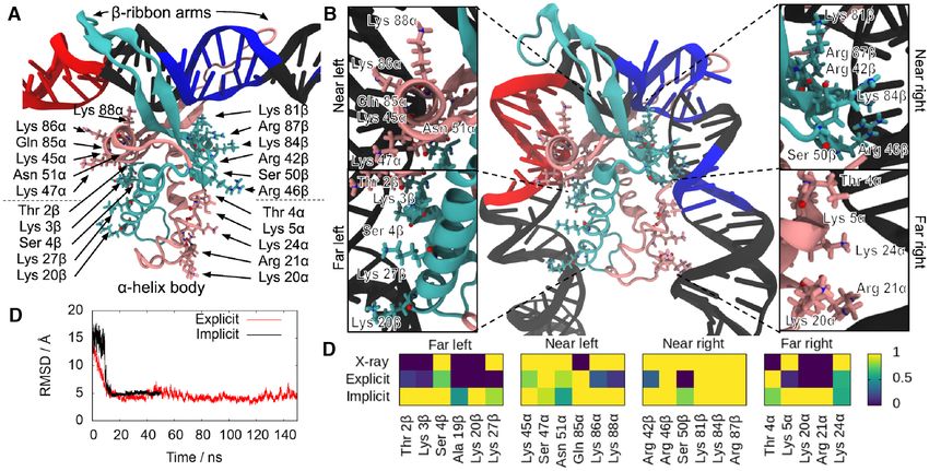
Nucleic Acids Research, 2021, Vol. 49, No. 15 8685
of extended −ribbon arms whose tip each contains a con- 37◦ C with shaking at 180 rpm to an OD600 ∼ 0.6. Overex-
served proline that intercalates between two base pairs (Fig- pression of IHF was induced by the addition of IPTG and
ure 1). These two intercalations stabilize strong bends 9 bp Arabinose to final concentrations of 1 mM and 0.2% (w/v)
apart and facilitate wrapping of two DNA ‘arms’ around respectively and growth was allowed to proceed for a fur-
the protein body, tightened by electrostatic interactions be- ther 3 h. Cells were harvested by centrifugation at 4000 rpm
tween the phosphate backbone and isolated cationic amino and 4◦ C in a Sorvall SLC6000 rotor. The pelleted cells were
acids on the protein’s surface, resulting in a binding site with then resuspended in 20 ml of 10 mM Tris pH 7.5, 10% su-
a total length of 35 bp and an overall bend angle of 160˚ (see crose (w/v) before being flash frozen in liquid nitrogen and
Figure 1), which has been supported by atomic force mi- stored at ––80◦ C.
croscopy (AFM) (23,24). IHF’s consensus sequence is lo- For purification stored cell pellets were thawed on ice and
cated on the right side of the binding region and is small the buffer was adjusted to contain 50 mM Tris pH 8.4, 150
compared to the total length of the wrapped DNA. How- mM KCl, 20 mM EDTA, 10 mM DTT and 0.2 mg ml–1
ever, most of the strongest IHF binding sites include an A- lysozyme. The resultant suspension was mixed by inversion
Downloaded from https://academic.oup.com/nar/article/49/15/8684/6342455 by guest on 17 December 2021
tract to the left-hand side (upstream of the specific sequence, and left on ice for 15 min before the addition of Brij58 to
see Figure 2) that increases affinity, the degree of bending 0.1% (w/v) and a further 15 min on ice. The suspension was
and the length of the attached DNA site (25). then clarified by centrifugation at 4◦ C and 38 000 rpm in a
Previous work has shown binding occurs through a two- Beckmann Ty70Ti rotor for 60 min. Polymin P was added
step process, a fast ∼100 s step that seems to be not to 0.075% (w/v) from a 1% stock in a dropwise fashion to
sequence-specific, and a slower millisecond step which is the supernatant whilst stirring at 4◦ C, stirring was contin-
site-specific (26). This second step seems to be associated ued for 10 min before centrifugation at 4◦ C and 16 000 rpm
with an activation energy of approximately 14 kcal/mol that in a Sorvall SS34 rotor for 20 min. The supernatant was
would be related with proline intercalation (27), but the free collected before being subjected to a 50% ammonium sul-
energy of the wrapping process (Figure 1) is found to be only fate (AmSO44 ) precipitation followed by an 80% AmSO44
around 3.6 kcal/mol (28). Recent studies have shown that precipitation. In each case. the sample was centrifuged as
IHF can bend DNA flexibly with a portion of the popula- above IHF remained soluble at 50% AmSO44 and precip-
tion that is only partially bent (i.e.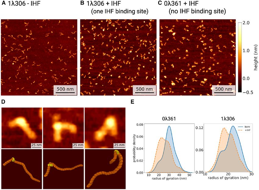
8686 Nucleic Acids Research, 2021, Vol. 49, No. 15
Downloaded from https://academic.oup.com/nar/article/49/15/8684/6342455 by guest on 17 December 2021
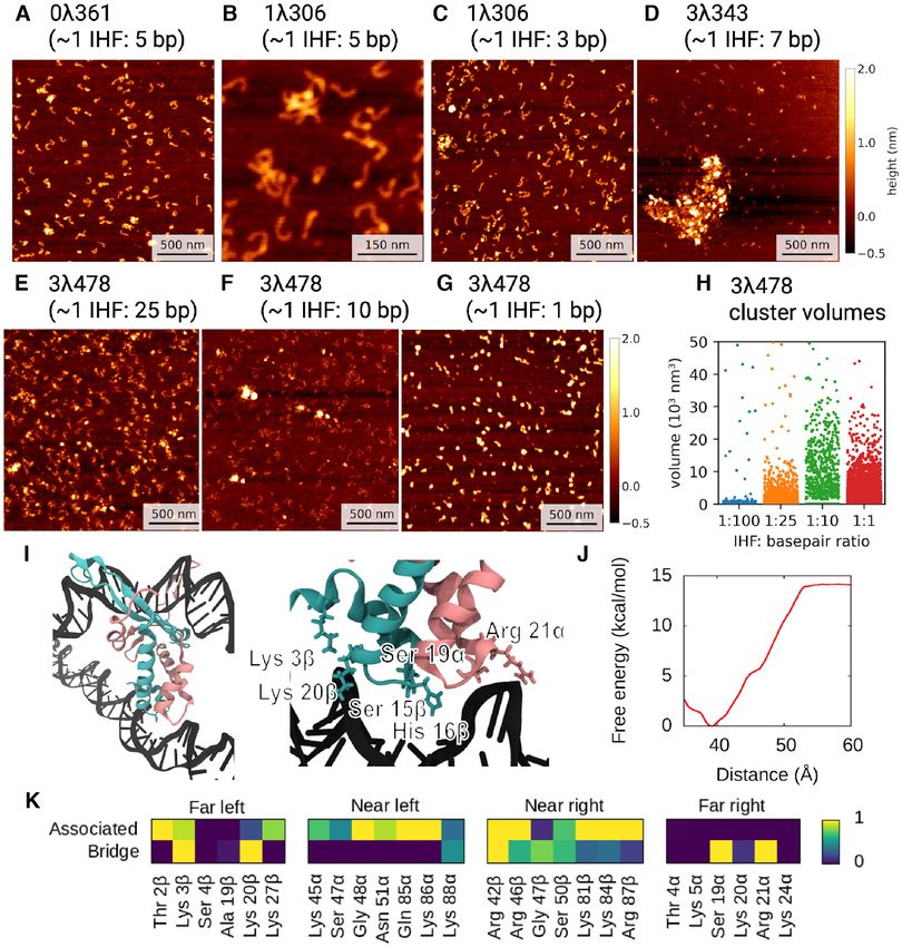
Figure 1. MD simulations of the process of DNA wrapping around IHF. Initial (A) and final (B) conformations of MD simulations in implicit solvent,
with the ␣ subunit shown in mauve and the  subunit in turquoise. The DNA is in black except for the consensus positions (in blue) and A-tract (in red).
The amino acids that interact with the lateral DNA arms in the 1IHF crystal structure are labelled and highlighted with atomic representation (N in blue, O
in red). (C) Time-evolution RMSD of trajectories in implicit and explicit solvent shows transition to a structure close to the PDB 1IHF. (D) DNA–protein
hydrogen bonds observed in the 1IHF crystal structure are also present in the trajectories on both solvent models. The number of hydrogen bonds is capped
at 1, so time-averages along simulations indicate the ratio of frames presenting interaction.
ments (S) defined by Ws = log2 (P(M)/P(B)), which eval-
uates the ratio of probabilities to find a particular sequence
in our PSSM (P(M)) with respect to a sequence background
(P(B)) (the whole E. coli genome in our case) (34). The
program also calculates the strength of a match using a
P-value, which evaluates the risk of false positives. We re-
covered the known specific binding sites with a P-value
cut-off of 0.0002 (see Figure 2). A more permissive scan-
ning, which considered all matches with Ws ≥ 1 or P(M) ≥
2P(B), revealed additional secondary sites, although prac-
tically none of them were in 0361 (see Supplementary Fig-
ure S1). This demonstrates the absence of any binding site
with a resemblance to the IHF consensus sequences in this
Figure 2. DNA constructs and IHF binding sites used in experiments.
Scheme for the different DNA fragments shown to relative scale with IHF
construct.
binding sites labelled (A) and their sequence specified (B). Only the sites
with P-values lower than 0.0002 on the PSSM scanning analysis are dis-
played by blue bars, which are sized according to the primary score (see Atomic force microscopy acquisition and analysis
Materials and Methods). The most conserved positions of the IHF spe-
cific binding sites are in the right-hand side (highlighted in bold) although Mica was freshly cleaved and then pre-treated by depositing
the presence of A-tracts in the left-hand side make the sequence more affine 20 l 0.01% (w/v) 1–5 kDa poly-L-lysine (Sigma-Aldrich,
(25). MO, USA), left for 5 min before washing with 1000 l fil-
tered milliQ H2 O and finally vacuum drying. The DNA
samples were prepared by adding a 20 l solution of 10 mM
3343 sequence to 11–351 position on 3478 (Supplemen- Tris, 50 mM KCl with 1 nM DNA (and 5–150 nM IHF)
tary Figure S1). to the pre-treated mica and leaving for ∼5 min. These were
The presence of sites with significant similarity to the then washed once more with 1000 l of filtered milliQ H2 O
IHF consensus sequence was evaluated by scanning the and vacuum dried before being imaged.
position-specific scoring matrix (PSSM) available at the All samples were imaged on a Bruker Bioscope Resolve
Regulon database (33) along our constructs using the pro- (Bruker, CA, USA) in tapping mode using TAP-300AI-
gram matrix-scan from the regulatory sequence analysis G probes (BudgetSensors, Bulgaria). Images were taken at
tools (RSAT) web server (http://rsat.ulb.ac.be/rsat/) (34). 512 × 512 px, with a pixel size of 3.9 nm. These were then
The algorithm assigns a score (Ws ) to all possible seg- loaded using pySPM (35)and preprocessed by using a first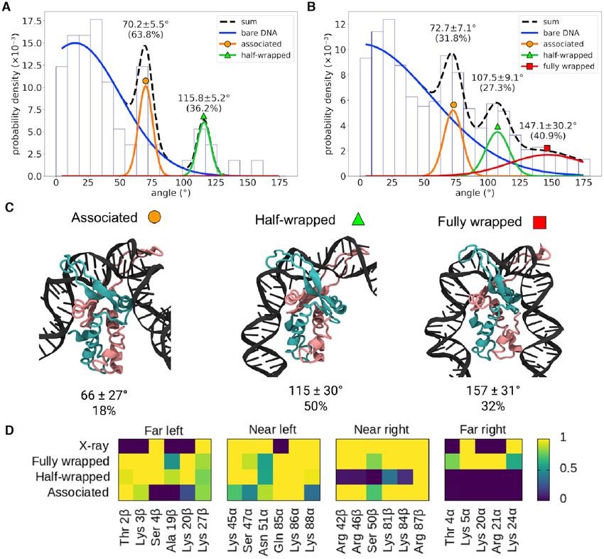
Nucleic Acids Research, 2021, Vol. 49, No. 15 8687
order line-by-line flattening and second order fits to flatten ing the simulations with unbent DNA just attached to the
the surface before filtering scars (36). extended −ribbon arms of IHF (Figure 1A). The complex
To characterize individual DNA strands, images were was then inserted at the relevant location of the linear DNA
then analyzed using custom code using the scikit-image molecule.
package (37) to threshold the image. The individual seg- A 61 bp section of the 1306 + IHF construct, centered
ments were then skeletonized to recover the DNA contour on the binding site, was explicitly solvated using a trun-
for further analysis. Segments whose length were not within cated octahedral TIP3P box and neutralized with a 0.2M-
50% of the anticipated contour length (such as debris, ag- equivalent concentration of K and Cl ions (42). The protein
gregates, ‘spot-like’ DNA or globular structures caused by and DNA were represented using the ff14SB (43) and BSC1
the deposition of poly-L-lysine in mica (38)) were discarded (44) force fields, respectively. Simulations were performed
for the subsequent analysis. They accounted for ∼15% of for 150 ns at constant T and P (300 K and 1 atm) following
the total. This heuristic threshold was sufficient to remove standard protocols (45). Only the last 10 ns sampled every
outlier objects (too short or too long) but did not result in 2 ps were used for the subsequent analysis with the idea to
Downloaded from https://academic.oup.com/nar/article/49/15/8684/6342455 by guest on 17 December 2021
any significant deletion of non-aggregated DNA molecules evaluate how similar the final state was to the original crys-
as indicated in Supplementary Figure S2. The average con- tallographic structure, which is the PDB entry 1IHF. It is
tour length per base pair was found to be around 0.30 nm worth noting that 5J0N was obtained via CryoEM and pos-
(10% smaller compared with the nominal B-DNA value of terior fitting based on 1IHF. The specific objective of this
0.34 nm), which is in agreement with the previous identi- simulation was to assess the appropriateness of the chosen
fied value for DNA absorbed on mica (39). The number of initial bound state to model the process of DNA wrapping
DNA molecules analyzed were n = 87/63 for 0361 ± IHF around IHF by reaching a comparable structure to X-ray
and n = 144/584 for 1306 ± IHF. The initial AFM ex- diffraction.
periments using the 0361 construct required front-loading Free MD simulations of the full DNA construct were
of lab access time for significant optimisation to get high done in implicit solvent for a direct comparison with AFM
quality imaging data, whereas later datasets could then images. The 1306 −/+ IHF was solvated using the implicit
use these optimised imaging and incubation conditions en- generalized Born model (46) at a salt concentration of 0.2
abling greater numbers of high quality data to be obtained. M with GBneck2 corrections, mbondi3 Born radii set and
One angle was measured per each individual DNA no cut-off for a better reproduction of molecular surfaces,
strand, first identifying the largest peak along the DNA salt bridges and solvation forces (47,48). Langevin dynam-
contour, which was IHF if present or random if not, and ics was employed for temperature regulation at 300 K with
then taking a 4 px (16 nm) vector either side of a 3 px win- a collision frequency of 0.01 ps–1 which reduces the effec-
dow around the peak. This heuristic method was designed tive solvent viscosity and, thus, accelerates the exploration
to account for the limited resolution in our AFM setup, of conformational space. Prolines were kept intercalated by
partially caused by the similar effective diameter of IHF restraining the distances between key atoms in the proline
compared to the width of the DNA. Once angular distri- side chain and the neighboring bases. Following minimiza-
butions were determined, goodness-of-fit tests were carried tion and equilibration, four independent replica simulations
out, with P > 0.99, where n = 87/63 for 0361 ± IHF and of 50 ns were performed of the 1306 + IHF construct
n = 584/144 for 1306 ± IHF (see Supplementary Figure starting from the same minimized structure. One replica of
S3 for more information). Each set of data was extracted the naked construct was also run for 50 ns starting with a
from a minimum of 3 independent samples being scanned straight B-form DNA. The last 45 ns sampled every 2 ps
at least five separated times. were used for the subsequent analysis with the objective to
For the analysis of clusters/aggregates the zero basis vol- characterize the different meta-stable states along the path-
ume (see Gwyddion documentation (40)) of each identi- ways of wrapping the DNA around the protein.
fied segment (before skeletonization) was calculated, filter-
ing any cluster smaller than 6 pixels.
Analysis of simulations
DNA bend angles were measured for the four independent
Unbiased molecular dynamics simulations
replicas containing IHF in implicit solvent using the soft-
All simulations were set up with the AMBER18 suite of pro- ware combo WrLINE/SerraLINE (36,49). The DNA he-
grams and performed using the CUDA implementation of lix axis was calculated for each frame using WrLINE (49).
AMBER’s pmemd program (41). A linear B-DNA molecule Then, SerraLINE was used to project WrLINE molecular
with a sequence of length 302 bp extracted from the xis gene contours onto the best-fit plane to approximate the experi-
of bacteriophage was generated using the NAB utility. mental methodology - and calculate bend angles (36). The
This sequence is highly homologous to the 306 bp exper- bend angle was defined as the angle between vectors join-
imental construct obtained using commercially available ing points 30 bp apart along the helix axis, where these
DNA (New England Biolabs), so both are herein referred to vectors were separated by a further 30 bp centered on the
as 1306 to minimise confusion. The both sequences have binding site. SerraLINE and WrLINE are freely available at
100% sequence identity apart from four additional base the repository github.com/agnesnoy. Following the obser-
pairs close to the ends, which represents
8688 Nucleic Acids Research, 2021, Vol. 49, No. 15
Hydrogen bonds were determined using cpptraj with a Restrained MD simulations in explicit solvent of DNA bridg-
distance cutoff of 3.5 Å and an angle cutoff of 120◦ , and the ing by IHF
time-average number of intermolecular hydrogen bonds in-
To investigate bridge formation, a second 61 bp piece of
volving each residue was calculated. In all plots, this value
DNA was pushed towards the initial IHF-DNA complex
was capped at 1, so time-averages could indicate the ratio
formed by unbent DNA of 61 bp bound just to the central
of frames from the simulation presenting interaction. The
part of the protein. This was positioned to lie perpendicu-
hydrogen bonds present in the crystal structure were deter-
lar to the main double helix in order to prevent repulsive in-
mined in the same manner based on the minimized 1IHF
teractions involving lateral DNA; other arrangements were
structure (10), so can only take integer values.
not as efficient in inducing realistic bridging for this reason
(Supplementary Figure S5). The distance between the back-
Umbrella sampling simulations in explicit solvent of DNA bone atoms closest to their centers of mass (an oxygen atom
wrapping around IHF from a phosphate group and Phe81␣ C␣) was gradually de-
Downloaded from https://academic.oup.com/nar/article/49/15/8684/6342455 by guest on 17 December 2021
creased using a one-sided harmonic potential with a spring
We performed a series of umbrella sampling (US) simula-
constant of 2 kcal mol–1 Å–2 . The asymmetric artificial po-
tions in explicit solvent to accurately calculate the free en-
tential, which was flat for nearer distances but harmonic for
ergy landscape of DNA wrapping around the lateral sides of
farer distances in relation to the target value, was used to
IHF. The initial structure was the same as used for previous
avoid biasing of the final bridged structure and, at the same
simulations in explicit solvent (i.e. unbent DNA molecule of
time, to avoid drifting of the second DNA strand away from
61 bp bound to IHF just through its protruding −ribbon
the protein−DNA complex. Following the formation of a
arms) and it was prepared as before. The reaction coordi-
bridge between the two segments of DNA, US as described
nates for the left- and right-hand sides were chosen as the
above was used to pull the second piece of DNA away from
distances between C␣ atoms of representative amino acids
the protein, with the reaction coordinate increased until a
from the protein interacting far sites (Pro18 and Ser19␣,
plateau was observed in the PMF. In all cases, the system
respectively) and the phosphorus atoms from the closest
was explicitly solvated and set up as before.
DNA base in the crystal structure. The reaction coordinates
were reduced over a series of 5 ns simulations in 2 Å incre-
ments from their positions in the minimized structure (Fig- RESULTS
ure 1A) until the PMF was observed to increase, resulting in
Modeling the process of DNA wrapping around IHF
a total simulation length of 150 ns for the left arm and 195 ns
for the right arm. The final frame of each US window was We created a structure with the non-curved H2 binding site
used as the starting structure for the next. The Grossfield attached to IHF via only its protruding −ribbon arms to
implementation of the weighted histogram analysis method simulate how DNA becomes wrapped around the protein
(WHAM) (50) was used to extract the potential of mean following the formation of an initial bound state (Figure
force (PMF). 1A). We embedded the complex in a DNA construct just
To account for shifts in the free energy landscape between over 300 bp for simulations in implicit solvent and subse-
simulations due to the flexibility of the two-atom reaction quent comparison with AFM images over the analogous
coordinate, a linear fit was performed in gnuplot to trans- construct. A shorter 61 bp DNA fragment was extracted
late the original distances to the distance between the cen- from the long piece of DNA with the idea of performing
ters of mass of the protein and the 10 bp region of DNA a more rigorous simulation in explicit solvent that would
in closest contact with each far side (Supplementary Figure evaluate the validity of the implicit solvent model and the
S4A). The free-energy offsets were estimated by comparison chosen initial bound state (see Materials and Methods). A
of the means in the plateau of the unbent state. continuum representation of the solvent reduces the com-
For each arm, two sets of US simulations were per- putational cost of simulations compared with the use of an
formed. In the first, the other arm was unrestrained and al- actual solvation box with discrete water molecules and ions,
lowed to bind to the protein. In the second, the other arm allowing the size of the system to be scaled up to what is
was held away from the protein by a potential with a spring workable for AFM experiments. However, non-bonded in-
constant of 2 kcal mol–1 Å–2 if its reaction coordinate fell teractions are not so accurately described on implicit sol-
Nucleic Acids Research, 2021, Vol. 49, No. 15 8689
Ser47␣ and Arg46 residues into the minor grooves of lat- case of 1306 and non-specifically for 0361. However, as
eral DNA (Figure 1B) and the additional interactions be- the radius of gyration and the rest of dimensional parame-
tween the DNA backbone and positively-charged and polar ters (Supplementary Figure S2) can only determine overall
amino acids. compaction, a more precise bending angle analysis was also
These interactions can be broadly divided into four re- performed.
gions based on their position relative to the center of the Our limited resolution prevented us from measuring
binding site and the protein subunit to which the involved DNA angles around IHF exclusively, so we considered
amino acid belongs. On the left-hand side (containing the bending distributions of naked DNA as a background (see
A-tract), the ␣ subunit is closer to the center and thus con- Materials and Methods). A large proportion of the total
stitutes the ‘near left’ side, while the  subunit is farther and values (∼60–70%) obtained with IHF were indistinguish-
composes the ‘far left’. On the right-hand side (containing able from the bending behavior of bare DNA molecules
the consensus sequence), the ␣ and  subunits are inversely (Figures S3A and B) as a result of the natural overlap be-
arranged, delimiting the ‘far right’ and ‘near right’ sides, re- tween the two angle distributions and the presence of some
Downloaded from https://academic.oup.com/nar/article/49/15/8684/6342455 by guest on 17 December 2021
spectively (see Figure 1B). unbound DNA, and this was accounted for in the subse-
There is generally strong agreement between the DNA- quent analysis.
protein hydrogen bonds presented by the X-ray diffraction The addition of IHF leads to a further two and three
structure and simulations irrespective of the solvent model peaks in the angular distributions for 0361 (Figure 4A)
used. We observed a more defined set of interactions at and 1306 (Figure 4B), respectively, compared to the sin-
the ‘near’ sites compared with the ‘far’ ones that could be gle peak found in bare DNA, according to the reduced chi-
caused by an increase of flexibility on the DNA part far- square goodness-of-fit tests (P-values > 0.99; see Figures
ther away from the main recognition side. In addition, the S3C and D). The two-sided Kolmogorov−Smirnov test was
shortness of the DNA at the crystal structure (35 bp versus applied to confirm the statistical difference of 1306 bend
61 bp on simulations) makes it difficult to capture some of distribution compared to 0361 in the presence of IHF (P-
the more distant interactions. value = 0.002) and from naked DNA (P-value = 0.008).
In general, our results demonstrate the validity of the The same test did not find statistically significant difference
molecular dynamics methodology for this system, as well between distributions of 0361 with/without IHF, proba-
as suggesting an explanation for the previously proposed bly due to the larger overlap and the larger amount of un-
two-step binding mechanism. In this two-step model, the bound DNA caused by the relatively low affinity, although
IHF arms would bind to DNA first and then the proline the presence of more than one peak was suggested by the
residues would intercalate to induce flexible hinges prior previous chi-squared test.
to wrapping. Although our simulations start from straight The peaks around 73 ± 7˚ and 107 ± 9˚ (mean ± s.d.
DNA with the prolines already intercalated, it is possible values from 1306) are common to both constructs, sug-
that some bending may occur following the initial recogni- gesting that they occur due to non-specific binding, whilst
tion that would facilitate the intercalation of prolines and the canonical large bending angle (147 ± 30˚), as seen in
the subsequent strong kink. In any case, the binding mech- the crystal structure (5), only appears in 1306 that con-
anism that we propose explains both the high activation en- tains one specific IHF binding site. The proportions of the
ergy of initial binding (27) due to proline intercalation and 73˚, 107˚ and 147˚ states (approximately 32%, 27% and
the smaller free energy of wrapping (28). 41%, respectively) show that all three binding modes are
present in roughly equal quantities for the construct with
a specific binding site (1306). However, the proportion
Multimodality of IHF specific and non-specific binding re-
of the 73˚ state (∼64%) to the 107˚ (∼36%) state is much
vealed by AFM
larger for 0361, suggesting the former state is mostly re-
To investigate the differences between specific and non- lated to non-specific binding. Some binding could still oc-
specific binding, two short sequences were amplified from cur as similar sequences to the left part of the specific bind-
phage , one with no IHF consensus sequence (0361) and ing site (an A-tract followed by AT base pairs downstream)
another with a single consensus sequence (1306) of lengths are present in the sequence of 0361, although they do
361 and 306 bp, respectively (Figure 2). The lack of se- not count towards the PSSM calculation (Supplementary
quences with similarity to the IHF specific recognition mo- Figure S1).
tif was confirmed by scoring the entire 0361 molecule to We then isolated the subpopulation of DNA molecules
the PSSM available at the Regulon database (33) (see Fig- with bend angles >54˚ and >126˚ in the presence of the pro-
ure 2, S1 and Materials and Methods). The two DNA frag- tein and we observed a reduction (mean ± s.e.) in the radius
ments were then imaged using AFM and compared to MD of gyration (19.1 ± 0.2 and 17.3 ± 0.4 nm, respectively) and
simulations to determine the different morphological be- end-to-end distance (51.5 ± 0.8 and 37.0 ± 1.5 nm, respec-
haviors. tively) compared with the construct in the absence of IHF
DNA contours were recovered by skeletonizing pre- (radius of gyration of 22.4 ± 0.4 nm and end-to-end dis-
processed AFM images (see Materials and Methods), with tance of 68.5 ± 1.4 nm), demonstrating the global effect of
qualitatively different behavior observed depending upon the IHF-induced bend.
the presence of IHF (Figures 3). Figure 3E shows a limited Our results are largely consistent with previous AFM
reduction of the radius of gyration for the two constructs studies where a broad distribution of angles was detected
if IHF is present, suggesting that the protein bends DNA between approximately 80–150˚ (18,19). The 73˚ state is one
to some extent in both cases, in a specific manner in the that has not been observed in AFM before, possibly as it8690 Nucleic Acids Research, 2021, Vol. 49, No. 15
Downloaded from https://academic.oup.com/nar/article/49/15/8684/6342455 by guest on 17 December 2021
Figure 3. Atomic force microscopy (AFM) images of DNA with and without IHF. (A) AFM image of DNA with one IHF binding site (1306) without
IHF. In the presence of IHF (1IHF:5 bp), DNA with one IHF binding site (1306) (B) shows more binding when compared to DNA without a binding site
(0361) (C), although globular artefacts from poly-L-lysine can be seen. (D) Three representative examples of IHF binding, with a comparison of AFM
(top) and molecular dynamics simulations (bottom). (E) Kernel density estimates of the radius of gyration of DNA ± IHF.
is within the expected range of angles for bare DNA and 59 ± 14 nm, −/+ IHF respectively) (Supplementary Fig-
could be excluded if not accounting for the bare DNA dis- ure S2), thus demonstrating the validity of our approach. In
tribution (Figures S3A and B) or due to the selection of an addition, we found that the deviation from planarity of the
H2 binding site. complexes was less than 10% on average, which indicated
small distortions on the complex due to surface absorption
The multiple IHF binding modes are confirmed by MD sim- (Supplementary Figure S6).
ulations All the frames of the replicas with IHF were merged to-
gether and were classified into three groups through hier-
To validate the states deduced from AFM, four independent archical agglomerative clustering using RMSD as the dis-
(replica) unrestrained MD simulations were performed of tance metric, finding groups with bend angles of 66 ± 27◦ ,
the 1306 + IHF construct in implicit solvent (Figure 3D, 115 ± 30◦ and 157 ± 31◦ , respectively, in good agreement
Supplementary Movies 1–4), starting from the bound but with the AFM results (see Figure 4). A representative frame
unbent state shown in Figure 1A. One unrestrained MD was selected for each group in Figure 4C and the mean bend
simulation was also performed of the naked 1306 molecule angle was calculated for each. The clusters are also char-
in implicit solvent for comparison. We imitated AFM by acterized by the hydrogen bonds between the protein and
projecting the simulations to the best fitted plane so we DNA (Figure 4D). In the ‘fully wrapped’ state (with a bend
could compare DNA bend angles (see Materials and Meth- angle of 157◦ ), both sides of the DNA form hydrogen bonds
ods). Averages and standard deviations of radius of gyra- with both subunits of the protein; in the ‘half-wrapped’ state
tion (25 ± 1 and 21 ± 1 nm, −/+IHF respectively) and (115◦ ), the left-hand side, which contains the A-tract, forms
end-to-end distances (81 ± 7 and 49 ± 8 nm, −/+IHF contacts with both subunits while the right-hand side forms
respectively) of the projected trajectories were found not no hydrogen bonds; and in the ‘associated’ state (66◦ ), only
to be significantly different from AFM (radius of gyration the ‘near’ subunits on the left and right sides form hydrogen
22 ± 5 and 20 ± 7 nm, end-to-end distance 68 ± 17 and bonds with the DNA.Nucleic Acids Research, 2021, Vol. 49, No. 15 8691
Downloaded from https://academic.oup.com/nar/article/49/15/8684/6342455 by guest on 17 December 2021
Figure 4. Analysis of different bending angles of IHF-bound DNA. Bending angle distribution obtained by AFM of 0361 (n = 87) (A) and 1306 (n = 144)
(B) with IHF. Distributions observed without IHF (blue lines, Figures S3A and B) are considered as background and disregarded. The remaining peaks
are well fitted by Gaussians (as shown by the reduced χ 2 in Figures S3C and D), representing two common states with bending angles around ∼70˚ and
∼110˚ and an additional state presented only by 1306 with an angle around ∼147˚, closer to the X-ray structure. The broadness of the peak around ∼147˚
is caused by resolution issues that make angles more difficult to measure as they get larger (23). (C) Simulations of the same construct (in implicit solvent)
can be classified into three clusters, the mean bending angles of which correspond well with the experimental data. Proportions for each state are obtained
via US simulations in explicit solvent (see Figure 5). Colour scheme is the same as in Figure 1. (D) The clusters (from implicitly solvated simulations) can
be characterized by the DNA–protein hydrogen bonds present in each state; while the fully wrapped DNA interacts with the protein on both sides, the
half-wrapped DNA interacts only on the left, and the associated state interacts primarily with the ‘near’ sites. Contact maps show the average number of
frames presenting at least one hydrogen bond between DNA and each residue using all frames belonging to each cluster or from the 1IHF crystal structure.
Bend angle distribution extracted from the replicas with Each individual replica presents a slightly different be-
IHF exhibits three peaks (68 ± 37◦ , 117 ± 4◦ and 149 ± 23◦ ; havior in terms of the time spent at the different states
see Supplementary Figure S4C) that align well with AFM and in the transitions between them (Supplementary Fig-
and with the previous structural classification, demonstrat- ure S4D). In the first one, the complex rapidly reaches the
ing the consistency of our methodology. However, we do fully wrapped state after briefly passing through the half-
not expect good agreement on the proportions of the dif- wrapped. Because this is the replica with the most canoni-
ferent states (4% associated, 42% half-wrapped, 54% fully cal behavior (more like the crystal structure), it is the one
wrapped) due to the limitations on the sampling. selected in Figure 1 for assessing the recovery of crystal-8692 Nucleic Acids Research, 2021, Vol. 49, No. 15
lographic interactions by means of implicitly solvated sim-
ulations. In the second replica, the complex oscillates be-
tween the half- and fully wrapped states, and, in the third
replica, it remains in the former. In the last replica, the
complex traverses the associated state before arriving to the
fully wrapped state. These simulations suggest a metastable
nature for the partially bent states (associated and half-
wrapped) in the course of DNA binding around IHF be-
fore reaching the global minimum occupied by the fully-
wrapped state.
To properly describe the free energy landscape of the
wrapping process and the proportion of the different states,
we performed restrained MD simulations (53,54) of a 61 bp
Downloaded from https://academic.oup.com/nar/article/49/15/8684/6342455 by guest on 17 December 2021
segment of the construct in explicit solvent (see Materials
and Methods). A more accurate representation of the sol-
vent was chosen on this occasion for describing DNA−IHF
interactions at a more rigorous level, enabling us to verify
the previous implicitly solvated simulations. The distance
between the bottom part of the protein and the interact-
ing DNA on each side was varied in a series of US simu-
lations, once with the other arm allowed to wrap and once
with the other arm held away from the protein. The PMF for
each simulation was calculated using the WHAM method
(50,55). By linearly interpolating between the two sets of re-
sults for each arm (Figure 5A and B) a two-dimensional free
energy landscape was constructed with the reaction coordi-
nates as orthogonal axes, as in Figure 5C.
The relative probabilities of the clusters were determined
by integrating over the relevant regions of the free en-
ergy landscape (Figure 5D and Supplementary Figure S4B).
This analysis predicts that the canonical fully wrapped
(157◦ ) state should be observed around 32% of the time,
the half-wrapped (115◦ ) state 50%, and the associated (66◦ ) Figure 5. Free energy of DNA bending by IHF. The free energy landscapes
state 18%; while these figures do not directly agree with determined through US simulations in explicit solvent differ for the left
AFM, probably due to the differences between the simu- arm (A), containing the A-tract, and the right arm (B), containing the
lation and experimental conditions, they do predict that all consensus sequence. The left arm presents a deep potential well regard-
three states should occur in significant proportions. Each less of whether the right arm is free to bind (red) or held away from the
protein (blue), while the right arm can bind fully only when the left arm
cluster corresponds to a minimum or plateau in the free en- is bind (red) and not held away (blue). (C) The 2D reconstruction of the
ergy landscape; the locations of these features, along with energy landscape via linear interpolation between arm’s positions shows
the observed paths between them (Supplementary Figure the striking asymmetry. (D) The observed clusters from implicitly solvated
S4D), are highlighted in Figure 5D. simulations correspond to minima and plateaus in the free energy land-
scape plotted here along with the approximate paths by which replicas tra-
versed. Fully wrapped is represented by a red square, partially-wrapped by
Asymmetry between DNA arms causes a mechanical switch a green triangle and associated state by orange circle. The white cross rep-
resents a sub-state from the fully wrapped form where left-hand side is fully
towards strong bent DNA bound and the right-hand side is partially bound. (E) Two superimposed
As discussed in the previous section, it is notable that the structures with the left arm bound (blue) and unbound (red) showing that,
when the left arm is unbound, the upper subunit protrudes on the right-
consensus sequence does not interact with IHF in the half- hand side, making it difficult for the DNA to interact with the bottom of
wrapped state and this state is also observed in the absence the protein.
of a specific binding site. The free energy surface, which is
described as a function of the two distances between the
bottom of the protein and the two DNA arms, reveals that While the free-energy landscape presented by the left-
there is a large activation barrier preventing the right-hand hand side appears to be independent of the position of the
side (which contains the consensus sequence) from binding right arm, the right-hand side is prevented from binding
before the left-hand side (which contains the AT-tract) (Fig- fully by a large barrier when the left arm is held away from
ure 5C). The form of the potentials is also qualitatively dif- the protein; this barrier is not present when the left arm
ferent. While the left-hand side presents a relatively deep is fully bound. This barrier is associated with a change in
potential with a minimum around 34 Å and an additional the structure of the protein. When the left arm is unbound,
small plateau at around 40 Å, implying that binding is en- the upper subunit of the protein on the right-hand side pro-
ergetically favorable (Figure 5A), the right-hand side has a trudes, so full binding of the right arm would require extra
much flatter shape with small minima around 31 and 43 Å, DNA bending apart from the flexible region. Binding of the
and appears to be dominated by thermal noise (Figure 5B). left arm flattens the surface presented to the right arm, re-Nucleic Acids Research, 2021, Vol. 49, No. 15 8693
moving this physical barrier and allowing the DNA on the ber of IHF/DNA clusters appear to form with a volume
right-arm to bind while remaining mostly straight (Figure of the order of 104 nm3 (Figure 6H) compared to volumes
5D). of around 103 seen for individual double-stranded DNA
This free energy landscape suggests that it is very energet- molecules (Supplementary Figure S2). At larger concentra-
ically favorable for the protein to bind the A-tract on its left- tions (1 IHF: 1 bp), IHF was numerous enough to coat the
hand side, but that it is less inclined to bind the consensus se- DNA reducing the size of visible clusters (Figure 6G and
quence, and that the A-tract must bind first (Supplementary H).
Movie S5). This is reflected in the observed clusters––there A shorter form of this construct (3343, 343 bp long)
is no cluster in which the left-hand side is unbound, while (Figure 6D) was seen to show larger aggregates, reaching
the position of the right-hand side distinguishes the fully volumes of order 105 –106 nm3 at 1 IHF:7 bp. The reason
and half-wrapped states (Figure 4C and D). The associated for the prominence of aggregation on the latter construct
state corresponds to the smaller local plateau or minimum could be related with the elimination of spare DNA, which
in each arm’s energy landscape (Figure 5), as seen when the might reduce electrostatic repulsion and steric extrusion be-
Downloaded from https://academic.oup.com/nar/article/49/15/8684/6342455 by guest on 17 December 2021
values of the reaction coordinates are plotted together for tween DNA duplexes. 3343 and 3478 are highly homolo-
each frame of a cluster as in Supplementary Figure S4B. gous apart from a tail of around 125 bp on one side of the
The lack of interaction of IHF with the consensus bases three binding sites in the longer construct that has a much
suggests that the half-wrapped and associated states are lower AT-content (52%) compared with 3343 (70%) and
forms of non-specific binding, a conclusion supported by thus presents lower affinity to IHF (Supplementary Figure
the occurrence of bend angles corresponding to these states S1). Although these conglomerates may be due to protein
in AFM observations of the 0361 construct (in which the aggregation, the difference between these and the earlier
consensus sequence does not appear). The consensus se- constructs demonstrates that it is not the case.
quence is therefore necessary only for the fully wrapped IHF-induced aggregation can be explained by the bridg-
binding mode, suggesting that initial binding of IHF to ing of two separate double-stranded DNA segments by a
DNA occurs without sequence specificity and a consensus single IHF dimer, a phenomenon observed in our MD sim-
sequence enhances bending. ulations (Figure 6I). These bridges result from non-specific
The free energy landscape features a significant minimum interactions involving positively charged and polar amino
that appears to correspond to another state (represented acids from the far binding sites of the protein and the back-
by a white cross in Figure 5D), in which the left-hand side bone of DNA (Figure 6K). US simulations (see Materi-
is fully bound and the right-hand side is partially bound. als and Methods) reveal that bridging is very energetically
However, the frames in this region are difficult to distin- favorable when a second double-stranded DNA is close
guish from those in the fully wrapped state as the bending enough, with a free energy difference on the order of 14
angles are very similar, and the corresponding minima in kcal/mol between the bridged and unbridged states (Figure
the free-energy landscape are separated by only a narrow 6J). The main DNA fragment, into which the prolines inter-
energy barrier of less than 1 kcal/mol. Since the system can calate, remains mostly unwrapped, probably due to the elec-
move freely between these two minima under the influence trostatic repulsion exerted between the two double-stranded
of thermal noise, it is more appropriate to consider this state DNA molecules (Figure 6I and Supplementary Figure S5).
an extension of the fully wrapped mode. The combination of AFM and MD suggests that the clus-
The asymmetric cooperative behavior observed between tering of constructs containing three binding sites is due to
arms in the presence of a specific sequence could serve for the capacity of IHF to bind more than one DNA strand at
reinforcing the prevalence of the fully-wrapped state, as it the same time. We observe that DNA·IHF·DNA bridging is
reduces the amount of accessible intermediate states that favourable even for low concentrations of IHF. The strong
the system would present in the case of both arms moving electrostatic repulsion between close DNA strands could
in a totally uncorrelated manner. This interconnection be- prevent the complete wrapping of DNA around the protein,
tween the two halves would help the IHF to sharply bend even on sequence-specific sites, facilitating the bridging in-
the DNA with a substantial probability in a similar way to teraction described above.
a mechanical switch.
DISCUSSION
DNA aggregation and bridging by IHF
Here, we have integrated advanced theory and experiments
At concentrations of 1 IHF:5 bp, occasional small clusters from physics to explore a basic phenomenon of life at the
of DNA/protein could be seen for both 0361 and 1306 single-molecule level (56), namely how individual proteins
(Figures 6). Further increases of the IHF concentration interact with DNA. Our findings enable new insights into
causes the formation of clusters for 1306 (Figure 6C). This the complex interactions between proteins and DNA. Both
behavior at a higher concentration (∼1 IHF:3 bp) has pre- AFM experiments and MD simulations show evidence of a
viously been seen (19) and shows how large cellular con- larger number of bending modes of the IHF/DNA complex
centrations of IHF––such as in the stationary phase––could than previously proposed. The presence of three states (fully
also increase genome compaction by bridging. wrapped, half-wrapped and associated) as well as the large
A construct with three binding sites (3478) behaves very proportion of unbound DNA suggests a multi-modular sys-
differently to the previous constructs that contain one or tem, where higher AT-content makes binding more proba-
no specific binding sites. Even at low concentrations, from ble (57,58) but in which a consensus sequence is needed to
around 1 IHF: 10–25 bp (Figures 6E and F), a greater num- fully bend DNA. We put forward a model for the mecha-8694 Nucleic Acids Research, 2021, Vol. 49, No. 15
Downloaded from https://academic.oup.com/nar/article/49/15/8684/6342455 by guest on 17 December 2021
Figure 6. IHF-bound DNA forms aggregates depending on protein concentration and on the type of DNA constructs. A large amount of IHF leads to
some aggregation on 0361 (A) and 1306 (B, C), with no or one binding site respectively. DNA constructs with three binding sites (3343 and 3478) start
to form clusters at lower concentrations. (D) Large aggregates are observed for 3343. (E, F) 3478 forms more numerous and/or denser clusters as IHF
concentration increases until saturation is achieved (G). (H) Cluster volumes for 3478 as function of IHF concentration (I) Bridge structure and zoom
from MD simulations with the interacting amino acids highlighted in atomic representation. Colour scheme is the same as in Figure 1. (J) Free-energy
landscape of bridging formation relative to an unwrapped state with a close additional DNA strand. (K) Contact maps are calculated as in Figures 1 and 3
revealing that the residues from the ‘far’ interacting regions are those which form the bridge. The DNA strand into which IHF intercalates remains mostly
unbent, interacting with the protein even less than in the associated state.
nism of IHF binding where full wrapping is sequence spe- from our simulations. This is a larger estimation compared
cific but initial binding is not (Figure 7). to single-molecule experiments (3.6 kcal/mol) (28) because
First, we provide structural insights into the two-step simulations were started with the most possible unbent con-
binding mechanism previously proposed (27), as the crys- figuration for DNA, so it is the upper limit of free energy
tallographic structure is reached by our simulations when relaxation associated to the wrapping process. Either the in-
started from a bound but unwrapped DNA (Figure 1). tercalation of prolines is followed by a rapid bending relax-
Thus, our results reinforce the idea that the protein first ation, or the first binding step favors some bending that fa-
binds DNA non-specifically via its extended arms; the pro- cilitates proline intercalations, being the two contributions
lines at the tips of the arms would then intercalate with (intercalation or bending) difficult to be neatly discrimi-
an activation energy around 14 kcal/mol (27); from there, nated from experimental data. In any case, simulations sup-
DNA wraps around the protein in a relatively downhill port a bind-then-bend mechanism that has also been ob-
process stabilized by up to 9 kcal/mol, a value calculated served in other DNA-flexing proteins (59).Nucleic Acids Research, 2021, Vol. 49, No. 15 8695
Downloaded from https://academic.oup.com/nar/article/49/15/8684/6342455 by guest on 17 December 2021
Figure 7. Model of IHF binding – bending and bridging. (A) IHF first recognizes DNA through its -arms and the prolines intercalate into a double-helix
structure that its straight in our model although it might already contain a certain bent. The first binding step appears to be either a loose association to
the DNA (73˚ binding) or a half-wrapped state (120˚). Both can then progress to the canonical fully wrapped state. As the initial state leaves the bottom
half of IHF unbound it can also bind to another strand of DNA non-specifically resulting in a bridge (transition conditioned to the second DNA strand
being nearby, dashed arrow). Free energy differences between states (F) were estimated via US simulations in explicit solvent. (B) A construct with a
single binding site can only form a single bridge whereas (C) multiple binding sites can form multiple bridges, leading to aggregation.
When IHF binds to DNA, it is highly likely that the left- tion of the fully-wrapped state was found to be higher in
hand side will bind to the DNA first, but not with the con- their experiments than in our results, but this difference
sensus part of the sequence. This results in the half-wrapped might be caused by the use of a different binding site (H’
state, which is ∼8 kcal/mol more stable than the initial versus H2, see Figure 2).
structure and in which the left-hand side is fully bound while The half-wrapped and associated states may not be the
the right-hand side is free. Alternatively, both sides may only conformations in which IHF binds non-specifically to
make interactions with the nearest subunit of the protein, DNA. Hammel et al. (60) obtained crystal structures of HU
resulting in a smaller bending angle, that we designate the where the DNA was bound across the ␣-helical body of the
associated state (favored by ∼5 kcal/mol). Transitions be- protein rather than between the extended -ribbon arms.
tween the associated and half-wrapped states were not ob- The similar electrostatic profiles on IHF and HU (see Sup-
served (Supplementary Figure S4D), and both appear to plementary Figure S7) suggest that this extra non-specific
be long-lived metastable states corresponding to plateaus binding mode could be possible in IHF. However, such state
or local minima in the free-energy landscape (Figure 5D). would need a different initial conformation for being ex-
As the associated and half-wrapped states are experimen- plored by simulations and we predict that it would not in-
tally seen in constructs both with and without a consensus duce a significant bend stronger than the typical angles for
sequence, this suggests a non-specific binding mechanism bare DNA, so this possibility was not explored in this work.
for these states. However, in the presence of a consensus se- The asymmetric allostery, by which DNA binds to the
quence, both of intermediate states lead towards the global right side only after binding to the left side, makes the
minimum (F ≈ −9 kcal/mol relative to the initial struc- fully wrapped state relatively more probable than both arms
ture), resulting in canonical binding. moving independently. This mechanism, thereby, constructs
Previous experiments (19,29,30) already indicated that a mechanical ‘switch’, since the ultimate structural configu-
IHF could bind DNA in more than one state, generally ration can switch between different states in a mechanically
in two states which were broadly described as a fully and dependent manner. The formation of such a sharp bend
partially bent. Magnetic tweezers experiments appeared to in one of these observed states is impossible to achieve in
show a smaller angle state ∼50˚ (19) (i.e. similar to our as- naked DNA, even for the most curved sequences (61,62) un-
sociated state). The lack of detection of a third state could less base stacking and complementary hydrogen-bond pair-
be due to the fact that the force applied on the DNA was ing are disrupted on the double helix generating kinks or
greater than the few tenths of piconewtons we predict is melting bubbles (36).
needed to overcome the potential barrier between the half- This regulatory behavior could be switched on or off
wrapped and fully wrapped states. By using fluorescence- by tension, structural influences upstream of the binding
lifetime-based FRET (29), Ansari and co-workers deduced site or by different levels of DNA supercoiling. The semi-
the presence of three binding modes and that two of these stable states observed would allow IHF to remain associ-
involved partially-bend DNA, in line with the results pre- ated with the DNA while retaining some flexibility, which
sented here, although they did not have the resolution could be important in the formation of higher-order nucle-
and/or the associated modelling to detect the structural oprotein complexes such as transcription regulatory loops.
properties of the non-canonical conformation. The propor- Similarly, other proteins in mammalian systems might ex-8696 Nucleic Acids Research, 2021, Vol. 49, No. 15
pect to behave in similar meta-stable ways when interacting DATA AVAILABILITY
with DNA under physiological levels of superhelical ten-
All relevant data is included in the main manuscript, the
sion. For example, nucleosomes present spontaneous un-
supplementary material and the University of York Data
spooling of the outer stretches of DNA causing multiple lev-
Repository (https://doi.org/10.15124/0b602b82-16e9-4fa8-
els of wrapping around the histones (63). These meta-stable
9a64-a15d7373f80e).
states are modulated by tension (64) or by the presence of
neighboring nucleosomes (65) and regulate the access to nu-
cleosomal DNA (66). SUPPLEMENTARY DATA
Remarkably, we observe that the more conserved bases
are within the region that is more dynamically bound to Supplementary Data are available at NAR Online.
IHF, which could illustrate the need for flexibility down-
stream of the protein or, as an alternative, it could reflect ACKNOWLEDGEMENTS
the difficulty of identifying consensus sequence motifs, and
Downloaded from https://academic.oup.com/nar/article/49/15/8684/6342455 by guest on 17 December 2021
therefore cognate binding sites, in the case of DNA shape We thank the Physics of Life (PoL) Group, University of
recognition (67). Similarly, it was found that the transcrip- York for providing pump-priming resources. We would also
tion factor GabR from Bacillus subtilis needs to recognize like to thank Alice Pyne for her assistance with the AFM
flexed DNA at a location distinct from its known binding experimental techniques and analysis.
sites (68).
Another interesting behavior observed was the FUNDING
bridging/clustering of DNA by IHF. At high concen-
trations of IHF (such as during periods of inactivity of cell Engineering and Physical Sciences Research Council (EP-
division and DNA replication when the bacterial nucleoid SRC) [EP/N027639/1, EP/T002166/1, EP/R029407/1,
is at its most compact in the cell cycle) this can occur EP/P020259/1]; Biology and Biotechnology Research
non-specifically, giving it a role in compaction. On the Council (BBSRC) [BB/R001235/1]; Leverhulme Trust
other hand, the constructs with multiple binding sites seem [RPG-2017-340]; V.V.-B. was funded by CONACYT agency
to preferably select for the bridging behavior over bending from Mexican government [291163]; calculations were per-
when DNA molecules are moderately concentrated. This formed on ARCHER, JADE, Cambridge Tier-2 and the lo-
behavior shows how the formation of bridges could be cal York facilities (Viking and YARCC clusters). Funding
driven by the screening of electrostatic repulsion between for open access charge: York Open Access Fund or EPSRC
neighboring DNA molecules thanks to positively-charged [EP/N027639/1].
architectural proteins (69). In this regard, hidden secondary Conflict of interest statement. None declared.
recognition sites causing DNA bridging by means of basic
amino acids have also been identified for other bacterial
REFERENCES
architectural proteins like Topoisomerase IB (70) and ParB
(71,72). This phenomenon could also be promoted by 1. Dame,R.T., Rashid,F.Z.M. and Grainger,D.C. (2020) Chromosome
steric hindrance or tension due to the presence of other organization in bacteria: mechanistic insights into genome structure
and function. Nat. Rev. Genet., 21, 227–242.
proteins preventing complete wrapping by each IHF, 2. Dillon,S.C. and Dorman,C.J. (2010) Bacterial nucleoid-associated
leaving the lower regions of the protein free to bind other proteins, nucleoid structure and gene expression. Nat. Rev.
DNA. Microbiol., 8, 185–195.
Our study not only explains the role of IHF in biofilms, by 3. Dorman,C.J., Schumacher,M.A., Bush,M.J., Brennan,R.G. and
Buttner,M.J. (2020) When is a transcription factor a NAP? Curr.
cross-bridging extracellular DNA, but undoubtedly shows Opin. Microbiol., 55, 26–33.
that the function of IHF is far more multifaceted than bend- 4. Verma,S.C., Qian,Z. and Adhya,S.L. (2019) Architecture of the
ing DNA. We observe that specific binding sites can be Escherichia coli nucleoid. PLos Genet., 15, e1008456.
simply modulated or extended by additional non-specific 5. Hacker,W.C., Li,S. and Elcock,A.H. (2017) Features of genomic
electrostatic-driven interactions between the protein and organization in a nucleotide-resolution molecular model of the
Escherichia coli chromosome. Nucleic Acids Res., 45, 7541–7554.
the DNA (69,73). In fact, positively charged patches on 6. Gruber,S. (2014) Multilayer chromosome organization through DNA
DNA-interacting proteins have been traditionally used for bending, bridging and extrusion. Curr. Opin. Microbiol., 22, 102–110.
predicting DNA-binding interfaces (74). We anticipate that 7. Stella,S., Cascio,D. and Johnson,R.C. (2010) The shape of the DNA
this could be a general mechanism used by other NAPs (75) minor groove directs binding by the DNA-bending protein Fis. Genes
Dev., 24, 814–826.
and eukaryotic chromatin-binding proteins (76,77) to en- 8. Skoko,D., Yoo,D., Bai,H., Schnurr,B., Yan,J., McLeod,S.M.,
able a variety of DNA bending and bridging modes. Promis- Marko,J.F. and Johnson,R.C. (2006) Mechanism of chromosome
cuous electrostatic interactions between negatively-charged compaction and looping by the Escherichia coli nucleoid protein fis. J.
DNA and positively-charged genomic architectural pro- Mol. Biol., 364, 777–798.
teins could be one of the primary molecular forces under- 9. Luscombe,N.M., Austin,S.E., Berman,H.M. and Thornton,J.M.
(2000) An overview of the structures of protein-DNA complexes.
pinning the physical organization of all kinds of chromo- Genome Biol., 1, REVIEWS001.
somes, including the formation of membrane-less phase- 10. Rice,P.A., Yang,S.W., Mizuuchi,K. and Nash,H.A. (1996) Crystal
separated condensates inside cells (78–80). Finally, the gen- structure of an IHF-DNA complex: a protein-induced DNA U-turn.
eral methods we have developed here for comparing AFM Cell, 87, 1295–1306.
11. Wei,J., Czapla,L., Grosner,M.A., Swigon,D. and Olson,W.K. (2014)
imaging with MD simulations have a utility that could be DNA topology confers sequence specificity to nonspecific
applied for many other protein-DNA interactions in the architectural proteins. Proc. Natl. Acad. Sci. U.S.A., 111,
chromosome, beyond just IHF. 16742–16747.You can also read