Loss of Cyclin C or CDK8 provides ATR inhibitor resistance by suppressing transcription-associated replication stress
←
→
Page content transcription
If your browser does not render page correctly, please read the page content below
Published online 30 July 2021                                                    Nucleic Acids Research, 2021, Vol. 49, No. 15 8665–8683
                                                                                                       https://doi.org/10.1093/nar/gkab628

Loss of Cyclin C or CDK8 provides ATR inhibitor
resistance by suppressing transcription-associated
replication stress
Rebecca L. Lloyd1 , Vaclav Urban2 , Francisco Muñoz-Martı́nez1 , Iñigo Ayestaran                                                       1
                                                                                                                                             ,
John C. Thomas1 , Christelle de Renty2 , Mark J. O’Connor2 , Josep V. Forment2 ,
Yaron Galanty1 and Stephen P. Jackson 1,*

                                                                                                                                                              Downloaded from https://academic.oup.com/nar/article/49/15/8665/6331679 by guest on 06 November 2021
1
 Wellcome/Cancer Research UK Gurdon Institute, and Department of Biochemistry, University of Cambridge, UK
and 2 Bioscience, Oncology R&D, AstraZeneca, Cambridge, UK

Received January 25, 2021; Revised July 04, 2021; Editorial Decision July 06, 2021; Accepted July 09, 2021

ABSTRACT                                                                        INTRODUCTION
The protein kinase ATR plays pivotal roles in DNA                               Ataxia telangiectasia and Rad3-related (ATR) is a funda-
repair, cell cycle checkpoint engagement and DNA                                mental DNA damage response (DDR) protein kinase in-
replication. Consequently, ATR inhibitors (ATRi) are                            volved in DNA double-strand break (DSB) signalling and
in clinical development for the treatment of can-                               cell cycle checkpoint engagement, and is also an apical reg-
cers, including tumours harbouring mutations in the                             ulator of the replication stress response (RSR) (1–3). ATR’s
                                                                                roles in the stabilisation and restart of stalled DNA replica-
related kinase ATM. However, it still remains un-                               tion forks are important for maintaining genomic integrity
clear which functions and pathways dominate long-                               since, in the absence of ATR function, stalled replication
term ATRi efficacy, and how these vary between                                  forks are converted into cytotoxic DSBs through replication
clinically relevant genetic backgrounds. Elucidating                            fork collapse. ATR also plays pivotal roles in unperturbed
common and genetic-background specific mecha-                                   S-phase by regulating dormant replication-origin firing and
nisms of ATRi efficacy could therefore assist in pa-                            controlling the S/G2 cell cycle transition (4). Importantly,
tient stratification and pre-empting drug resistance.                           replication stress (5) has been identified as a hallmark of
Here, we use CRISPR–Cas9 genome-wide screen-                                    cancer (6). While this may be in-part due to faster prolifer-
ing in ATM-deficient and proficient mouse embryonic                             ation rates and nucleotide shortages in S-phase, amplifica-
stem cells to interrogate cell fitness following treat-                         tion of the oncogenes CCNE1 and MYC specifically induce
ment with the ATRi, ceralasertib. We identify factors                           replication stress by shortening G1 and promoting firing
                                                                                of intragenic origins that would otherwise be repressed by
that enhance or suppress ATRi efficacy, with a sub-                             near-completed transcription (7). This also increases con-
set of these requiring intact ATM signalling. Strik-                            flicts between DNA replication and transcription (8), with
ingly, two of the strongest resistance-gene hits in                             such collisions causing genome instability through replica-
both ATM-proficient and ATM-deficient cells encode                              tion fork collapse (9,10). Replication stress, particularly in
Cyclin C and CDK8: members of the CDK8 kinase                                   the absence of replication fork protection, can also lead
module for the RNA polymerase II mediator com-                                  to exhaustion of replication protein A (RPA). This results
plex. We show that Cyclin C/CDK8 loss reduces                                   in insufficient RPA to coat the single-stranded DNA (ss-
S-phase DNA:RNA hybrid formation, transcription-                                DNA) that arises during replication fork stalling or the un-
replication stress, and ultimately micronuclei forma-                           coupling of the DNA helicase and DNA polymerase, ulti-
tion induced by ATRi. Overall, our work identifies                              mately leading to global replication fork collapse and repli-
novel biomarkers of ATRi efficacy in ATM-proficient                             cation catastrophe (11). Importantly, ATR’s fundamental
                                                                                roles in the RSR, including limiting origin firing and pro-
and ATM-deficient cells, and highlights transcription-
                                                                                moting nucleotide synthesis, act to prevent RPA exhaustion
associated replication stress as a predominant driver                           and replication catastrophe, and likely present a key survival
of ATRi-induced cell death.                                                     mechanism for cancer cells with high endogenous replica-
                                                                                tion stress.

* To   whom correspondence should be addressed. Tel: +44 1223334102; Email: s.jackson@gurdon.cam.ac.uk
Present address: Stephen P. Jackson, Wellcome/Cancer Research UK Gurdon Institute, University of Cambridge, Cambridge CB2 1QN, UK.


C The Author(s) 2021. Published by Oxford University Press on behalf of Nucleic Acids Research.
This is an Open Access article distributed under the terms of the Creative Commons Attribution License (http://creativecommons.org/licenses/by/4.0/), which
permits unrestricted reuse, distribution, and reproduction in any medium, provided the original work is properly cited.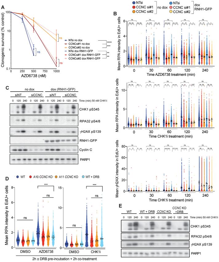
8666 Nucleic Acids Research, 2021, Vol. 49, No. 15
Exploiting this concept, ATR inhibitors (ATRi) are in A549 ATM-knockout cell lines were generated as de-
clinical development for the treatment of cancers (12) scribed before (37,38). mESC Cdc25a KO clones #4/5 were
including those with mutations in the related kinase kindly provided by Oscar Fernández-Capetillo (24) and
ataxia telangiectasia mutated (ATM) (13–16), or high en- U2-OS T-Rex GFP-RNase H1(D210N) (RNH1(D210N)-
dogenous replication stress (17–20). The latter is also GFP) or GFP-RNase H1 (RNH1-GFP) cells by Pavel Jan-
a biomarker for hypersensitivity towards inhibitors of scak (39). mESCs were cultured on 0.1% gelatin-coated
CHK1 and WEE1 kinases, both of which also play key tissue culture flasks, and Cas9-expressing cell lines were
roles in the RSR (17). Recent studies using CRISPR– maintained in blasticidin (10 g/ml mESC and U2-OS, 5
Cas9 and siRNA screening methodologies identified loss g/ml HAP-1). AZD6738 and AZD1775 were made by As-
of POLE3/4, RNASEH2A/B, and ERCC1 as additional traZeneca. DRB (5,6-dichloro-1-beta-ribo-fuanosyl benz-
biomarkers of ATRi hypersensitivity (21–23). Similarly, imidazole), actinomycin D, aphidicolin, hydroxyurea, car-
loss of the cyclin-dependent kinase (CDK)-regulator phos- boplatin and etoposide were obtained from Sigma-Aldrich.
phatase CDC25A was found to promote ATRi resistance, XL413 (S7547), VE-822 (S27102), LY2603618 (S2626) and
Downloaded from https://academic.oup.com/nar/article/49/15/8665/6331679 by guest on 06 November 2021
implicating abrogation of the G2/M checkpoint as a major LY2606368 (S7178) were obtained from SelleckChem. CX-
driver of ATRi efficacy (24). However, these studies have 5461 (509265) was obtained from Merck Millipore, and
thus far been performed in wild-type (WT) or TP53 knock- BRD-6989 (6438) from Tocris. Final assay DMSO concen-
out cells to facilitate screen performance. This may mean trations were normalised to 1:1000 as required. Recombi-
that identified hits do not fully reflect the drivers of cell nant human IFN␥ was obtained from Peprotech (300–02)
death in tumours harbouring specific genetic defects, such and used at a final concentration of 2.5 ng/ml.
as mutations in ATM, that are likely to be targeted with
ATR inhibitors in the clinic. In particular, the hypersensi-
In vitro growth and cell viability assays
tivity of ATM-mutated cancers to ATR inhibition (13–16)
could be driven by over-lapping and redundant functions MTT cell proliferation assays. For each biological repli-
of ATM and ATR kinases, given that there are over 700 cate (n = 3) cells were seeded as technical replicates at 5000
suggested ATM/ATR substrates harbouring S/T-Q motifs cells per well of a 96-well plate, 16 h prior to drug treatment.
(25–27). ATM and ATR signalling are already known to Following treatment, medium was replaced with 50 l 0.5
crosstalk to regulate processes including DNA repair, cell mg/ml MTT (3-[4,5-dimethylthiazol-2-yl]-2,5- diphenylte-
senescence and apoptosis, and cell cycle checkpoint con- trazolium bromide; thiazolyl blue) in growth media and
trol (28–31), with roles for ATM in regulating DNA replica- cells incubated for a further 4 h. 50 l 10% SDS was then
tion having also been reported (32–35). It is therefore likely added to the cells and incubated at 37◦ C overnight before
that drivers of ATRi sensitivity and resistance will differ reading absorbance at 595 nm.
between ATM-proficient and ATM-deficient cells, mean-
ing that elucidating both common and genetic background- Clonogenic survival assays. For each biological replicate
specific mechanisms of ATRi efficacy will be critical for ac- (n = 3), 16 h prior to treatment cells were seeded in trip-
curately stratifying patients and pre-empting innate or ac- licate in 6-well plates at 500 cells/well (mESC, A549, U2-
quired drug resistance. OS, HAP-1) or 1500 cells/well (FaDu). ATM KO FaDu
To address the above issues, in this study we exam- cells were seeded in conditioned media. Cells were incu-
ine genetic drivers of ATRi efficacy by performing pooled bated with the drug for 6 (mESC, HAP-1) or 9 days (A549,
CRISPR–Cas9 screens in Atm WT and Atm knockout (KO) FaDu, U2-OS) before methanol fixation (mESC) and crys-
mouse embryonic stem cells (mESCs) using the ATRi, cer- tal violet staining. The number of colonies (defined as con-
alasertib (AZD6738) (36). Our ensuing data reveal how taining > 30 cells) were counted blind and normalised to
drivers of ATRi efficacy depend on functional ATM ex- the untreated conditions to account for variations in plating
pression, thereby providing further insight into how ATR efficiencies. For short-term treatments, cells were washed
and ATM cooperate to maintain genomic integrity. Fur- three times with fresh drug-free media following treatment.
thermore, by focusing on the strongest resistance-gene hits
in both ATM-proficient and ATM-deficient cells, we in- Incucyte cell confluency. For each biological replicate, cells
vestigate the mechanism by which loss of Cyclin C or were seeded as technical replicates at 5000 cells per well of
CDK8, members of the CDK8 kinase module of the RNA a 96-well plate. Live-cell imaging was acquired at 10× mag-
polymerase II (RNAPII) mediator complex, promote resis- nification every 2 h, and percentage phase confluency was
tance to ATR and CHK1 inhibition by limiting DNA:RNA quantified using Incucyte ZOOM 2018A software (Essen
hybrid formation in S-phase and transcription-associated Bioscience). Doubling times were calculated in GraphPad
replication stress. These findings also suggest new therapeu- prism V.8 during the exponential growth phase using the
tic opportunities for ATR inhibitors and provide insights exponential growth equation.
for better stratifying patients and pre-empting innate or ac-
quired drug resistance. CRISPR–Cas9 screen for resistance and sensitivity to
AZD6738. 1 × 10⁸ Atm WT or KO mESCs were indepen-
MATERIALS AND METHODS dently infected with a pre-packaged lentiviral library at a
MOI (multiplicity of infection) of 0.1, giving a library cover-
Cell lines and compounds
age of ∼1000×. The Kosuke Yusa murine v2 sgRNA library
Cell line origins and their cell growth media can be found was used (40). After 48 h, 1 × 10⁸ cells/genotype were col-
in Supplementary Methods Table S1. mESC, FaDu and lected for sequencing and a further 1 × 10⁸ cells treated with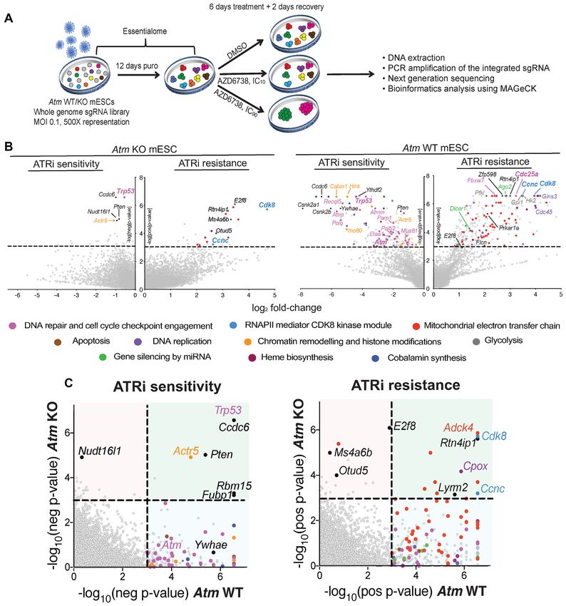
Nucleic Acids Research, 2021, Vol. 49, No. 15 8667 puromycin (2 g/ml) for 12 days to select for cells with the ses, sgRNAs with DMSO read counts of 0 in any 2 repli- stably integrated sgRNA cassette. Following establishment cates and
8668 Nucleic Acids Research, 2021, Vol. 49, No. 15
previously reported (42) before antibody staining. For an- 20 min, and DNA was counterstained with 1 g/ml DAPI.
tibody staining, primary antibodies were incubated at 4◦ C Image acquisition was performed on a WiScan Hermes
O/N using the antibodies listed in Supplementary Meth- system (IDEA Bio-Medical) equipped with 40×/0.75 NA
ods Table S2. Cells were washed 3× in PBST before incuba- air objective, and nuclear GFP foci counts were analysed
tion with Alexa Fluor secondary antibodies for 1 h at RT in using integrated WiSoft Athena software.
the dark, followed by DAPI staining (1 g/ml) for 20 min
at RT. Plates were imaged using an Opera Phenix spinning Stable cell line generation. Lentivirus-based CDK8-GFP
disc confocal microscope (PerkinElmer) at 40× magnifica- and CDK8(D173A)-GFP plasmids were obtained from
tion. All data were imported into Harmony image analysis VectorBuilder, packaged in HEK293-lentiX cells using sec-
software (PerkinElmer) for subsequent analyses. ond generation packaging plasmids, and the lentivirus used
to infect HAP-1 cells. Forty-eight hours later, cells with the
Defining and counting nuclei. Nuclei were defined based on integrated plasmids were selected in puromycin (0.5 g/ml)
DAPI intensity. Border objects were removed from analyses. for 7 days prior to single-cell plating and clonal expan-
Downloaded from https://academic.oup.com/nar/article/49/15/8665/6331679 by guest on 06 November 2021
sion. For gene knockouts using CRISPR, synthetic crRNA
Defining and counting micronuclei. Micronuclei were ini- (CRISPR RNA) and tracrRNAs (trans-activating RNA)
tially defined based upon the DAPI stain using Harmony’s were obtained from IDT (Supplementary Methods Table
‘find micronuclei’ function, and further refined based upon S4), duplexed and transfected into Cas9-expressing cells us-
the following parameters: fraction of nucleus area 0.75, area >2 m2 , fraction of nucleus inten- tions. Pooled editing efficiencies were assessed after 72 h and
sity >0.25, intensity DAPI CV (%)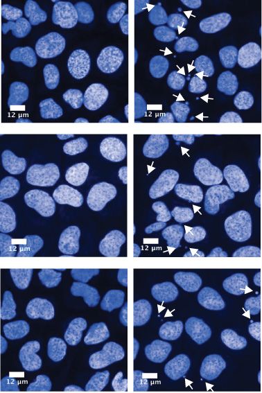
Nucleic Acids Research, 2021, Vol. 49, No. 15 8669
ing as required. 1.5 M AZD6738 had been optimised to momatic (version 0.38 (46)), specifying the TruSeq3-
kill ∼95% of non-edited control cells in this assay format, SE.fa:2:30:10 adapters in the ILLUMINACLIP argument.
thereby strongly enriching for resistant cells. A sgRNA tar- The trimmed sequences were then aligned to the reference
geting Lrrc29 was used as a negative control as our screen Mus musculus genome and annotation (build GRCm38.p6,
outcomes did not predict this gene to influence ATRi sen- NCBI:GCA 000001635.8) using HISAT2 (version 2.1.0
sitivity in Atm WT mESCs. Genomic DNA from surviv- (47)) with default settings. Gene expression levels were then
ing cells, alongside from non-transfected control cells, was quantified from the aligned reads using featureCounts (ver-
extracted (Invitrogen, K1820) and the sgRNA-target loci sion 2.0.1 (48)) with the options –ignoreDup and -s 2. Ref-
PCR amplified using the associated primers in Supplemen- erence annotation GTF file was also obtained from genome
tary Methods Table S4. The forward primer was used for build GRCm38.p6. Lastly, the R package DESeq2 (49) was
Sanger sequencing of the PCR product. Sequence traces be- used for exploratory data analysis and visualisation, as well
tween transfected and non-transfected cells were compared as differential expression analyses. Reference genome se-
using TIDe software (3.2.0) (44), and used to calculate the quence and annotation files were obtained from Ensembl
Downloaded from https://academic.oup.com/nar/article/49/15/8665/6331679 by guest on 06 November 2021
% of each InDel (–50 bp to + 50 bp) per sample. To ac- (50).
count for in-frame mutations likely having minimal impact
on protein expression, InDels were classified as in-frame or Statistical analyses. Statistical tests were performed as in-
out-of-frame based on whether the size of the InDel was a dicated in the figure legends to determine statistical sig-
multiple of 3. Undefined InDels could be the result of noise nificance in replicate comparisons. A P-value < 0.05 was
in the Sanger sequencing trace, or InDels outside the range deemed statistically significant.
–50 bp to +50 bp.
RNA-seq. RNA sequencing (RNA-seq) was performed in RESULTS
mESCs using two WT biological replicates and three inde-
Identification and validation of factors influencing ATRi sen-
pendent Ccnc KO and Cdk8 KO clones. Cells were treated
sitivity in ATM-proficient and ATM-deficient cells
for 4 h with either DMSO or 900 nM AZD6738 prior to
sample collection. A 4-hour timepoint gave the biggest win- To systematically interrogate drivers of ATRi efficacy in a
dow in Ccnb1 mRNA expression, which has been shown clinically relevant genetic background, we performed whole
to be prematurely upregulated upon ATR inhibition (4) genome CRISPR–Cas9 genetic screens in an isogenic pair
(data not shown). RNA was extracted using a RNeasy kit of Atm WT and KO mESCs with a view to finding gene-
with on column DNase digestion according to the manufac- products that, when lost, caused either hypersensitivity or
turer’s protocol (Qiagen, 74106). RNA integrity was con- resistance to the ATRi AZD6738 (36) (Figure 1A). We
firmed using Agilent Tapestation RNA screentape, show- chose mESCs as our cell model in part because, compared
ing two sharp peaks representing the 18S and 28S ribo- to other cell models that we tested, ATM loss had mini-
somal subunits, with a 28S:18S ratio of approximately 2 mal impact on cell proliferation rates (Supplementary Fig-
(RINe > 9). Using 5 g input RNA, mRNA was iso- ure S1A), thereby facilitating the comparison of hits ob-
lated from ribosomal RNA using the NEBNext Poly(A) tained between each genotype. Furthermore, the faster pro-
mRNA magnetic isolation module (E7490) according to the liferation rates of mESCs ensured that cells underwent mul-
manufacturer’s instructions. Following two rounds of en- tiple rounds of replication and cell divisions in a 6-day
richment, successful isolation and sample integrity (RINe treatment window. This was important because ATRi ef-
2–3) were confirmed using high sensitivity Agilent Screen ficacy is likely to be influenced by S-phase drug exposure
tape. Library preparation was performed using the NEB- and subsequent mitotic entry (Supplementary Figure S1A).
Next Ultra II directional library prep kit (E7760) accord- To enhance our prospects for uncovering both resistance
ing to the manufacturer’s instructions for ‘purified RNA or and sensitivity factors, and to facilitate comparisons be-
clean RNA’, and using NEBnext multiplex oligos (E7335). tween both genetic backgrounds, we performed parallel
cDNA concentrations were measured using the Qubit ds- screens at doses of AZD6738 that we had determined to
DNA assay kit (Q32854), and molar concentrations calcu- equate to the IC10 and IC90 values for each cell line (as ex-
lated using the average DNA fragment size following anal- pected, Atm KO mESCs were significantly more sensitive to
yses on the Agilent Tapestation with high sensitivity DNA AZD6738 treatment than their WT counterparts; Supple-
screentape (D1000). Equal molar concentrations were mul- mentary Figure S1B). Following successful quality control
tiplexed at a final concentration of 10 nM and sequenced (QC) of our screens (Supplementary Figure S1C–H), ensu-
using single-ended reads on an Illumina Hiseq4000 system. ing MAGeCK (51) bioinformatics analyses revealed a vari-
Due to limitations in the number of samples that could be ety of factors and pathways predicted to contribute to ATRi
combined in a single multiplex for sequencing, WT sam- hypersensitivity or resistance (Figure 1B, Supplementary
ples were independently multiplexed with Ccnc KO samples, Figure S2A, Supplementary Table S1). Furthermore, pre-
and again with Cdk8 KO samples, in order to eliminate any viously reported genes modulating sensitivity in Atm WT
sequencing-run bias. cells served as positive controls, indicating the reliability of
our screens (Cdc25a for resistance (24); Atm and Trp53 for
RNA-seq data analysis. FASTQ files were first checked sensitivity (14–16,52)). Notably, some gene-products whose
with FASTQC (version 0.11.5, http://www.bioinformatics. inactivation was recently reported to hypersensitise cells
babraham.ac.uk/projects/fastqc/ (45)) for quality control. to ATR inhibition (RNASEH2A, RNASEH2B, ERCC1,
The adaptor sequences were then removed using Trim- POLE3 and POLE4 (21–23)) behaved as essential in our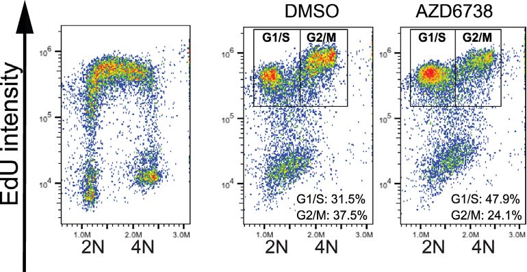
8670 Nucleic Acids Research, 2021, Vol. 49, No. 15

                                                                                                                                                                 Downloaded from https://academic.oup.com/nar/article/49/15/8665/6331679 by guest on 06 November 2021
Figure 1. CRISPR–Cas9 screens using AZD6738 in Atm WT and KO mESCs. (A) Schematic of CRISPR–Cas9 screens performed in this study. (B) Drop-
out and enrichment analyses following AZD6738 treatment in Atm WT and KO mESCs. Genes were statistically ranked using MAGeCK analysis software,
and top hits with a P-value < 0.001 classified into related pathways and complexes. Dashed line = P-value of 0.001. Two MAGeCK analysis methods were
performed for both IC10 and IC90 doses, and all analyses can be found in Supplementary Table S1 and Supplementary Figure S2A. The analyses that
provided the greatest number of significant drop-out or enrichment hits (FDR < 0.1) for each genotype are presented. (C) Drop-out and enrichment
analyses following AZD6738 treatment from Figure 1B, comparing MAGeCK P-values for each gene between Atm WT and Atm KO mESCs. A P-value
of 0.001 is indicated by the dotted line. Genes in the upper right quadrant were hits in both cells lines, upper left quadrant were hits only in Atm KO cells,
and those in the bottom right quadrant were hits only in Atm WT cells. Top hits in one or both cell lines are colour coded based on the same classification
as Figure 1B.
Nucleic Acids Research, 2021, Vol. 49, No. 15 8671
Downloaded from https://academic.oup.com/nar/article/49/15/8665/6331679 by guest on 06 November 2021
Figure 2. Validation of hits identified through our CRISPR–Cas9 screens. (A) Competition assays between CRISPR–Cas9 edited and non-edited cells in
response to AZD6738 treatment. Cas9-expressing mESCs were transfected with synthetic sgRNAs against the target loci, resulting in a mixed pool of edited
and non-edited cells after 7 days. Each pooled population was treated for 5 days with DMSO or 1.5 M AZD6738, with a high dose strongly enriching
for resistant cells. Genomic DNA was extracted and the sgRNA-targeted loci PCR amplified to allow editing efficiencies and % InDels (–50 bp to +50 bp)
to be calculated using TIDe software (3.2.0) (44). The control sgRNA targeted Lrrc29 which was not predicted to influence ATRi sensitivity in Atm WT
mESCs according to our screening data. (B) Results of MTT cell proliferation assays in Atm WT and KO mESCs following siRNA-depletion of YWHAE,
in response to two chemically distinct ATR inhibitors (AZD6738 and VE-822). Error bars = mean ± SEM (biological n = 3). (C) Clonogenic survivals
of ATM WT and KO A549 and FaDu cells following siRNA-depletion of CCDC6, in response to two chemically distinct ATR inhibitors (AZD6738 and
VE-822). Error bars = mean ± S.D (biological n = 3). FaDu ATM KO cells are inherently extremely sensitive to doses of VE-822 which effectively inhibit
ATR kinase activity, therefore precluding analysis of further reductions in clonogenic survival upon CCDC6 depletion.
screening conditions in mESCs, likely explaining our inabil- ple genetic backgrounds. Using CRISPR-based cell-growth
ity to detect these (Supplementary Figure S2B). To more competition assays (44,53), we provisionally validated both
easily compare hits obtained through our screens in Atm genes alongside various other resistance-gene hits from the
KO versus WT mESCs, we directly compared the P-values Atm WT screens that commonly acquire missense muta-
for each gene between both genotypes (Figure 1C) and gen- tions and/or reduced expression in cancer (Prkar1a (54–
erated a comprehensive list of genes predicted to enhance or 56), Flcn (57,58), E2f8, Ccnc (59) and Cdk8 (60)) (Figure
suppress ATRi efficacy in both ATM-deficient and ATM- 2A). We therefore speculate that such mutations might rep-
proficient cells (Supplementary Table S3). resent secondary mutations in tumours that may appear un-
Two of the strongest resistance gene hits in the Atm KO connected to DNA replication and repair yet limit ATRi
mESCs were Ccnc and Cdk8, which encode two compo- efficacy in the clinic.
nents of the same complex that regulates RNAPII-mediated Notably, we identified substantially more sensitiser and
transcription. Single guide-RNAs (sgRNAs) targeting both resistance hits in the Atm WT background than in the Atm
genes were also strongly enriched in the screen outputs KO mESCs (Figure 1C). Importantly, this was not due to
from Atm WT cells, thereby highlighting the importance an inability to detect changes in cell fitness in Atm KO
of this pathway in driving AZD6738 efficacy across multi- cells upon gene editing, since genes encoding DNA-PK8672 Nucleic Acids Research, 2021, Vol. 49, No. 15
(Prkdc) and components of the Fanconi anaemia pathway loss of Cyclin C or CDK8 appeared specific to inhibition of
(FA) had a greater impact on cell fitness in Atm KO ver- the RSR but did not act more generally in response to other
sus Atm WT mESCs in accordance with existing literature sources of replication stress or DNA damage. Notably, Cy-
(37,61,62) (Supplementary Figure S2C). Moreover, we val- clin C and CDK8 have been reported to upregulate the tran-
idated the ATM-status dependency of ATRi sensitivity us- scription of a subset of p53-dependent genes (64). However,
ing small interfering RNAs (siRNAs) against Ywhae (14– rather than causing resistance, p53 loss caused sensitivity
3–3ε) (one of the strongest dropouts in WT cells only) and to AZD6738, consistent with previous reports (15,16,52).
Ccdc6 (one of the strongest dropouts in both Atm WT and Given that CKM positively regulates the p53 downstream
KO cells) in various ATM-deficient models, using two in- responses, this is unlikely to underpin the mechanism of
dependent ATR inhibitors (AZD6738 and VE-822; Figure ATRi resistance driven by CKM loss. To further support
2B and C). These data also supported the relevance of our this notion, we deleted CCNC or CDK8 in human U2-
screening outputs in mESCs for understanding ATRi ef- OS and HAP-1 cells respectively (Supplementary Figure
ficacy in human cancer cell models. Mechanistically, hits S3K-L)––two cell lines that display dysfunctional p53 sig-
Downloaded from https://academic.oup.com/nar/article/49/15/8665/6331679 by guest on 06 November 2021
that we selectively detected in Atm WT mESCs could in- nalling (Supplementary Figure S3M). Loss of Cyclin C or
dicate proteins involved in ATM-regulated pathways that CDK8 in these cell lines also caused AZD6738 resistance
are therefore epistatic with ATM loss. Alternatively, finding (Figure 3D and E), thereby supporting a p53-independent
fewer resistance hits in Atm KO compared to WT cells may mechanism and corroborating our findings in human can-
highlight the existence of numerous pathways that are re- cer cell lines. Importantly, unperturbed Cyclin C- or CDK8-
sponsible for ATRi hypersensitivity in ATM-deficient cells. deficient cells showed no discernible difference in cell cycle
This in turn would suggest that patients with tumours har- profiles or growth kinetics compared to their WT counter-
bouring ATM defects will be less likely to possess innate or parts in all three cellular backgrounds (Supplementary Fig-
readily acquire resistance to AZD6738 treatment through ure S4A–C).
downregulation of, or loss-of-function mutations in, a sin- To assess whether CDK8 kinase activity is required for
gle other gene. AZD6738 treatment response, we complemented CDK8
Taken together, these data underscored successful sys- KO HAP-1 cells with either a WT CDK8-GFP construct,
tematic identification of factors and pathways promoting or one bearing a D173A mutation that inactivates CDK8
sensitivity and resistance to AZD6738, and highlighted how catalytic activity (65). We observed that the D173A mutant
these vary depending on the cell’s ATM status. provided ATRi resistance comparable to complete CDK8
loss, suggesting that CDK8 kinase activity is critical for
its function in regulating ATRi sensitivity (Figure 3F, Sup-
Loss of Cyclin C or CDK8 provides ATRi resistance
plementary Figure S5A-B). Accordingly, we observed that
For ensuing mechanistic follow up studies, we focussed on CDK8 kinase activity was abrogated to a similar extent
two of the strongest resistance gene hits that were com- in both Cyclin C and CDK8 deficient cells, as measured
mon genetic modulators of ATRi efficacy in both Atm KO by probing for phosphorylation of CDK8 targets STAT1
and WT backgrounds: the genes encoding Cyclin C (Ccnc) (pS727) and STAT5 (pS726) (66,67) (Supplementary Figure
and cyclin-dependent kinase 8 (Cdk8), which comprise part S5C; as expected, CDK8 activity was also restored by com-
of the CDK8 kinase module (CKM) for the RNAPII me- plementing cells with WT, but not D173A mutant, CDK8).
diator complex (Figure 1C). De novo CRISPR-mediated
gene KO of either Ccnc or Cdk8 in both Atm WT and
Cyclin C/CDK8 loss modulates ATRi sensitivity indepen-
KO mESCs (Supplementary Figure S3A–C) produced re-
dently of CDC25A and G2/M checkpoint function
sistance to AZD6738 treatment, thereby further validat-
ing our screening results (Figure 3A, Supplementary Fig- CDC25A loss has been reported to provide ATRi resis-
ure S3D). Importantly, concomitant knockout of Ccnc and tance by opposing the G2/M checkpoint override and pre-
Cdk8 yielded no additive resistance, suggesting that they mature mitotic entry caused by ATRi treatment (24). We
function in a common pathway (Figure 3B, Supplemen- therefore sought to establish whether Cyclin C and CDK8
tary Figure S3E). Supporting this, we noticed that Cdk8 KO were epistatic with CDC25A regarding ATRi sensitivity.
cells also had reduced Cyclin C levels (Supplementary Fig- Firstly, we assessed Cdc25a transcript levels in Cdk8 KO
ure S3A, B), consistent with a previous report showing that and Ccnc KO mESCs, and found no change compared with
CDK8 is required for Cyclin C stability, but not vice-versa the WT cells (Figure 4A). Furthermore, de novo CRISPR-
(63). mediated knockout of Cdc25a in Ccnc WT or Ccnc KO
Furthermore, we found that loss of Cyclin C or CDK8 backgrounds demonstrated a clear additive effect of inac-
conferred resistance to a clinical ATRi, VE-822, that is tivating each gene on AZD6738 resistance, consistent with
chemically distinct from AZD6738, as well as towards the Cyclin C and CDC25A acting in different pathways to pro-
CHK1i (LY2603618) or WEE1i (AZD1775), but not to- mote AZD6738 resistance (Figure 4B). To test whether loss
wards various DNA-damaging agents that we tested: ion- of Cyclin C/CDK8 suppressed AZD6738-induced G2/M
ising radiation (IR) which causes various forms of DNA checkpoint abrogation via an alternate pathway, we per-
damage, the strong apoptosis inducers etoposide and carbo- formed pulse-chase experiments with the nucleotide ana-
platin, as well as hydroxyurea (HU) or aphidicolin which in- logue EdU (Figure 4Ci). As expected, incubation with
duce DNA replication stress by impairing DNA polymerase AZD6738 caused faster progression of cells though mitosis
processivity (Figure 3C, Supplementary Figure S3F–J). and into G1 phase, as indicated by the boxes in Figure 4Ci,
This indicated that the resistance mechanism afforded by and quantified in Figure 4Cii-iii). Importantly, progressionNucleic Acids Research, 2021, Vol. 49, No. 15 8673
Downloaded from https://academic.oup.com/nar/article/49/15/8665/6331679 by guest on 06 November 2021
Figure 3. Loss of Cyclin C/CDK8 provides ATRi and CHK1i resistance. (A) Clonogenic survivals of Ccnc/Cdk8 WT/KO Atm WT/KO mESCs treated
with AZD6738, represented as AUCs (areas under the curve). Error bars = mean ± S.D (biological n = 3). Survival curves can be found in Supplementary
Figure S3D. (B) Clonogenic survivals of WT and Ccnc/Cdk8 single and dual KO mESCs treated with AZD6738. Data also represented as AUCs. Error
bars = mean ± SD (biological n = 3). (C) Clonogenic survivals of Ccnc/Cdk8 WT/KO Atm WT/KO mESCs treated with the CHK1 inhibitor LY2603618,
represented as AUCs. Error bars = mean ± SD (biological n = 3). Survival curves can be found in Supplementary Figure S3F. (D) Clonogenic survivals of
WT and CCNC KO U2-OS cells in response to AZD6738 treatment. A3/B24 clones were confirmed as WT by immunoblot analyses and genotyping after
single-cell expansions of sgRNA-transfected cells. Data also represented as AUCs. Error bars = mean ± SD (biological n = 3). (E) Clonogenic survivals
of WT and CDK8 KO HAP-1 cells in response to AZD6738 treatment. Data also represented as AUCs. Error bars = mean ± SD (biological n = 3). (F)
Clonogenic survivals of WT and CDK8 KO HAP-1 cells, complemented with WT or kinase dead (D173A) CDK8-GFP, treated with AZD6738. Data
also represented as AUCs. Error bars = mean ± SD (biological n = 3). Additional clones are shown in Supplementary Figure S5A and B. All statistical
analyses were performed using a one-way ANOVA test with multiple comparisons. P-values < 0.05 (*), 0.01 (**), 0.001 (***) and 0.0001 (****) were
deemed statistically significant.
rates from G2 to G1 were not discernibly different between age that persists until mitosis, wherein it leads to increased
WT, Ccnc KO and Cdk8 KO mESCs, when assessed in micronuclei formation.
either the presence or absence of AZD6738 (Figure 4Cii–
iii). However, despite the G2/M checkpoint being equally Cyclin C loss reduces basal and ATRi/CHK1i-induced
defective, we noted that micronuclei levels following 24 DNA:RNA hybrids
h treatment with AZD6738 were significantly reduced in
CCNC KO U2-OS cells as compared to WT controls (Fig- CDK8 kinase activity is primarily associated with phospho-
ure 4D). These data therefore suggested that when ATR is rylating the C-terminal domain (CTD) of RNAPII and var-
inhibited, Cyclin C/CDK8 promote increased DNA dam- ious transcription factors (68), therefore pointing towards8674 Nucleic Acids Research, 2021, Vol. 49, No. 15
Downloaded from https://academic.oup.com/nar/article/49/15/8665/6331679 by guest on 06 November 2021
Figure 4. Loss of Cyclin C/CDK8 promotes ATRi resistance independent of CDC25A or G2/M checkpoint regulation. (A) Cdc25a mRNA transcript
levels in WT and Ccnc/Cdk8 KO mESCs as assessed by RT-qPCR using two independent primer pairs. Transcript levels are normalised to the house-keeping
gene Gapdh. Cdc25a KO mESCs (24) were used as a control. Error bars = mean ± SEM (biological n = 4 WT, Cdk8 KO, Ccnc KO; n = 2 Cdc25a KO). (B)
Clonogenic survivals of isogenic WT and de novo generated Ccnc/Cdc25a single and dual KO mESCs treated with AZD6738. Error bars = mean ± SD
(biological n = 3). (C) Actively replicating S-phase Ccnc and Cdk8 WT and KO mESCs were pulse-labelled with 10 M EdU for 30 min prior to release
into media containing DMSO or 900 nM AZD6738 for 6 h. Error bars = mean ± SEM (biological n = 3 Ccnc KO, n = 4 Cdk8 KO). (i) Schematic of
experimental design and gating strategy using FACS profiles in WT mESCs. (ii) Cell cycle distributions based on DAPI histograms. iii) Distribution of
EdU positive cells between G1/S and G2/M 6 h after removal of EdU. DNA content 2N = G1, 4N = G2/M. EdU positive cells were actively replicating
at the time of EdU labelling. (D) Mean number of micronuclei/cell after 24 h treatment with DMSO or 1.5 M AZD6738 in U2-OS CCNC WT and
KO cells. Error bars = mean ± SD (biological n = 3). Arrows indicate micronuclei in the representative images. Representative images were taken at 40×
magnification, scale bars = 12 m. All statistical analyses were performed using a one-way ANOVA test with multiple comparisons, except in Figure 3B
where a two-way ANOVA test was used. P-values < 0.05 (*), 0.01 (**), 0.001 (***) and 0.0001 (****) were deemed statistically significant.
transcriptional control as the mechanism by which Cyclin supported the reliability of our data (Supplementary Fig-
C/CDK8 loss promotes ATRi resistance. Previous stud- ure S6G–J), we did not identify any transcripts that were
ies on CKM in mammalian cells have established that its significantly differentially regulated upon AZD6738 treat-
depletion leads to transcriptome changes but not a genome- ment in both Ccnc and Cdk8 KO, but not WT, cells (Sup-
wide reduction in RNAPII activity (69–71). In line with this, plementary Table S4). Similarly, Gene Ontology analyses of
we observed no consistent reduction in mRNA synthesis or proteins whose transcripts were upregulated or downregu-
abundance of elongating RNAPII in our Cyclin C/CDK8- lated in a treatment-independent manner in both Cdk8 and
deficient cells as compared to controls (Supplementary Fig- Ccnc KO clones compared to WT mESCs did not provide
ure S6A-F). We therefore probed for specific changes in the a clear explanation why the Ccnc/Cdk8 KO cells are ATRi
transcriptome by performing RNA-seq in WT, Cdk8 KO resistant (Supplementary Table S4).
and Ccnc KO mESCs following DMSO or AZD6738 treat- Instead, we speculated that loss of Cyclin C/CDK8 may
ment. While quality control metrics and biological controls reduce collisions between the transcription and replicationNucleic Acids Research, 2021, Vol. 49, No. 15 8675
Downloaded from https://academic.oup.com/nar/article/49/15/8665/6331679 by guest on 06 November 2021
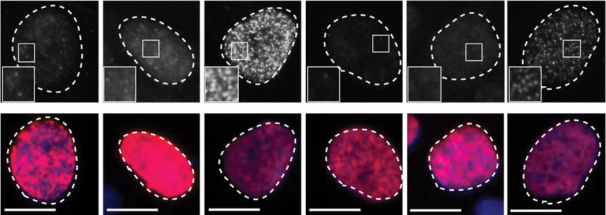
Figure 5. Cyclin C loss supresses DNA:RNA hybrid formation in response to ATRi and CHK1i. (A) Mean number of RNH1(D210N)-GFP foci/nuclei
normalised to untreated NT siRNA levels in response to AZD6738 or CHK1i (LY2606368) treatment, following siRNA-depletion of Cyclin C. Error
bars = mean ± SD (biological n = 2). Representative images are of RNH1(D210N)-GFP foci in EdU positive cells, following treatment with DMSO,
AZD6738 for 24 h, or CHK1i for 4 h. Scale bars = 12 m. A full field of view is provided in Supplementary Figure S7C which also highlights the
heterogeneity of foci induction in Cyclin C-depleted cells treated with CHK1i. SiRNAs targeting CDK8 were also tested but resulted in the accumulation
of cells in G1 which may bias the experimental outcome (Supplementary Figure S7A and B). (B) Clonogenic survivals of RECQL5-depleted WT and
CCNC KO U2-OS cells treated with AZD6738. Error bars = mean ± SD (biological n = 3). Statistical analyses were performed using a two-way ANOVA
test by comparison to CCNC WT cells transfected with the control (NT) siRNA. P-values < 0.05 (*), 0.01 (**), 0.001 (***) and 0.0001 (****) were deemed
statistically significant.
machineries upon ATR inhibition, whereby transcription- posed to promoting DNA:RNA hybrid resolution. Sup-
replication conflicts are associated with DNA:RNA hybrid porting this, RECQL5, which is reported to limit and re-
formation and genome instability, primarily through caus- solve transcription-replication conflicts (73,74), was iden-
ing replication fork collapse (9,72). We therefore used a tified as an ATRi sensitiser in our CRISPR–Cas9 screens
reporter system employing catalytically dead RNase H1 (Figure 1B). Depletion of RECQL5 enhanced sensitivity
(RNH1(D210N) (39))––which binds to but does not pro- to AZD6738 and rescued the ATRi resistance observed
cess DNA:RNA hybrids - to quantify DNA:RNA hybrid upon Cyclin C loss, with CCNC-knockout RECQL5-
levels in response to ATR or CHK1 inhibition following de- depleted cells showing equivalent sensitivity to WT cells
pletion of Cyclin C. Since CDK8 depletion caused signifi- transfected with a control siRNA (Figure 5B). Taken to-
cant accumulation of U2-OS cells in G1 and also reduced gether, these results showed that Cyclin C loss counteracts
Cyclin C protein levels (63) (Supplementary Figure S3A-B DNA:RNA hybrid formation caused by ATR and CHK1
and Supplementary Figure S7A and B), we focused largely inhibition in S-phase, suggesting that counteracting or re-
on Cyclin C loss for further studies. Strikingly, this revealed ducing transcription-replication conflicts improved cell sur-
that both basal and ATRi/CHK1i-induced DNA:RNA hy- vival.
brid levels were markedly reduced in cells depleted of Cy-
clin C (Figure 5A; as shown in Supplementary Figure S7C,
Loss of Cyclin C suppresses replication stress in response to
DNA:RNA hybrids formed in each case in S-phase cells).
ATR or CHK1 inhibition
In light of these data, we hypothesised that Cyclin C loss
alleviated the rate of transcription-replication encounters Both transcription-replication conflicts and DNA:RNA hy-
that are enhanced upon ATRi or CHK1i treatment, as op- brids have been established as sources of genome instabil-8676 Nucleic Acids Research, 2021, Vol. 49, No. 15
ity, largely through promoting replication fork stalling and as early or late S-phase based on EdU and DAPI intensity.
collapse into single-ended DSBs (seDBSs) (9,10). In the ab- This revealed that CHK1i-treated CCNC WT cells accumu-
sence of replication fork protection, replication stress can lated with DNA damage in early S-phase, whereas in CCNC
also cause RPA exhaustion, leading to nuclease processing KO cells ␥ H2AX formed only at later time points and in
of exposed ssDNA, global replication fork breakage and both early and late S-phase (Figure 6F, Supplementary Fig-
ATM activation (11). Importantly, ATR and CHK1 play ure S8I). In CCNC WT cells, this was associated with a dra-
pivotal roles in counteracting this phenomenon, known matic reduction in EdU incorporation by 240 minutes, indi-
as replication catastrophe, by limiting global origin firing cating that any cells capable of entering mitosis would do so
and promoting nucleotide synthesis. We therefore assessed with an under-replicated genome. Conversely, despite accu-
whether Cyclin C loss decreased replication stress and ul- mulating DNA damage (␥ H2AX) by 240 min, CCNC KO
timately prevented replication catastrophe in the presence cells appeared more likely to enter mitosis with a near-fully
of ATRi or CHK1i. To assess this, we monitored RPA32 replicated genome, therefore supporting a model in which a
chromatinisation and histone H2AX Ser-139 phosphory- delay in replication catastrophe is sufficient to promote cell
Downloaded from https://academic.oup.com/nar/article/49/15/8665/6331679 by guest on 06 November 2021
lation (␥ H2AX) over time in CCNC WT and KO U2-OS survival. Together, these data suggested that Cyclin C loss
cells treated with ATRi or CHK1i. RPA32 hyper-positive in S-phase alleviates replication stress induced by ATR or
cells were indicative of replication stress, while pan-nuclear CHK1 inhibition, and that this enhances cell survival.
RPA32 and ␥ H2AX dual-positive cells detected at later
time points suggested replication catastrophe (11) (Fig-
Cyclin C loss alleviates transcription-associated replication
ure 6A). As expected, we found that RPA32 and ␥ H2AX
stress
accumulation occurred primarily in EdU positive cells, con-
firming that active DNA replication in S-phase is required Although DNA:RNA hybrid formation and transcription-
for DNA damage generation. We observed that Cyclin C replication conflicts are often linked, they are not a single
loss reduced replication stress (RPA chromatinisation) in- event or interchangeably defined, with both being able to
duced by either ATR or CHK1 inhibition (Figure 6Bi; promote genome instability through replication fork col-
Supplementary Figure S8A and B). In CCNC KO cells, lapse. Furthermore, DNA:RNA hybrids and transcription-
ATRi-induced replication stress remained below that ob- replication conflicts can be both caused by, or cause, the
served in WT cells for the full 24 h treatment window tested other, and various sources of DNA:RNA hybrids have been
(Supplementary Figure S8C), with this reduction presum- identified (76). We therefore deemed it important to dis-
ably sufficient to limit under-replicated DNA from enter- tinguish whether the Cyclin C associated replication stress
ing mitosis as indicated by the reduced micronuclei forma- arising from ATRi or CHK1i treatment was caused by col-
tion in CCNC KO cells at 24 h (Figure 4D). On the other lisions between transcription and replication machineries,
hand, CHK1i-induced replication stress resulted in replica- the DNA:RNA hybrids, or both. To investigate whether the
tion catastrophe, consistent with the more potent induction DNA:RNA hybrids were themselves the cause, we assessed
of DNA:RNA hybrid formation following treatment with the impact on ATRi/CHK1i-induced replication stress of
CHK1i compared with ATRi treatment. In agreement with overexpressing RNase H1 which degrades DNA:RNA hy-
the reduction in DNA:RNA hybrids observed in Cyclin C- brids (Supplementary Figure S9A). Through these exper-
depleted cells, we observed a delayed induction of replica- iments, we observed no significant impact of RNase H1
tion catastrophe in CCNC KO compared to WT cells upon overexpression on cell survival, replication stress or repli-
CHK1 inhibition (Figure 6Bi-ii). In line with this, in CCNC cation catastrophe following CHK1i or ATRi treatment in
KO cells we observed delayed kinetics of RPA32 phospho- the presence or absence of Cyclin C (Figure 7A–C, Supple-
rylation on Ser-4/8, which has been associated with repli- mentary Figure S9B). These data therefore indicated that
cation fork collapse and seDSB (single-ended DSB) forma- the DNA:RNA hybrids themselves were likely not directly
tion (11,75), as well as delayed ATM activation detected by responsible for ATRi or CHK1i-induced replication stress
its auto-phosphorylation on Ser-1981 (Figure 6C). This oc- and cell death, but instead suggested that the upstream
curred in a dose-dependent manner, with CCNC KO cells physical collisions between transcription and replication
experiencing a delayed onset of DNA damage by several machineries were promoting genome instability. Further-
hours at lower CHK1i doses (Supplementary Figure S8D more, Cyclin C depletion similarly rescued the impact of
and E). A similar delay in ␥ H2AX formation and Ser-4/8 ATRi or CHK1i treatment in both RNase H1 un-induced
RPA32 phosphorylation was observed in Ccnc and Cdk8 and RNase H1 overexpressing cells, thereby supporting the
KO mESCs as compared to WT control cells (Supplemen- idea that Cyclin C loss acts upstream to limit DNA:RNA
tary Figure S8F). Importantly, given that Cyclin C loss only hybrid formation rather than acting at the level of their res-
delayed the onset of CHK1i-induced replication catastro- olution.
phe, we assessed whether this was sufficient to increase cell Finally, to assess a role for transcription-replication col-
survival. Indeed, short-term treatment with either ATRi or lisions in Cyclin C-associated replication stress, we pre-
CHK1i still induced cell death in CCNC WT cells, whereas incubated cells with the transcription inhibitor DRB (7,77–
CCNC KO cells retained the resistance phenotype observed 79) at a dose and time-points that reduced transcription-
with continuous ATRi or CHK1i treatment (Figure 6D and replication collisions as indicated by a proximity ligation
E, Supplementary Figure S8G and H). assay (PLA) between RNAPII and the DNA replication
To assess at which stage in S-phase DNA damage was component PCNA, yet had minimal or no impact on the
occurring in our studies, we probed for ␥ H2AX following cell cycle (Supplementary Figure S9C–E). Accordingly, we
CHK1 inhibition using flow cytometry and gated the cells found that DRB pre-treatment reduced ATRi- and CHK1i-Nucleic Acids Research, 2021, Vol. 49, No. 15 8677
Downloaded from https://academic.oup.com/nar/article/49/15/8665/6331679 by guest on 06 November 2021
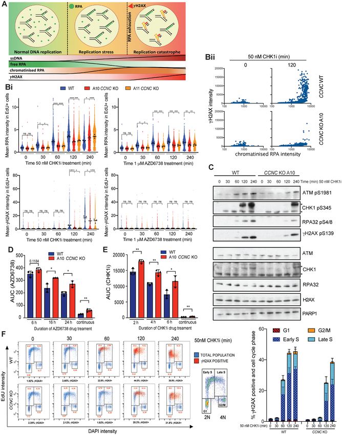
Figure 6. Cyclin C loss supresses replication stress in response to ATRi and CHK1i. (A) Schematic of replication stress and replication catastrophe, which
can be monitored by increased RPA32 chromatinisation (replication stress), followed by detection of ␥ H2AX in RPA32 hyper-positive cells at later time8678 Nucleic Acids Research, 2021, Vol. 49, No. 15
induced RPA chromatinisation in CCNC WT cells to levels single protein is in most instances insufficient to promote
similar to those observed in CCNC KO cells (Figure 7D, ATRi resistance in ATM-deficient cells, as cell death can
Supplementary Figure S9F). We also observed a similar de- still be driven through deregulation of another critical path-
lay in the kinetics of ␥ H2AX, CHK1 pSer-345 and RPA way. By highlighting that it may be difficult to generate re-
pSer-4/8 following CHK1i treatment in DRB pre-treated sistance phenotypes in this setting, our findings therefore
CCNC WT cells comparable with CCNC KO cells (Fig- support the targeted use of ATRi against tumours with im-
ure 7E). Notably, Cyclin C loss or DRB pre-treatment did paired ATM function, and propose the attraction of inhibit-
not impact on IR-induced DNA damage as indicated by ing ATR rather than other DDR factors such as PARP in
similar levels of ␥ H2AX, CHK1 pSer-345 and ATM pSer- ATM-deficient tumours, where resistance can be imparted
1981 (Supplementary Figure S9G). This is consistent with by single inactivation of various genes due to one primary
our extended cell survival data (Supplementary Figure S3J) mode of cell death (38). Alternatively, certain hits arising
and underlines a specific impact of Cyclin C on replication in our screens that increased sensitivity in Atm WT but not
stress-induced DNA damage. Overall, these data supported Atm KO cells may encode proteins that act in the same path-
Downloaded from https://academic.oup.com/nar/article/49/15/8665/6331679 by guest on 06 November 2021
a major role for transcription-associated replication stress way as ATM to modulate ATRi efficacy, and therefore ex-
in driving ATR and CHK1 inhibitor efficacy and highlight hibit an epistatic relationship with ATM loss. Such compo-
Cyclin C and CDK8 as key factors capable of modulating nents represent candidates for identifying ATM-mediated
this response. back-up pathways that can compensate in the absence of
functional ATR. We note however that, in some cases, an
DISCUSSION increased impact on cell fitness in Atm KO mESCs may
have affected our ability to detect drug-dependent changes
ATR inhibitors are in clinical trials for the treatment of in the sgRNA abundances of certain genes in Atm KO
cancers (12), with patient selection hypotheses including cells. We therefore encourage the cross-referencing of ATM-
ATM deficiency (13–16) and high replication stress (17– dependant hits with our essential-gene screen outcomes
20). To identify mechanisms of ATRi efficacy in cells with when prioritising candidates for further study. Neverthe-
clinically relevant mutations, we performed the first whole- less, through validating several such factors and their ATM-
genome ATRi CRISPR–Cas9 screens in paired isogenic status dependency, we believe that our data provide a re-
ATM-proficient and ATM-deficient cells. Through this ap- source to inform future studies into interplays between the
proach, we identified known as well as novel factors which, ATR and ATM kinases, as well as the clinical use of ATR
when lost, provided either sensitivity or resistance to ATRi inhibitors.
in one or both genetic backgrounds. Of these hits, sev- Our studies have shown that loss of Cyclin C or CDK8
eral modulated ATRi sensitivity independent of ATM ex- provides ATRi resistance in both ATM-proficient and
pression, including Trp53 and Ccdc6 (sensitivity) and Ccnc ATM-deficient cells. Cyclin C and CDK8 form part of
and Cdk8 (resistance). These genes highlight mechanisms of the CDK8 kinase module of the RNAPII mediator com-
ATRi efficacy that may drive cell death across multiple tu- plex, and deregulation of either gene has functional con-
mour types. Notably, we identified substantially fewer gene sequences for cancer development (59,60,80,81). We found
hits in Atm KO compared to WT cells, which may reflect that Cyclin C- or CDK8-deficient cells were selectively re-
the numerous interplays between ATR and ATM to reg- sistant to inhibitors of the RSR (ATR, CHK1 and WEE1)
ulate DNA repair, DNA replication and cell cycle check- but did not function via regulation of CDC25A or re-
point engagement. It is therefore plausible that loss of a
←−−−−−−−−−−−−−−−−−−−−−−−−−−−−−−−−−−−−−−−−−−−−−−−−−−−−−−−−−−−−−−−−−−−−−−−−−−−−−−−−−−−−−−−−−−−
points (replication catastrophe). (B) Chromatinised RPA32 and ␥ H2AX intensities in EdU positive nuclei measured by immunofluorescence in CCNC
WT and KO U2-OS cells. S-phase cells were labelled with 10 M EdU for 30 min prior to pre-extraction and fixation. Representative images can be found
in Supplementary Figure S8A. (i) Mean intensities normalised to non-treated WT cells, following 0–240 min treatment with 50 nM CHK1i (LY2603638)
or 1 M AZD6738 (biological n = 3). Mean intensities for each replicate are displayed as black triangles and were used for the overall mean calculations
and statistical analyses. Mean intensities for each individual cell were normalised to the mean intensity of non-treated WT cells in each replicate, and all
three replicates overlaid in blue (CCNC WT), red (CCNC KO A10) or orange (CCNC KO A11) for visualisation of single-cell data. These data are also
normalized to the non-treated conditions of each individual cell line (Supplementary Figure S8B) to highlight that the suppression of replication stress upon
ATRi/CHK1i in CCNC KO cells is independent of any differences in basal intensities. Statistical analyses were performed using one-way ANOVA analyses
with multiple comparisons. P-values < 0.05 (*), 0.01 (**), 0.001 (***) and 0.0001 (****) were deemed statistically significant. ii) Representative example
of replication catastrophe occurring in CCNC WT, but not KO, cells following 120 min CHK1i treatment. Mean chromatinised RPA32 and ␥ H2AX
intensities for each EdU-positive cell are presented as a scatter plot. (C) Immunoblots for markers of DSB formation, indicative of replication catastrophe.
CCNC WT and KO U2-OS cells were treated for 0–240 min with 50 nM CHK1i (LY2603638) prior to lysis. *Both bands are modified forms of CHK1
as indicated by the shift in total protein. (D) Clonogenic survivals of CCNC WT and KO U2-OS cells treated with AZD6738 for 6, 16 or 24 h prior to
drug wash-out, represented as AUCs. Error bars = mean ± SD (biological n = 3). Survival curves can be found in Supplementary Figure S8G. Statistical
analyses were performed using a two-tailed unpaired Student’s t-test. P-values < 0.05 (*), 0.01 (**), 0.001 (***) and 0.0001 (****) were deemed statistically
significant. (E) Clonogenic survivals of CCNC WT and KO U2-OS cells treated with CHK1i LY2603638 for 2, 4 or 6 h prior to wash-out, represented as
AUCs. Error bars = mean ± SD (biological n = 3). Survival curves can be found in Supplementary Figure S8H. Statistical analyses were performed using
a two-tailed unpaired Student’s t-test. P-values < 0.05 (*), 0.01 (**), 0.001 (***) and 0.0001 (****) were deemed statistically significant. (F) Representative
FACS plots gated for cell-cycle phase using EdU versus DAPI intensity (blue), overlaid with ␥ H2AX positive cells (red) after 0–240 min treatment of
CCNC WT and KO U2-OS cells with 50 nM CHK1i (LY2603638). DNA content 2N = G1, 4N = G2/M. S-phase cells are EdU positive following 30 min
treatment with 10 M EdU, and are gated into early (2N–3N) or late (3N–4N) S-phase. Numbers in red indicate the % of the total population that are
␥ H2AX positive and in the associated cell-cycle phase. A representative gating strategy for ␥ H2AX positive cells is shown in Supplementary Figure S8I.
Quantification of % cells that are ␥ H2AX positive and in the associated cell-cycle phase in CCNC WT and KO cells following CHK1i treatment is also
provided. Error bars = mean ± SD (biological n = 3).Nucleic Acids Research, 2021, Vol. 49, No. 15 8679
Downloaded from https://academic.oup.com/nar/article/49/15/8665/6331679 by guest on 06 November 2021
Figure 7. Cyclin C-associated replication stress is transcription-dependent. (A) Clonogenic survivals of U2-OS cells ± Cyclin C depletion ± doxycycline
induction of RNH1-GFP, treated with AZD6738. Error bars = mean ± SD (biological n = 3). Statistical analyses were performed using two-way ANOVA
analyses. P-values < 0.05 (*), 0.01 (**), 0.001 (***) and 0.0001 (****) were deemed statistically significant. (B) Mean chromatinised RPA32 and ␥ H2AX
intensities in EdU positive nuclei measured by immunofluorescence in WT or Cyclin C-depleted U2-OS cells, ± doxycycline-induced RNH1-GFP ex-
pression, treated for 0–240 min with 50 nM CHK1i (LY2603638) or 1 M AZD6738. S-phase cells were labelled with 10 M EdU for 30 min prior to
pre-extraction and fixation. Mean intensities for each replicate are displayed as black triangles and were used for the overall mean calculations and sta-
tistical analyses. Mean intensities for each individual cell were normalised to the mean intensity of non-targeted siNT cells in each replicate, and all three
replicates overlaid for visualisation of single-cell data. Statistical analyses were performed using one-way ANOVA analyses with multiple comparisons.
P-values < 0.05 (*), 0.01 (**), 0.001 (***) and 0.0001 (****) were deemed statistically significant. (C) Immunoblots for markers of DSB formation, in-
dicative of replication catastrophe. U2-OS cells were siRNA-depleted of Cyclin C, or transfected with the control NT siRNA, and RNH1-GFP expression
induced by the addition of doxycycline 24 h prior to treatment with 50 nM CHK1i (LY2603638) for 0, 2 or 4 h. CCNC siRNA #1 was used here, and the
results obtained using siRNA #2 are provided in Supplementary Figure S9B. (D) Mean chromatinised RPA32 intensities in EdU positive nuclei measured
by immunofluorescence in CCNC WT and KO U2-OS cells, normalised to non-treated WT cells (biological n = 3). Cells were pre-incubated with DMSOYou can also read


