Phenotypic heterogeneity in persisters: a novel 'hunker' theory of persistence
←
→
Page content transcription
If your browser does not render page correctly, please read the page content below
FEMS Microbiology Reviews, fuab042, 46, 2022, 1–16

                                                                                https://doi.org/10.1093/femsre/fuab042
                                                                                Advance Access Publication Date: 6 August 2021
                                                                                Review Article

REVIEW ARTICLE

Phenotypic heterogeneity in persisters: a novel
‘hunker’ theory of persistence

                                                                                                                                                       Downloaded from https://academic.oup.com/femsre/article/46/1/fuab042/6343042 by guest on 04 July 2022
J. Urbaniec1 , Ye Xu1 , Y. Hu3 , S. Hingley-Wilson1, *,† and J. McFadden1,2,‡
1
 Department of Microbial Sciences and University of Surrey, Guildford, Surrey, GU27XH, UK, 2 Quantum
biology doctoral training centre, University of Surrey, Guildford, Surrey, GU27XH, UK and 3 Farnborough
Sensonic limited, Farnborough road, GU14 7NA, UK
∗
  Corresponding author: Department of Microbial Sciences, University of Surrey, Guildford, Surrey, UK. Tel: (44) 1483 684380; E-mail:
s.hingley-wilson@surrey.ac.uk
One sentence summary: This review discusses recent developments in the field of antibiotic persistence, and aims to provide a novel, comprehensive
‘hunker’ theory of persister cell formation based on intrinsic heterogeneity of bacterial growth and metabolism.
Editor: Justin Nodwell
†
  S. Hingley-Wilson, https://orcid.org/0000-0002-6514-1424
‡
  J. McFadden, https://orcid.org/0000-0003-2145-0046
Urbaniec and Xu should be noted as joint first authors
Hingley-Wilson and McFadden should be noted as joint last author

ABSTRACT
Persistence has been linked to treatment failure since its discovery over 70 years ago and understanding formation, nature
and survival of this key antibiotic refractory subpopulation is crucial to enhancing treatment success and combatting the
threat of antimicrobial resistance (AMR). The term ‘persistence’ is often used interchangeably with other terms such as
tolerance or dormancy. In this review we focus on ‘antibiotic persistence’ which we broadly define as a feature of a
subpopulation of bacterial cells that possesses the non-heritable character of surviving exposure to one or more antibiotics;
and persisters as cells that possess this characteristic. We discuss novel molecular mechanisms involved in persister cell
formation, as well as environmental factors which can contribute to increased antibiotic persistence in vivo, highlighting
recent developments advanced by single-cell studies. We also aim to provide a comprehensive model of persistence, the
‘hunker’ theory which is grounded in intrinsic heterogeneity of bacterial populations and a myriad of ‘hunkering down’
mechanisms which can contribute to antibiotic survival of the persister subpopulation. Finally, we discuss antibiotic
persistence as a ‘stepping-stone’ to AMR and stress the urgent need to develop effective anti-persister treatment regimes to
treat this highly clinically relevant bacterial sub-population.

Keywords: antibiotic persistence; antibiotic; antimicrobial resistance (AMR); Escherichia coli; Mycobacterium; microbial
heterogeneity

INTRODUCTION                                                                    small fraction of Streptococcus cells was able to survive penicillin
                                                                                treatment that proved lethal to the rest of the isogenic popu-
Discovery of persistence                                                        lation (Hobby, Meyer and Chaffee 1942). Shortly after, in 1944,
The phenomenon of antibiotic persistence was first observed by                  the phenomenon was named ‘bacterial persistence’ by an Irish
an American microbiologist Gladys Hobby who observed that a                     academic Joseph Bigger who observed that a small minority of

Received: 15 March 2021; Accepted: 4 August 2021

C The Author(s) 2021. Published by Oxford University Press on behalf of FEMS. This is an Open Access article distributed under the terms of the

Creative Commons Attribution License (http://creativecommons.org/licenses/by/4.0/), which permits unrestricted reuse, distribution, and
reproduction in any medium, provided the original work is properly cited.

                                                                                                                                                  1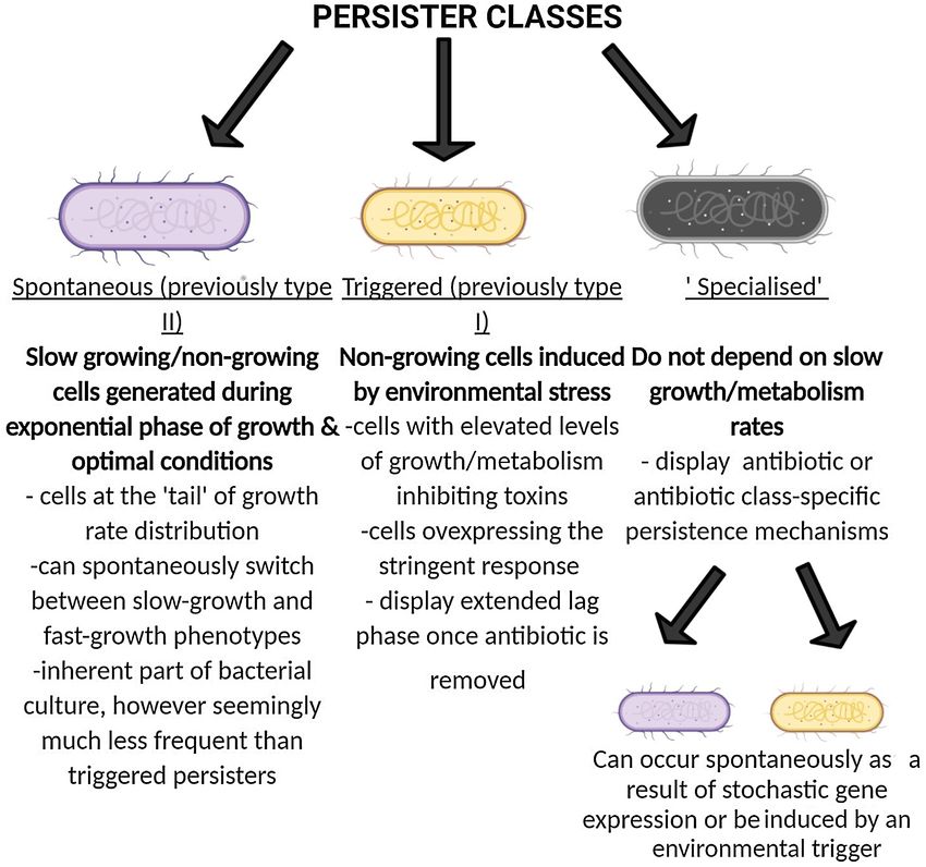
2      FEMS Microbiology Reviews, 2022, Vol. 46, No. 1

Table 1. Terms used commonly in persistence research.

antibiotic persistence                                A phenotypic feature defining a subpopulation of isogenic bacterial cells which display
                                                       a greatly reduced killing rate to antibiotic(s) when compared to the entire population;
                                                       the reduction of the killing rate is largely independent of the antibiotic concentration
                                                            (i.e. no change in minimum inhibitory concentration (MIC) to the antibiotic is
                                                        observed); also referred to as subpopulation tolerance and heterotolerance (Balaban
                                                                                                  et al. 2019)
antibiotic tolerance                                      A feature defining an entire population of bacterial cells which display decreased
                                                          killing rate to antibiotic(s); similarly to persistence the reduction of killing rate is
                                                                largely independent of the antibiotic concentration (Balaban et al. 2019)
antibiotic/antimicrobial resistance                        A feature of the entire population of microorganisms which possess a genetic
(AMR)                                                    resistance mechanism that allows them to overcome the harmful effect of a given
                                                      antibiotic; AMR is concentration dependent and characterised by an increase in MIC for
                                                                              a given antibiotic (Cantón and Morosini 2011)

                                                                                                                                                     Downloaded from https://academic.oup.com/femsre/article/46/1/fuab042/6343042 by guest on 04 July 2022
persistent (chronic) infection                         Any infection (bacterial/viral/fungal) which persists in the host for a prolonged period
                                                                            (Centre for Disease Control and Prevention 2019)

cells (about 1 in 106 ) in cultures of Staphylococcus aureus, iso-        growth of the entire population. In this sense, persisters could
lated from patients, could not be eradicated even using high              also be described as being hetero-tolerant, i.e. a subpopulation
doses of penicillin. Bigger also hypothesised that persistence            of antibiotic tolerant cells within a population of mostly sensi-
was induced by environmental stresses, such as pH, and might              tive cells (Balaban et al. 2019). Persistence is also a term used
be caused by slow growth rate of persister cells rather than a            to refer to infections that persist in the host without causing
heritable resistance (Bigger 1944). It would be another forty years       disease (Centre for Disease Control and Prevention 2019) and
before the phenomenon of persistence was further investigated             without reference to antibiotic persistence. Although it has often
through molecular techniques, largely due to the difficulty of            been speculated that these ‘infection persisters’ are the same
studying minority subpopulations using traditional microbio-              kind of cells as antibiotic persisters, this remains to be estab-
logical methods. Alongside molecular advances, increasing evi-            lished. To avoid confusion of terms, Balaban has recently pro-
dence has emerged implicating antibiotic persistence as a cause           posed that persisters, meaning a subpopulation of tolerant cells,
of treatment failure (Cohen, Lobritz and Collins 2013) and as a           should, at least initially, be referred to as ‘antibiotic persisters’
‘stepping-stone’ for genetic antibiotic resistance (Windels et al.        (Balaban et al. 2019) to distinguish them from infection persis-
2019; Liu et al. 2020). Antibiotic persistence is noted in multi-         ters. A summary of these terms is shown in Table 1. Another
ple clinically relevant pathogens from Mycobacterium tuberculosis         distinct subpopulation of cells that can be observed in bacte-
(Manina, Dhar and McKinney 2015; Vilcheze and Jacobs 2019),               rial culture are viable but non-culturable cells (VBNCs/ sleeper
Pseudomonas aeruginosa (Nguyen et al. 2011), Escherichia coli (Bal-       cells) that remain intact and metabolically active after exposure
aban et al. 2004) and Methicillin-Resistant S. aureus (MRSA) (Kim         to an antibiotic but, unlike persisters, do not resume growth
et al. 2018) and is likely to be a cornerstone to disease control         after removal of the antibiotic (Dong et al. 2020). It has been
and to combatting AMR.                                                    shown that, if incubated for long enough, some VBNCs of sev-
                                                                          eral bacterial species may eventually resume growth (Dong et al.
                                                                          2020) and could thereby represent a persister population with a
What is a persister?
                                                                          very long lag phase. This assumption is supported by transcrip-
In this review, the term ‘persistence’ will refer to a subpopula-         tomics, where it has been demonstrated in an innovative study
tion of bacterial cells that possess the non-heritable character          that E. coli ‘sleepers’ display similar levels of expression of sev-
of surviving prolonged exposure to an antibiotic that kills iso-          eral persistence-related genes to persister cells (Bamford et al.
genic sister cells. The key features that distinguish persistence         2017). However, for the purposes of this review, we will retain
from genetic resistance are that it involves only a subpopula-            the term persisters to cells that both remain viable and eventu-
tion of isogenic cells that do not carry resistance genes, so the         ally regrow after removal of antibiotic. The link between persis-
phenomenon is sometimes termed as phenotypic, rather than                 ter formation and persister subclasses within biofilms is another
genetic, resistance. It also tends to not be antibiotic-specific; or,     intriguing avenue of research and one which may require further
at least, persister cells can be detected that are tolerant to a wide     nomenclature changes in the future.
variety of antibiotics, suggesting that the majority of persistence           Note that the above persister definition encompasses cells
is a general, rather than antibiotic-specific, phenomenon. How-           that survive exposure to antibiotics not only in vitro, but also
ever, it appears that there is a subclass of persisters that are          during treatment of infections in humans or animal models.
antibiotic-specific, which we will also describe further in this          The existence of such cells was established as early as 1956 by
review.                                                                   McCune and Tompset who used a mouse-infection tuberculo-
    Persisters are also sometimes described as being tolerant to an       sis (TB) model to demonstrate that antibiotic-sensitive M. tuber-
antibiotic, rather than genetically resistant, with the term ‘tol-        culosis bacilli could be cultured from one third of experimen-
erance’ indicating that the capability of surviving exposure to           tal animals 90 days after successful treatment with isoniazid,
antibiotics is reversible and non-heritable. In contrast to persis-       para-aminosalicylic acid (PAS) and streptomycin (as determined
tence, antibiotic tolerance can be a feature of an entire popula-         by culture, microscopy and sub-inoculation) (McCune, Tompset
tion of cells when, for example, it is exposed to stress, starvation      and Mcdermott 1956). A shortened revival time was detected in
or stationary phase growth, which are conditions that restrict            immuno-compromised mice, suggestive of immune-regulation
Urbaniec et al.      3

of persistence (McCune et al. 1966). Pathogens might survive in          Can antibiotic persistence facilitate the emergence of
a treated host due to lack of penetration of the antibiotic into         genetic antibiotic resistance?
the persister bacilli or in tissue sites (Ray et al. 2015), cell types
(Greenwood et al. 2019) or through an inadequate host immune             It has recently been hypothesized that this already clinically rel-
response, as in in utero infections of bovine viral diarrhoea virus      evant subpopulation may be a reservoir for genetic antibiotic
(Khodakaram-Tafti and Farjanikish 2017). However, these expla-           resistance. For example, prolonged exposure to DNA-damaging
nations are unlikely to account for the persistence of the infec-        fluoroquinolones and hydroxyl radicals produced as a result of
tion in multiple body sites after prolonged treatment, as in the         exposure to β-lactams, fluoroquinolones and aminoglycosides
above mouse model. It seems more likely, and thereby it is gen-          (Kochanski et al. 2007) is likely to lead to a degree of DNA dam-
erally assumed, that the subpopulation of cells that survive pro-        age, and as a result activation of the SOS response. The SOS
longed antibiotic treatment in patients or animal models are             response activation in turn causes induction of expression of
indeed the same kind of persister cells that survive prolonged           error-prone DNA polymerases such as DNA polymerase IV &
antibiotic exposure in vitro. This is an important assumption as it      V, which increases rates of adaptive mutation (McKenzie et al.
justifies the relevance of in vitro persistence models to in vivo sys-   2000), provided that the cell survives the antibiotic exposure (i.e.

                                                                                                                                                Downloaded from https://academic.oup.com/femsre/article/46/1/fuab042/6343042 by guest on 04 July 2022
tems and clinical disease, however to our knowledge it remains           is a persister). Indeed, there is an increasing array of evidence
unproven.                                                                suggesting that persistence (& whole population antibiotic toler-
    The McCune and Tompset experiments also revealed another             ance) could accelerate genetic resistance development (Vogwill
important aspect of persistence: it is not the same for all antibi-      et al. 2016; Mok and Brynildsen 2018; Liu et al. 2020). For exam-
otics. Although persister cells survived 12 weeks of treatment           ple, there is in vitro evidence that persistence can be a contribut-
with isoniazid, PAS and streptomycin in the mouse model, no              ing factor to the emergence of resistant strains of M. tuberculosis,
viable bacteria could be recovered from animals treated for the          possibly as a result of increased levels of mutagenesis-inducing
same period with just pyrazinamide and isoniazid. This suggests          hydroxyl radicals found in M. tuberculosis persisters (Sebastian
that pyrazinamide may be more effective at killing TB persisters,        et al. 2017).
however the exact mechanism through which this occurs is cur-                Interestingly, persistence to one antibiotic could not only
rently unknown. The observation that antibiotics differ in their         facilitate the emergence of genetic resistance to that antibi-
relative efficacy against persisters vs the bulk population of cells     otic, but other antibiotics as well. For instance, rifampicin (RIF)-
demonstrates that persisters are physiologically distinct from           persisters in M. tuberculosis which carried elevated levels of
the bulk population. This provides the rationale for the long-           hydroxyl radicals were found to carry de novo acquired muta-
term aim of identifying novel compounds that target these phys-          tions not only associated with RIF-resistance mutations (rpoB),
iologically distinct antibiotic-refractory cells in order to shorten     but also moxifloxacin-resistant mutations (gyrA) (Sebastian et al.
treatment regimes.                                                       2017). Increased mutagenesis rates were also identified in mox-
                                                                         ifloxacin persisters of Mycobacterium. smegmatis, which devel-
                                                                         oped moxifloxacin resistance, as well as resistance to etham-
                                                                         butol and isoniazid (Swaminath et al. 2020). A similar mecha-
What are the evolutionary origins of antibiotic
                                                                         nism has also been identified in S. aureus where exposure to
persistence?
                                                                         ciprofloxacin resulted in the induction of error prone DNA poly-
Although persistence is non-heritable, the frequency of persis-          merases that increased the number of resistant mutants 30-fold
ter cells in a population is a heritable trait, as has been shown        when compared to a SOS response or error-prone polymerase
by several studies that demonstrate gene mutations that either           deficient knock-out control (Cirz et al. 2007).
increase or decrease that frequency (Moyed and Bertrand 1983;                Finally, analysis of clinical isolates of MRSA carried out by
Wolfson et al. 1990; Hingley-Wilson et al. 2020). Being heritable,       Liu and colleagues (Liu et al. 2020) has also demonstrated that
the frequency of persistence in the population would be visible          certain combination therapies which are effective in preventing
to natural selection and likely evolved as a ‘bet hedging’ strategy.     emergence of genetic AMR through a mechanism called ‘sup-
‘Bet hedging’ is an evolutionary survival strategy first described       pressive action’ fail to do so if tolerance to the primary (non
in 1974 (Slatkin 1974). It proposes that in uncertain changing           ‘suppressive’) drug is established prior or during the course of
environments it is evolutionarily favourable to introduce phe-           treatment. Suppressive action is a mechanism by which a com-
notypic heterogeneity into an isogenic population to be better           bination of two drugs is less effective than the single drug alone,
prepared for unpredictable environmental perturbations, such             for example in the case of treatment with any β-lactam antibi-
as encountering antibiotics (Slatkin 1974). The theory is sup-           otic (which most effectively targets dividing cells) and chlo-
ported by mathematical models in which populations generat-              ramphenicol (which slows down growth) (Jawetz, Gunnison and
ing higher persister cell levels have an increased fitness when          Speck 1951). This paradoxically decreases the survival rate of
repeatedly exposed to bactericidal antibiotics (Johnson and              chloramphenicol resistant mutants and thereby decreases their
Levin. 2013). It also seems to be supported by recent evidence           frequency in the population. Pre-establishment of tolerance to
that laboratory exposure of E. coli to repeated dual-antibiotic          the non-suppressive antibiotic negates its anti-AMR effect, pro-
regimes results in significant enrichment of the persister frac-         viding in-vivo evidence of a direct link between tolerance (&
tion, through convergent mutations in proteins involved in tRNA          persistence), resistance and clinical treatment failure (Liu et al.
synthetases, as well as methionine synthetase metG (Khare and            2020). This study suggests that targeting persisters and inhibit-
Tavazoie 2020). An alternative perspective is that persistence           ing the persistence state could thereby inhibit the development
is an inevitable consequence of ‘errors’ during cell cycle and           of genetic AMR.
division that introduce phenotypic heterogeneity, a hypothe-
sis sometimes known under the acronym ‘PASH’ (persistence
                                                                         How is antibiotic persistence quantified?
as stuff that happens) (Levin, Concepcion-Acevedo and Udekwu
2014). It could be that different persister classes as described         The “gold standard” experimental system used to demonstrate
below have different evolutionary origins.                               the presence of persisters and estimate their abundance is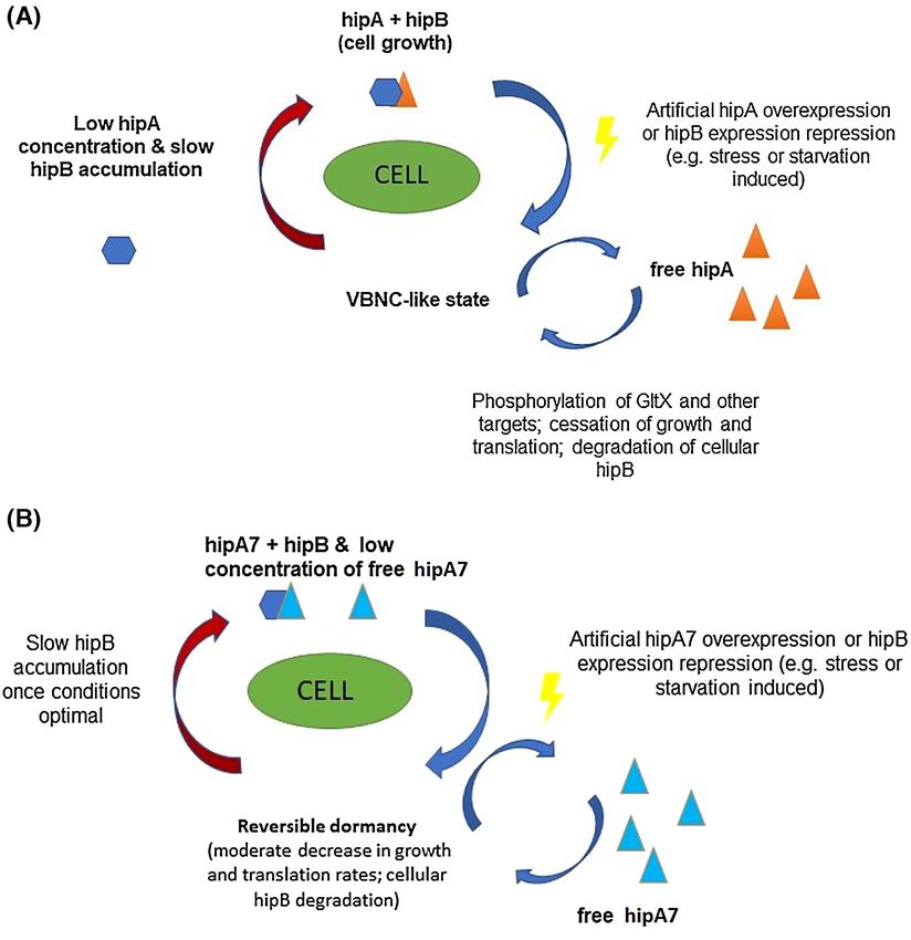
4      FEMS Microbiology Reviews, 2022, Vol. 46, No. 1

the antibiotic time-kill experiment, which generates the clas-           and then subjected to three rounds of enrichment for mutants
sic biphasic kill curve from the average kill (Fig. 1). Typically,       that survived repeated exposure to 100 μg of ampicillin, the third
an exponentially-growing population of isogenic bacteria is              exposure being for 20 h. Hip mutants were identified as strains
exposed to antibiotic and samples are taken at regular time              that showed a higher survival after exposure to ampicillin, com-
points for determination of colony forming units (cfu, on a log          pared to the wild-type. Mutants that were genetically resistant
scale) as a function of time. Under conditions where persis-             or unable to grow on minimal media were discarded. It should
tence is detectable, the kill rate of is made up of two compo-           be noted that mutants with substantially reduced growth of
nents: a fast initial kill characteristic of the susceptible popula-     the whole population were also discarded. Note that techniques
tion and a slower kill characteristic of the persister subpopula-        to examine growth of sub-populations were not yet available.
tion. If the slow killing phase is extrapolated to zero time then        Nonetheless, all the hip mutants identified showed some degree
where it crosses the y axis provides an estimate of the persister        of growth impairment in rich media (Lysogeny broth commonly
fraction in the initial population (Balaban et al. 2019). Figure 1       known as LB), providing a link between mutations increasing
shows an ‘ideal’ difference in kill curve shapes between suscep-         persistence and slow growth (Moyed and Bertrand 1983). How-
tible, highly-persistent, genetically-resistant and tolerant popu-       ever, two mutants, HM7 and HM9, grew as rapidly or faster than

                                                                                                                                               Downloaded from https://academic.oup.com/femsre/article/46/1/fuab042/6343042 by guest on 04 July 2022
lations. For instance, a typical time-kill curve of M. tuberculosis      the parental strain in minimal media yet continued to display
using front-line antibiotics such as isoniazid follows a biphasic        1000-fold higher frequencies of persisters than the wild-type in
curve, as highlighted in this comprehensive review detailing TB          this media, proving that slow growth of the bulk population is
persisters (Vilcheze and Jacobs 2019).                                   not a requirement of high levels of persistence. The researchers
    It should, however, be emphasized that kill curves are               performed batch time-kill assays for wild type and both the
extraordinarily sensitive to the precise conditions of the exper-        hipA7 and hipA9 mutants. Examination of the curves obtained
iment and particularly the growth state of the initial popula-           suggested a population of around 0.1% persisters in the wild-
tion, which can lead to well-known reproducibility issues. This          type and about ten times this in both mutants. The researchers
experimental and stochastic variation in each experiment can             went on to utilize classical microbial genetic approaches to map
obscure data from the relatively minute persister subpopula-             the genetic loci for both HM7 and HM9 (Moyed and Bertrand
tion. Different batches of antibiotic or media, culture age, varia-      1983) to the closely-linked loci hipA7 and hipA9.
tion in storage or growth conditions and the presence of resident            Later studies (Korch, Henderson and Hill 2003) established
phages can also influence the shape of kill curves (Harms et al.         that the hipA7 gene encoded the toxin component of one of sev-
2017). Culture conditions can also profoundly influence pheno-           eral of toxin-antitoxin systems present in the E. coli genome. To
typic heterogeneity (Smith et al. 2018) which, as we will later dis-     our knowledge the hipA9 gene remains uncharacterised. Toxin-
cuss, plays a key role in persistence. It also appears that selec-       antitoxin modules are widely distributed in bacteria and consist
tion and outgrowth may also affect reproducibility where growth          of genes encoding a stable toxin and an unstable antitoxin (Ezaki
regulation is affected (Urbaniec, unpublished results).                  et al. 1991). The wild-type hipA gene encodes the toxin, hipA,
    Since, as illustrated by the biphasic kill curve (Fig. 1), persis-   which is neutralized by its antitoxin, hipB, encoded by the adja-
tence is a property of only a minority of cells, traditional micro-      cent hipB gene. If not neutralized, hipA acts as a serine-threonine
biological, cell biological or molecular biological analysis tech-       kinase that inhibits protein synthesis by phosphorylating (and
niques that examine bulk populations are unsuitable for study-           therefore inactivating) glutamyl-tRNA synthetase (GltX), respon-
ing the phenomenon. Two approaches have been most produc-                sible for attachment of glutamine to tRNA. The protein inhi-
tively applied to study persistence: microbial genetics and single       bition results in accumulation of uncharged tRNA which indi-
cell microscopy studies. Indeed, single cell studies from our lab-       rectly increases levels of (p)ppGpp (guanosine pentaphosphate)
oratory show just how heterogenous both microbial populations            in the cell. (p)ppGpp is an alarmone (i.e. a signaling molecule
and persisters family are (Fig. 2). It should be observed in this fig-   produced in unfavourable conditions) and component of the
ure that persisters themselves are only noted on here if they are        stringent response in response to amino acid starvation. Down-
non-growing (blue line on left hand side) as division time is not        stream effects of its increase include inhibition of RNA synthesis
measured in these studies following antibiotic administration.           and initiation of growth arrest to induce a state of “dormancy”,
    The majority of antibiotic persistence studies have been per-        understood as a reversible state of little-to-no growth and lower
formed on the microbial workhorse, E. coli, which will be dis-           metabolic activity (Semanjski et al. 2018). ppGpp levels are them-
cussed first, particularly with reference to the canonical hipA7         selves regulated by proteins collectively referred to as RSH (RelA
system from which much of the theory of persistence has been             SpoT homologs) that are upregulated by various stress-related
derived. After examining both genetic and single cell studies of         molecules as the aforementioned uncharged tRNA or cAMP. The
this system we will go on to outline a general theory of persis-         hipAB system thereby connects with a wide range of cellular
tence and then examine how well the theory accounts for the              physiological systems (Atkinson, Tenson and Hauryliuk ).
evidence from a variety of systems.                                          Evidence for hipA toxin-induced growth arrest has been
                                                                         obtained from several systems. For example, Korch and col-
                                                                         leagues discovered that over-expression of hipA from a plas-
ANTIBIOTIC PERSISTENCE IN ESCHERICHIA                                    mid induces a VBNC-like state in approximately 95% of the pop-
COLI                                                                     ulation that continued for up to 72hrs with continuous hipA
                                                                         expression. A fraction of cells, from 1.8% to 20% resumed growth
Genetic studies to identify E. coli hip mutants
                                                                         following the 72hrs of continuous induction decreasing with
The first significant advance in the study of persisters was the         increasing induction time. Additionally, in cultures overexpress-
application of mutagenesis and microbial genetic studies in the          ing HipA a significant (10 000-fold) increase in the number of
1980’s to identify ‘high-persister’ or hip mutant strains of E.          persisters to ampicillin was observed (Korch and Hill 2006). It
coli K12 strain, including the intensively-studied hipA7 strain          was hypothesized that the much lower fraction of persisters in
(Moyed and Bertrand 1983). In this study, an E. coli K12 strain          the wild-type population is accounted for by lower levels of hipA
was exposed to untargeted chemical mutagenesis in rich media             expression, similar to that of the HipB antitoxin.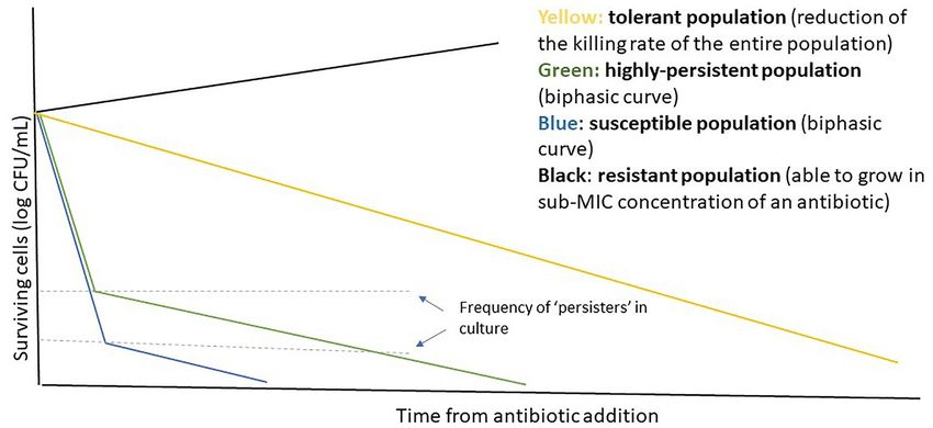
Urbaniec et al.           5

                                                                                                                                                                                Downloaded from https://academic.oup.com/femsre/article/46/1/fuab042/6343042 by guest on 04 July 2022
Figure 1. The highly heterogenous persister cell can be observed at batch level. Biphasic killing at batch level: an example time-kill curve shape of resistant, susceptible,
high-persister and tolerant cultures. The resistant population is able to grow in sub-inhibitory concentrations of an antibiotic, whereas tolerant & persister cells are
eventually killed by the antibiotic, but at a much slower rate. The higher number of persister cells observed in high-persistent strains ensures that the number of cells
surviving the initial ‘rapid-killing’ phase of antibiotic exposure is greater than in the susceptible parental strain.

Figure 2. The growth rate of E. coli and the persister’s family is highly heterogenous. Single cell microfluidics study showing division time in minutes throughout
experimental frames (1 frame = 69 seconds) of the whole population of E. coli HipQ cells prior to antibiotic administration (number of cells = 157). Different colours
represent different persister ‘families’ (cells from the same lineage). The time point of 160 minutes represents the final time point and also includes non-growing cells
(i.e. the blue line represents a non-growing persister cell).

    The hipA7 high-persistence phenotype is conferred by point                           persistence’ phenotype (Korch, Henderson and Hill 2003). The
mutations at two separate sites in the hipA gene (hipA7 allele),                         non-toxicity is thought be a consequence of a reduced inabil-
both of which are necessary for the hip phenotype (Moyed and                             ity of the hipA7 protein to phosphorylate targets such as GltX
Bertrand 1983). How these mutations lead to high levels of per-                          and several other proteins involved in regulation of transcrip-
sistence is not fully clear (Fig. 3A and B). One appears to ren-                         tion (Semanjski et al. 2018). It also has a weaker binding affinity
der the hipA protein non-toxic since its overexpression only                             to its cognate antitoxin. However, and rather paradoxically over-
moderately inhibits growth & translation in comparison to wild-                          expression of the hipA7 allele causes a 12-fold higher phospho-
type hipA, while the other is required for the observed ‘high-                           rylation of GltX when compared to the wild-type toxin (Seman-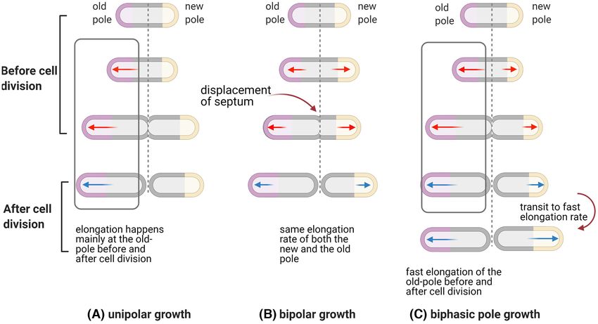
6      FEMS Microbiology Reviews, 2022, Vol. 46, No. 1

                                                                                                                                                                          Downloaded from https://academic.oup.com/femsre/article/46/1/fuab042/6343042 by guest on 04 July 2022
Figure 3. Schematic representation of A. hipA and B. hipA7-induced changes in cellular metabolism. (A) hipA phosphorylates GltX, as well as several other proteins
involved in growth & translation regulation, inducing a HipA/HipB ratio-dependent VBNC-like state in cells containing unbound hipA. Growth can resume only after
hipB binds to and inactivates free hipA. (B) The mutant hipA7 protein (lower panel) can exist in the unbound form at higher levels in the cell without inducing a
VBNC-like state as it only phosphorylates GltX and with lower efficiency than the wild-type protein. High levels of free hipA7 in the cell induce reversible “dormancy”
which is proposed to be responsible for the high-persistence phenotype.

jski et al. 2018). A model has been proposed to account for                           induced to overexpress the wild-type hipA from a plasmid (Ger-
these apparently contradictory findings, in which chromosoma-                         main et al. 2013). This observed ‘high-persister’ phenotype to flu-
lly expressed HipA7 exists more abundantly in the unbound                             oroquinolones is presumably a result of toxin-induced extended
form when compared to hipA, allowing phosphorylation of more                          lag phase of hipA/hipA7 persisters, discussed further in section
GltX. This results in the slower growth rate of hipA7 express-                        three of this review.
ing strain (however this phenotype is lost whenever hipA is
artificially overexpressed). Additionally, hipA also phosphory-
                                                                                      Single cell studies of the hipA7 mutant
lates other targets which are proposed to contribute to its toxi-
city (Semanjski et al. 2018) and ability to induce the VBNC state                     Although characterization of hip mutants provided insights into
(Korch, Henderson and Hill 2003). Interestingly, naturally occur-                     factors that might increase or decrease the frequency of per-
ring hipA7 variants has been identified in urinary tract infec-                       sistence, the nature of the persister cells remained a matter of
tions suggesting that the locus may also induce persistence in                        conjecture until pioneering studies performed by Balaban and
vivo (Schumacher et al. 2015).                                                        colleagues who developed microfluidic systems to perform live
    It is important to note that although both hipA and hipA7                         cell imaging of bacterial populations before, during and after
alleles have primarily been investigated in relation to persis-                       antibiotic exposure. The focus of much of their studies was the
tence to ampicillin, there is a evidence that these toxins con-                       hipA7 mutant as its characteristic high frequency of persister
tribute to multidrug tolerance. For instance, recently identified                     cells (1000 x more than wild-type frequencies in their studies)
E. coli clinical isolate carrying the HipA7 allele on the bacterial                   made the phenomenon much more experimentally tractable.
chromosome has been shown to display up to ∼100-fold higher                           The character of persistence was found to be associated with
persistence to fluoroquinolone ciprofloxacin when compared to                         cells that, prior to antibiotic exposure, were either slow-growing
its parental strain (Schumacher et al. 2015). A similar effect has                    or non-growing, prompting the hypothesis that persistence is
also been observed previously in lab-adapted E. coli MG1655 cells                     due to pre-existing phenotypic heterogeneity associated with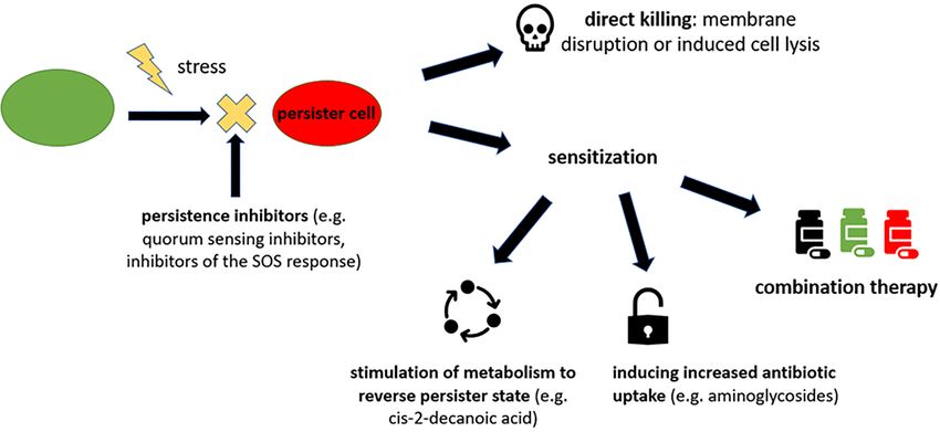
Urbaniec et al. 7
slow growth rate in the starting population (Balaban et al. 2004). inducing persistence (Gerdes and Maissoneuve 2012). Although
The established association between HipA7 and toxin-antitoxin discovery of a single ‘persistence switch’ (in this case Lon-
systems prompted the proposal that stochastically variable lev- mediated toxin accumulation) would certainly allow for a more
els of expression of the hipA toxin and consequent low or zero systematic approach to persistence research, the ‘real-life’ sce-
growth in a sub-fraction of cells might be a source of the pre- nario is likely more complex. The model proposed by Gerdes and
existing variation (Korch, Henderson and Hill 2003; Keren et al. colleagues was based on the observations of a decreasing num-
2004) and origin of persistence. Thereafter, many other high- ber of persisters following deletions of 10 subsequent TA mod-
persistent mutants in numerous bacterial strains and species ules. Unfortunately, some of the data on which this model was
have been described (Torrey et al. 2016; Wilmaerts et al. 2018). based may have been confounded by the presence of resident
Additionally, low persister mutants that have a lower frequency prophages whose lytic cycle can be induced by an antibiotic (in
of persisters than wild-type cells have been identified, eg E. coli this case ciprofloxacin) resulting in cell lysis and reduced num-
ybaL (Hingley-Wilson et al. 2020). Both types of mutants iden- ber of surviving cells, irrespective of persistence mechanisms
tify genes that influence the frequency of persisters in bacterial (Harms et al. 2017). This led to the re-evaluation of the role of
populations and thereby provide clues as to the mechanisms of TA modules in persistence with some groups reporting a link
Downloaded from https://academic.oup.com/femsre/article/46/1/fuab042/6343042 by guest on 04 July 2022
persistence, or, at least, mechanisms involved in the generation between TA modules and persistence irrespective of ppGpp lev-
of persisters. els in E. coli (Chowdhury, Kwan and Wood 2016) & M. smegmatis
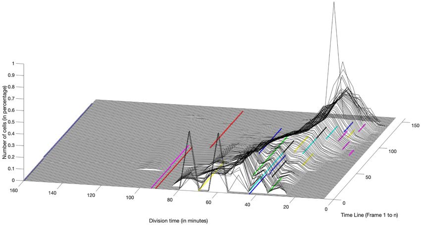
Moreover, Balaban observed two distinct types of persister (Bhaskar et al. 2018) and others reporting no link between persis-
cells: triggered (previously type I) persisters which were non- tence levels and TA module deletion in E. coli (Goormaghtigh et al.
growing cells in stationary phase that displayed extended lag 2018). The role of TA modules in persistence thereby appears to
phase upon inoculation into fresh media (presumably caused by be more complex than initially reported.
nutrient starvation) and stochastic (previously type II) persisters Following Moyed and Bertrand’s landmark publication, a sig-
which were slow-growing or non-growing cells generated dur- nificant amount of research has focussed on the (p)ppGpp alar-
ing exponential phase of growth seemingly without an environ- mone system. (p)ppGpp levels are upregulated as a result of var-
mental trigger (Balaban et al. 2004; Balaban et al. 2019). Persister ious environmental stressors such as nutrient starvation, acid
sub-populations, detected with dual-reporter phages in combi- stress or heat shock (Abranches et al. 2009), which could act
nation with single cell imaging, were also noted in M. tubercu- as a trigger in the formation of persister cells, at least in E.
losis following INH treatment as (Jain et al. 2016). Interestingly, a coli where a link has been established between elevated ppGpp
potential third class of persisters (referred to here as ‘specialised levels in persister cells and nutrient starvation or oxidative
persisters’) which are not slow-growing prior to antibiotic expo- stress (Radzikowski et al. 2016; Brown 2019). Experiments on P.
sure and often display antibiotic specific persistence mecha- aeruginosa, both in vitro and in a mouse infection model, have
nisms has also been observed (Wakamoto et al. 2013; Goormath- implicated RelA/SpoT, which synthesize and degrade (p)ppGpp,
igh and Van Melderen 2019), as shown in Fig. 4. respectively. A mutant lacking the genes (and therefore inca-
pable of initiation of (p)ppGpp-mediated stringent response)
produced 100-fold fewer persisters compared to the wild-type
MOLECULAR MECHANISMS INVOLVED IN strain (Nguyen et al. 2011). The frequency of persisters to multi-
PERSISTER CELL FORMATION ple antibiotics was also altered in deletion mutants of transcrip-
Toxin-antitoxin systems and induction of the stringent tion factors sigE or sigB (Pisu et al. 2017). SigE regulates the tran-
response are only one of the many pathways leading to scription of relMtb , which in turn regulates (p)ppGpp (Avarbock
et al. 2005). Interestingly, deletion of relMtb resulted in reduced
persistence
(p)ppGpp synthesis and decreased infection persistence of M.
Inspired by the discovery of the hipA system multiple other tuberculosis in a murine model (Dahl et al. 2003).
TA systems have been linked to the establishment of persis- Taken altogether these findings point towards the hypothesis
tence. HokB toxin expression in E. coli is upregulated by the that persistence is a complex phenomenon potentially involv-
Obg GTPase which is involved in stress response as a regula- ing multiple mechanisms (Fig. 5), both passive (such as growth
tor of cellular energy levels and whose overexpression results arrest as a result of toxin accumulation) and active (such as
in pore formation in the membrane and ATP leakage from the response to oxidative stress). This makes the effort of establish-
cell (Wilmaerts et al. 2018). TisB toxin, also present in E. coli, is ing a comprehensive persistence model a challenging task.
involved in a multi-drug tolerance phenotype under the regula-
tion of the SOS response pathway (Dorr, Vulic and Lewis 2010).
Another well researched toxin-antitoxin module, MazF-MazE, is
Specialised persisters
induced under stressed conditions and disrupts protein synthe-
sis in M. tuberculosis leading to degradation of selected mRNA It seems likely that the phenomenon and mechanisms of per-
targets and induction of the stress response operons resulting sistence are much more varied than toxin-antitoxin systems &
in growth arrest (Tiwari et al. 2015). ppGpp-induced growth arrest. For example, ‘specialised’ persis-
However, a fair amount of uncertainty surrounds the effort ter cells are generated during exponential growth, but are not
of developing a unified persistence model based on toxin accu- associated with slow individual cell growth, and are specific to
mulation, with conflicting results often reported by different a particular antibiotic (Wakamoto et al. 2013; Goormagthigh and
research groups. One notable example is a model which aimed van Melderen 2019) and Fig. 4. The antibiotic isoniazid is an inac-
to provide a ‘blanket’ pathway on how persistence is estab- tive prodrug, which becomes activated inside the mycobacterial
lished in the model organism E. coli. This neat model proposed cells through cleavage by the bacterial enzyme KatG (catalase-
that increasing levels of ppGpp as a response to external stress peroxidase). Single-cell level observations have shown that, dur-
causes accumulation of polyphosphate, activation of Lon pro- ing exposure to isoniazid, the growth/ division rate of a cell
tease and Lon-mediated accumulation of toxin components of does not correlate with persistence. Instead, slowly growing cells
TA systems which in this model acted as effector molecules were as likely to die as fast-growing cells. Instead, intrinsic noise8 FEMS Microbiology Reviews, 2022, Vol. 46, No. 1
Downloaded from https://academic.oup.com/femsre/article/46/1/fuab042/6343042 by guest on 04 July 2022
Figure 4. Proposed addition of specialised persisters to classification of persister cells (Balaban et al. 2019) (figure created with Biorender.com). Both spontaneous
and triggered persisters are slow/non-growing prior to antibiotic addition and often display a general, rather than antibiotic-specific, persistence phenotype. It is,
however, important to note that not all slow/non-growing cells are persisters. In contrast, ‘specialised’ persisters do not depend on slow growth/metabolic rates
to survive antibiotic exposure, but instead display antibiotic-specific persistence mechanisms. They can occur spontaneously, for example, in Mycobacteria, through
stochastically low levels of expression of the enzyme catalase-peroxidase that activates isoniazid (Wakamoto et al. 2013) or be induced by a stress-signal, for example
ciprofloxacin persisters which are induced by exposure of E. coli to this antibiotic (Dorr et al. 2009; Goormaghtigh and van Melderen 2019).
in gene expression leads to fluctuations in the level of tran- DNA damage prior to growth resumption. This made them much
scription and translation of all cellular genes resulting in cell- more likely to survive exposure to DNA-damaging antibiotics,
to-cell variation of individual gene expression levels (Raj and such as ciprofloxacin (Mok and Brynildsen 2018). A similar effect
van Oudenaarden 2008). In mycobacteria, intrinsic noise in katG was observed when E. coli cells were exposed to sub-MIC concen-
gene expression gave rise to a pulsing of intracellular enzyme trations of ofloxacin, which significantly (1200-fold) increased
activity that appeared to be associated with persistence spe- the number of persisters upon subsequent exposure to a high
cific to isoniazid. Even a small increase in katG expression was concentration of this antibiotic, when compared to cultures
shown to lead to a huge decrease in the number of persister cells only exposed to high concentrations of ciprofloxacin. A pro-
and persister cells tended to express less ‘pulsing’ of KatG lev- posed model behind this involves ‘priming’ of the SOS response
els than non-persister cells (Wakamoto et al. 2013). Similarly, a through low-levels of DNA damage, resulting in the overexpres-
single-cell imaging study carried out on E. coli has demonstrated sion of the TisB toxin and subsequent toxin-induced growth
that survival to fluoroquinolones in actively growing cells, prior arrest (Dorr, Lewis and Vulic 2009). Both MazF and TisB persisters
to exposure to the antibiotic, requires appropriate timing of also displayed a multidrug tolerance phenotype and were toler-
the SOS system induction during the recovery & repair phase ant to ampicillin, presumably as a result of their arrested growth
and hence, prior to growth resumption. This study demon- state (Dorr, Lewis and Vulic 2009; Mok and Brynildsen 2015).
strated that fluoroquinolone persisters are highly heteroge- However, there is also supporting evidence that persistence to
nous and are not always slow-growing or SOS-induced prior to fluoroquinolones is governed by more than the presence of TA
antibiotic addition as discussed below (Goormaghtigh and Van systems and is not only a passive-by product of arrested growth
Melderen 2019). state (Bernier et al. 2013), as described above (Goormaghtigh
and Van Melderen 2019). It is plausible to assume that both
Persistence as a result of extended lag phase types of growth arrested and growth-independent (specialist)
fluoroquinolone persisters could co-exist in an isogenic culture
Duration of the lag phase can be extended by elevated levels and that their ratio would fluctuate depending on environmen-
of growth-inhibiting toxin components of the TA systems (such tal/culture conditions.
as MazF or hipA) and was found to be an important mecha-
nism for establishment of persistence to fluoroquinolones in E.
Transporter-linked persistence
coli. Fluoroquinolones damage the DNA of both actively grow-
ing and non-growing cells and therefore, through extension of Increasing intracellular antibiotic uptake has been shown to
their lag phase E. coli persister cells were able to repair incurred lead to elimination of persister cells is some systems (Allison,Urbaniec et al. 9
Downloaded from https://academic.oup.com/femsre/article/46/1/fuab042/6343042 by guest on 04 July 2022
Figure 5. Simplified network displaying the role of the TA systems and the stringent response in establishment of persistence. Cells sense changes in the external
environment through receptors such as two-components systems (histidine kinases), thermosensor molecules, pH sensing molecules or GltX inhibition through
accumulation of free radicals or phosphorylation by HipA/HipA7. Downstream signaling causes accumulation of stress-related molecules in the cells such as cAMP,
uncharged tRNA, heat shock proteins etc. This is sensed by RelA/SpoT proteins which in turn convert ATP/GTP into the (p)ppGpp alarmone. (p)ppGpp affects cellular
metabolism directly (by interacting with RNA polymerase) and indirectly (by depleting cellular GTP levels) and has been linked to establishment of the persister state.
Toxin components can also directly inhibit crucial metabolic processes such as protein synthesis or replication or affect cellular membrane potential. Depending on
the free/bound toxin ratio, this can either result in cell death or temporary growth arrest (persistence).
Brynildsen and Collins 2011). Subsequently, it was demon- old & misfolded protein aggregates at the ‘old pole’, as previously
strated that, when treated with β-lactam antibiotics, E. coli hypothesized (Rang, Peng and Chao 2011; Lapinska et al. 2019).
persister cells show higher efflux pump activity compared to Whatever its origin, since asymmetric cell division appears to be
non-persister cells and accumulate lower levels of drug intra- a source of asymmetry in growth rate, it is potentially a source
cellularly (Pu et al. 2016). Heterogeneity of uptake mechanisms of persistence.
and efflux pumps may thereby be another source of persisters. Mycobacteria appear to undergo asymmetric cell division
Several time-kill studies have also confirmed that increasing to an even greater extent than E. coli. In a pioneering single
efflux pump activity can decrease the number of persisters (de cell imaging study, M. smegmatis cells were demonstrated to
Steenwinkel et al. 2010; Caleffi-Ferracioli et al. 2016; Pu et al. 2016). grow faster and bigger in the old-pole inheritor (i.e. from the
‘grandmother’) and slower and smaller in the new-pole daugh-
ter. However, susceptibility appeared to be antibiotic-specific,
Intrinsic asymmetry of cell division and persistence
with shorter, slower cells exhibiting increased tolerance to cell
Bacteria reproduce asexually through binary fission, during wall synthesis inhibitors such as cycloserine or meropenem and
which a ‘mother’ cell first duplicates its genetic material, then old pole larger, faster cells exhibiting enhanced tolerance to the
moves a copy of the bacterial chromosome to each cell pole and RNA-polymerase inhibitor rifampicin (Aldridge et al. 2012). This
afterwards splits in half into two ‘daughter’ cells by synthesis heterogeneity was determined to be dependent upon key pro-
of new cell wall. This mechanism of cell division results in an teins, such as the scaffold protein Wag31 and loss of asym-
intrinsic asymmetry since each daughter cell inherits an ‘old metry protein Lam (Rego, Audette and Rubin 2017), the role of
pole’ (from the mother cell) and a ‘new pole’ (synthesized dur- which, along with others, have been expertly reviewed recently
ing division). Further fission events will then result in some cells (Longsdale and Aldridge 2018). Asymmetric distribution during
accumulating ‘old poles’ (‘old pole daughters’ of ‘old pole moth- cell division of irreversibly oxidized proteins (IOPs) in both M.
ers’) (Lapinska et al. 2019). Research on E. coli has shown that smegmatis and M. tuberculosis also resulted in heterogeneity of
these old pole cells tend to have lower growth & glucose accumu- growth rate, with daughter cells inheriting more IOPs grow-
lation rates when compared to their ‘new pole’ sisters and that ing more slowly and showing increased susceptibility to antibi-
this effect is cumulative until the ‘old pole’ lineage reaches an otics (Vijay et al.2017). Moreover, as mentioned above, suscep-
apparent asymptote (in other words the change accumulation tibility to the pro-drug isoniazid was determined to be inde-
with subsequent generations reaches near-zero). The underly- pendent of the growth rate of single cells and instead, related
ing mechanism behind this phenomenon is currently unknown, to stochastic expression of the KatG gene, which encodes an
but does not appear to simply be caused by an accumulation of isoniazid activating catalase peroxidase (Wakamoto et al. 2013).10 FEMS Microbiology Reviews, 2022, Vol. 46, No. 1
Alongside antibiotic-specific responses, the growth medium or fission, from a single mother cell, so would be expected to epi-
conditions (i.e carbon source) may also affect asymmetry in a genetically inherit cellular constituents controlling the mother’s
manner dependent upon pole age (Priestman et al. 2017). Other growth rate, such as copy number of key enzymes, number of
studies found that growth rate is independent of pole age and ribosomes etc. However, whatever the cause, cell division is
suggest that cell-size at birth (L0 ) is key with both cells elongat- clearly an engine for the continuous generation of cells with
ing at the same rate post division (Santi et al. 2013; Wakamoto widely varying growth rates, including very slow growing cells
et al. 2013). These models are diagrammatically represented in that might be spontaneous or triggered persisters (i.e. growth
Fig. 6 and include a recent biphasic pole growth model which rate dependent).
centres upon the variable lag phase providing growth hetero- The source of epigenetic variation in growth rate is currently
geneity (Hannebelle et al. 2020). Regardless of the model, it is unknown, but it is well established that a myriad of genes can
clear that a considerable degree of cell asymmetry, and hence influence growth rate, as is apparent from the commonplace
growth rate and size, is characteristic of mycobacteria and may observation that mutation or induced changes in gene expres-
provide a reservoir for the phenotypic diversity that generates sion very often lead to altered growth rates. It thereby seems
persistence. Moreover, the clinical relevance of heterogeneity likely that stochastic variation in expression of many genes will
Downloaded from https://academic.oup.com/femsre/article/46/1/fuab042/6343042 by guest on 04 July 2022
has also been demonstrated in bacilli grown from TB patient cause stochastic variation of growth rate. Note also that, in the
sputum samples with larger, faster cells exhibiting increased mutational screen that led to the isolation of the original E. coli
tolerance to rifampicin and oxidative stress (Vijay, Vinh and Hai, hip mutants including the hipA7 strain (Moyed and Bertrand
2017). It appears that in mycobacteria, persisters may be antibi- 1983), all of the putative hip mutants grew slower than the wild-
otic specific and the high heterogeneity resulting from asymme- type in the rich media. As already described, the phenotype of
try may be one of the reasons for the high antibiotic persistence the intensively studied hipA7 mutant is caused by a mutation
noted in mycobacterial infections. in a toxin-antitoxin system whose expression depresses growth
rate.
The hypothesis that persistence is due to epigenetic variation
in growth rate is supported by recent results from our own lab-
ORIGIN OF PHENOTYPIC HETEROGENEITY OF
oratory, which has identified control of epigenetic inheritance
BACTERIAL POPULATIONS AS A SOURCE OF as a potential source of persisters (Hingley-Wilson et al. 2020).
ANTIBIOTIC PERSISTENCE The research studied the hipQ mutant of E. coli, isolated in dur-
Noise in gene expression levels leads to phenotypic ing mutagenesis screen in 1990, which displays increased spon-
taneous persisters when exposed to norfloxacin and ampicillin
heterogeneity
(Wolfson et al. 1990). Single cell growth studies across several
It has been apparent since the advent of single cell studies generations established that the high persister phenotype of
that clonal populations of cells exhibit stochastic variation, also hipQ is associated with the novel phenotype of reduced pheno-
known as noise, in the expression of genes (Elowitz et al. 2002; typic inheritance (RPI), identified as reduced correlation of growth
Cai, Friedman and Xie 2006; Levine and Hwa 2007). The causes parameters such as division time, size at birth or cell elongation
of this variation are largely unknown, but the amount of noise rate, either between mothers and daughter cells, or between sis-
in any system is inversely proportional to the copy number of ter cells. The results suggested that genes influencing epigenetic
molecules controlling the system, so very low copy numbers inheritance play a role in persister cell formation. The study also
of many cellular components such as regulatory molecules and identified the locus of the hipQ phenotype as a mutation in a
structural molecules (Guptasarma, P. 1995) will lead to high noise gene, ydcI, that encodes a putative transcription factor (Hingley-
levels in phenotypes controlled by low copy number molecules. Wilson et al. 2020).
Random partitioning of low copy number molecules at cell divi-
sion is also a source of phenotypic variation (Huh and Pauls-
A hunker theory of persistence
son 2011). Moreover, ergodicity-breaking, essentially incomplete
mixing of the cell so that it does not sample all its possible Aforementioned studies tend to argue against a single mecha-
microstates within one division cycle, has also been proposed nism of persistence prompting a more general theory of persis-
to be a factor responsible for phenotypic heterogeneity (Rocco, tence. However, growing slowly, or not at all (”dormancy”) has
Kierzek and McFadden 2013). been shown to induce tolerance, particularly to cytotoxic antibi-
Heterogeneity may be present in any cell phenotype but, otics. For example, Toumanen and colleagues determined that
given the strong association between growth rate and persis- the rate of killing of E. coli cultures exposed to β-lactams was
tence, phenotypic variation that influences growth rate is highly directly proportional to their growth rate, with slower growing
likely to influence the frequency of persisters in a population. cultures exhibiting enhanced tolerance (Tuomanen, Cozens and
That growth rate heterogeneity exists is apparent from many Tosch 1986).
studies and is illustrated in Fig. 2 which shows the distribution of For antibiotics to reach their target, they need to penetrate
growth rate in a population of E. coli cells taken from a microflu- the cell and bind to their target. Cells that, for a variety of rea-
idics experiment performed in our own laboratory. sons, hunker down by growing more slowly, metabolising more
Growth rate not only varies widely within the population but slowly, transporting materials across their cell membranes more
also, and rather strikingly, along cell lineages, including the per- slowly or activating antibiotics more slowly, will be killed more
sister family lineages as can be seen in Fig. 2, which illustrates slowly leading to a state of persistence.
growth rate of single E. coli cell lineages tracked across multi- This hunker theory of persistence is consistent with the
ple generations. As can be seen, the growth rate of daughter finding that persisters are not necessarily slow growing, as in
cells varies markedly from growth rate of their mother cell. In the specialised class of persisters (Fig. 4). Clearly, not all slow-
fact, the correlation of growth rate between mother and daugh- growing cells are persisters; and not all persisters are slow grow-
ter cells is very close to zero (Hingley-Wilson et al. 2020). This is ing. Slow growth may predispose towards the state of persis-
remarkable since each pair of daughter cells is derived, by binary tence; but it is neither sufficient nor necessary. Just as there areUrbaniec et al. 11
Downloaded from https://academic.oup.com/femsre/article/46/1/fuab042/6343042 by guest on 04 July 2022
Figure 6. Elongation and division of M. smegmatis produces asymmetric bacterial cells (created with Biorender.com) (A) In the unipolar growth model, cells with the
new pole are smaller and grow slower than those that inherited the old pole (Aldridge et al. 2012). (B) In the bipolar growth model, the old pole daughter has a similar
growth rate prior to and after division, but is a larger size due to displacement of the septum at cell division (Santi et al. 2013; Wakamoto et al. 2013). (C) In the biphasic
growth model, the new pole inheritor grows slower than the old pole daughter and transits to fast-growth before the next cell division due to variable duration of lag
phase (Hannebelle et al. 2020 ). Red arrow: growth rate of cell pole at cell division, Blue arrow: growth rate of cell pole after cell division.
many possible ways of hunkering down to survive a storm (shel- which takes 14 weeks on average (Hu et al. 2019). DLM inhibits
tering in the basement, shutting the windows, reinforcing the the synthesis of mycolic acids, which disrupts the formation
roof), there may be many ways to survive the onslaught of antibi- of cell wall and facilitates drug penetration into mycobacte-
otics. Indeed, it seems likely that any stochastically-varying fac- rial cells (Liu et al. 2018). DLM also showed bactericidal activity
tor that varies growth, metabolism, drug penetration or activ- against non-growing cells (although at higher concentrations)
ity in individual cells, is likely to be capable to initiating a per- and shortened treatment duration of chronic TB disease in the
sister state and that these may be antibiotic specific (i.e. the guinea pig model (Chen et al. 2017).
SOS response after removal of ofloxacin) (Goormaghtigh and Van Since discovery of entirely new drugs is notoriously diffi-
Melderen 2019). cult, repurposing antibiotics has been proposed as an alternative
strategy to identify effective drugs. Some regimens which are
used to treat other diseases have also been shown to be effective
IS IT POSSIBLE TO DEVISE THERAPIES THAT in the killing of persisters. Mitomycin C, a former cancer drug,
TARGET THE PERSISTENCE STATE? was shown to be effective on both actively growing cells and
Clinical application of persister-targeting treatment non-growing cells through spontaneous cross-linking of DNA in
regimes bacteria. Mitomycin C is more effective than ciprofloxacin and
ampicillin in killing persisters of enterohaemorrhagic E. coli, S.
Until recently, little attention had been given specifically to the aureus and P. aeruginosa within laboratory culture as well as in
elimination of persisters in the clinical setting, however some the Lubbock chronic wound pathogenic biofilm model, a model
antibiotics which were designed to treat drug resistance have which closely represents growth conditions of polymicrobial
also been found to shorten treatment duration, suggesting that infections (Kwan, Chowdhury, and Wood 2015). Therefore, mito-
the elimination of persisters in clinical infections can play a role mycin C could potentially be a broad-spectrum compound used
in enhancing treatment efficacy. Two new licensed drugs for TB to eliminate persisters in the treatment of recalcitrant infections
treatment - bedaquiline (BDQ) and delamanid (DLM) which have (Kwan et al. 2015). Unfortunately, like many cancer drugs, mito-
been used to treat multidrug-resistant M. tuberculosis since 2012 mycin C treatment can cause multiple side-effects, notably hair
and 2014 (Zumla et al. 2014), also showed potential in eliminat- loss, nausea and vomiting, leukopenia & thrombocytopenia or
ing persistence in clinical treatment. BDQ binds to the a and c in some cases pneumonitis and haemolytic-ureaemic syndrome
subunits of the F0 domain of the ATP synthetase, thus inhibit- (NICE 2020) and therefore development of less ‘invasive’ persis-
ing ATP synthesis and causing cell death in both replicating ter treatment regimens is still urgently needed.
and metabolically-active non-growing mycobacteria (Goulooze, Although only a few compounds specifically targeting persis-
Cohen and Rissmann 2015). A BDQ-modified regimen could ters have been used in a clinical setting, several potential strate-
achieve total organ CFU count clearance in the M. tuberculosis- gies of persister cell elimination have been proposed, including
infected mice after 8 weeks, much faster than the standard direct killing of persisters, sensitization of persisters to antibi-
regimen (rifampicin, isoniazid, pyrazinamide and ethambutol) otics and inhibition of persister cells formation (Fig. 7). TheYou can also read