MECHANISMS FOR THE FORMATION OF MEMBRANOUS NANOSTRUCTURES IN CELL-TO-CELL COMMUNICATION # - De Gruyter
←
→
Page content transcription
If your browser does not render page correctly, please read the page content below
CELLULAR & MOLECULAR BIOLOGY LETTERS
                                    http://www.cmbl.org.pl

Received: 16 August 2008                                            Volume 14 (2009) pp 636-656
Final form accepted: 18 June 2009                                DOI: 10.2478/s11658-009-0018-0
Published online: 25 June 2009                        © 2009 by the University of Wrocław, Poland

                                                                                       Review
      MECHANISMS FOR THE FORMATION OF MEMBRANOUS
     NANOSTRUCTURES IN CELL-TO-CELL COMMUNICATION #
   KARIN SCHARA1,2, VID JANŠA1, VID ŠUŠTAR1, DRAGO DOLINAR1,2,
JANEZ IVAN PAVLIČ3,4, MARUŠA LOKAR4, VERONIKA KRALJ-IGLIČ1,
                      PETER VERANIČ5 and ALEŠ IGLIČ4*
   1
    Laboratory of Clinical Biophysics, Institute of Biophysics, Faculty of Medicine,
     University of Ljubljana, Lipičeva 2, SI-1000 Ljubljana, Slovenia, 2University
     Medical Centre Ljubljana, Zaloška 9, SI-1000 Ljubljana, Slovenia, 3Faculty of
Health Studies, University of Ljubljana, Poljanska 26a, SI-1000 Ljubljana, Slovenia,
 4
  Laboratory of Physics, Faculty of Electrical Engineering, University of Ljubljana,
      Tržaška 25, SI-1000 Ljubljana, Slovenia, 5Institute of Cell Biology, Faculty
     of Medicine, University of Ljubljana, Lipičeva 2, SI-1000 Ljubljana, Slovenia

Abstract: Cells interact by exchanging material and information. Two methods
of cell-to-cell communication are by means of microvesicles and by means of
nanotubes. Both microvesicles and nanotubes derive from the cell membrane
and are able to transport the contents of the inner solution. In this review, we
describe two physical mechanisms involved in the formation of microvesicles
and nanotubes: curvature-mediated lateral redistribution of membrane
components with the formation of membrane nanodomains; and plasma-
mediated attractive forces between membranes. These mechanisms are clinically
relevant since they can be affected by drugs. In particular, the underlying
mechanism of heparin’s role as an anticoagulant and tumor suppressor is the
suppression of microvesicluation due to plasma-mediated attractive interaction
between membranes.
#
 The content of this Review was first presented in a shortened form at the 12th Mejbaum-
Katzenellenbogen Seminar “Membrane Skeleton. Recent Advances and Future Research
Directions”, June 15-18, 2008, Zakopane, Poland. Publication cost was partially covered
by the organizers of this meeting.
* Author for correspondence. e-mail: ales.iglic@fe.uni-lj.si, tel.: +386 1 4768 825
Abbreviations used: cardiolipin – 1,1’,2,2’-tetraoleoyl cardiolipin; FITC – fluorescein
isothiocyanate; GPVs – giant phospholipid vesicles; HLA, -B, -C – human leukocyte
antigens of class I; MHC – major histocompatibility complex; MVs – microvesicles;
POPC – 1-palmitoyl-2-oleoyl-snglycero-3-phosphocholine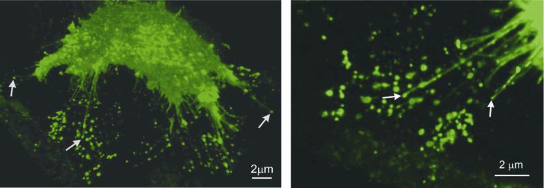
CELLULAR & MOLECULAR BIOLOGY LETTERS                                              637

Key words: Membrane nanostructures, Cell-to-cell communication, Microvesicles,
Nanotubes, Trousseau syndrome, Heparin

INTRODUCTION
The main subject of this review is cell-to-cell communication mediated by the
transport of microvesicles [1-4] that are free to travel to distal cells and fuse with
them [5] or by nanotubes that connect neighboring cells [6-9]. Both
microvesicles and nanotubes derive from the cell membrane and are able to
transport membrane constituents and the contents of the inner solution. The
nanotubes and microvesicles are small enough to be classified as nanostructures,
with their radius ranging from fifty to a few hundred nm. An important feature
of the formation mechanisms for both is that they are preceeded by lateral
reorganization of the membrane into rafts and nanodomains [10-15]. As this
process is mediated by the local curvature of the membrane, which is in turn
determined by the local composition of the constituents, it is the intrinsic shape
of the constituents and their interactions with the neighboring molecules
[9, 11, 15-20] that determine whether microvesiculation or tubulation of the
membrane occurs [9, 10, 13-15]. In this review, we present some recent results
on the formation of microvesicles and nanotubes, and interpret them in terms of
curvature-mediated lateral redistribution of membrane nanodomains.

MICROVESICULATION
Microvesiculation is a major yet underappreciated mechanism playing a vital
role in the development of cells and organisms. Microvesicles (MVs) derived
from cell membranes as the final products of the budding process (see also [4])
were found to have important roles in vascular haemostasis [21-23], the
promotion of cancer [24], and inflammation [2, 25]. Increased levels of
circulating MVs were found in patients suffering from cardiovascular disorders
[26], cancer [28], infection [29] and autoimmune diseases [30-34], and were
associated with an increased risk of thromboembolic events.
The abundance of MVs reflects the extent of the membrane pool available for
budding and vesiculation, the efficiency of the clearance mechanisms, the
properties of the solution surrounding the membrane, and those properties of the
membrane that render it more or less likely to bud and vesiculate. The ions,
soluble molecules and macromolecules (e.g. proteins, carbohydrates, lipids) in
the solution not only enter into chemical reactions with the membrane-bound
receptors, but may affect the budding and vesiculation process via different
physical mechanisms [13, 35-40]. The majority of MVs isolated from the
peripheral blood derive from the platelet membrane [41]; however, assuming
that the essential physical properties of all biological membranes are the same
[42], the relevant mechanisms can be studied in simpler systems, such as
erythrocytes and giant phospholipid vesicles (GPVs), which can be observed live
under an optical microscope.
638                                 Vol. 14. No. 4. 2009       CELL. MOL. BIOL. LETT.

Membranes can be induced to vesiculate by manipulating the solution that is in
contact with the outer layer of the membrane (Fig. 1). In erythrocytes, there is no
internal cytoskeleton, so the shape of the cell is determined by the properties of
the membrane with its underlying dense fibrillar skeleton of spectrin and
anchoring proteins. If detergents are added to a suspension of erythrocytes,
minutes later, the membrane folds either outwards or inwards, depending on the
type of detergent added [43].

Fig. 1. The budding of the erythrocyte membrane (left), and isolated microvesicles (right).
The budding and vesiculation process was induced by adding different detergents to
a suspension of erythrocytes. Dodecylzwittergent induced predominantly tubular buds and
microvesicles (A), while dodecylmaltoside induced predominantly spherical buds and
microvesicles (B). Adapted from [46] with permission from Medicinski razgledi.

According to the bilayer couple model [35-37], this folding is caused by
a change in the area difference between the outer and the inner membrane layers.
If the substance added causes an increase of the outer membrane layer with
respect to the inner membrane layer, the membrane folds outwards and the
erythrocytes transform from a discocytic shape to an echinocytic shape. Further,
buds devoid of membrane cytoskeleton form on top of the echinocyte spicules
(Fig. 1, left), and are eventually pinched off the mother cell to become free
microvesicles (Fig. 1, right). The shape of the buds (microvesicles) depends on
the properties and interactions of the intercalated molecules and the membrane
constituents [44]. It was suggested that such intercalated detergents induce the
formation of membrane inclusions [45], which may be isotropic or anisotropic
with respect to the axis perpendicular to the membrane normal. Isotropic
inclusions give rise to spherical microexovesicles (Fig. 1B). In anisotropic
inclusions, different orientations within the membrane plane are energetically
non-equivalent, if the membrane is anisotropically curved (such as in tubules).
CELLULAR & MOLECULAR BIOLOGY LETTERS                                                639

The orientational ordering of inclusions is energetically favorable, and therefore
stabilizes tubular structures [45].

Fig. 2. The heat-induced budding of a giant phospholipid vesicle membrane composed of
cardiolipin and POPC (A). Upon the addition of phosphate buffer saline, the protrusion
became bead-like (B, C), eventually detached from the mother membrane (D), and
decomposed into spherical vesicles (E), which were free to move away from the mother
vesicle (F). The white arrows indicate the mother vesicle, the black arrows indicate the
daughter vesicles. Bar = 20 μm. Adapted from [38] with permission from Elsevier.

Fig. 3. The plasma protein β2 glycoprotein I was added to a solution with a budding GPV
composed of cardiolipin and POPC (A-C). Thereafter, the bud adhered to the mother
membrane (D-F). Vesiculation was prevented by protein-mediated interactions between
the membranes of the bud and mother vesicle. The white arrows indicate the daughter
vesicle. Bar = 20 μm. Adapted from [38] with permission from Elsevier.
640                               Vol. 14. No. 4. 2009      CELL. MOL. BIOL. LETT.

In studies with giant phospholipid vesicles, budding can be induced by
increasing the temperature. Vesiculation may be also triggered by adding
phosphate buffer saline to a suspension containing GPVs (Fig. 2). However, the
presence of certain plasma proteins (in particular, beta2-glycoprotein I and
antiphospholipid antibodies) in the suspension may cause buds to adhere to the
mother membrane [38, 47] and thereby prevent them from becoming free
vesicles (Fig. 3). The accompanying theoretical studies on protein-mediated
interactions between membranes indicate that plasma proteins can indeed induce
attractive interactions between membranes even when they are like-charged, due
to the positional and orientational ordering of protein molecules with spatially
distributed charges [47].

MEMBRANE NANOTUBES
The results of some recent studies indicate that in many cases, vesicular
transport between cells and between cell organelles over longer distances is not
random, and that it takes place between specific surface regions of the cell
organelles [6-9]. Such organized transport may be achieved via nanotube-
directed transport involving carrier vesicles, or via direct transport through
nanotubes.
It was recently found that cells can communicate with each other by direct
interactions over distances of several cell diameters [7]. The connecting
structures, called nanotubes, can bridge distances of more than 100 μm,
depending on the cell type [48, 49]. They are versatile in ultra-structure and
formation, and consequently also in function [reviewed in 50-53]. Our recent
results revealed that the nanotubes in epithelial cells can be divided into two
distinct types with respect to their formation, stability and cytoskeletal content [9].
Type I nanotubes are shorter, usually not longer than 30 μm, are more dynamic,
and contain actin filaments. They are formed when a cell explores its
surroundings through a tubular membrane protrusion extended from the cell
surface in order to make contact with another cell. The same type of actin-
containing nanotubes bridge cells at distances of less than 30 μm, and are most
likely derived from the adherence cell-cell contacts of cells that move apart. In
terms of characteristics, the retraction fibers observed in migrating and mitotic
cells most resemble the type I nanotubes, and thus can be included in this
category [54].
Type II nanotubes can be longer, up to several 100 μm, and are more stable and
contain cytokeratin filaments. They are formed when two already connected
cells start to move apart. Type II nanotubes differ from all previously found
nanotubes: they have no actin filaments but have cytokeratin filaments, which
are probably responsible for their stability [55] and longer life span. They do not
seem to be involved in transport between cells, but rather in providing a
positional effect to connected cells by sensing their direction of migration and
enabling reverted movement along the intercellular tether [9].
CELLULAR & MOLECULAR BIOLOGY LETTERS                                                   641

Type I nanotubes largely outnumber type II nanotubes, which usually connect
cells as single tethers. The two types of nanotubes also differ with respect to the
proteins of the intercellular junctions that connect the cytoskeletal filaments of
each kind between neighbouring cells (in preparation).
The protruding actin-containing nanotubes of type I start growing from the cell
surface as filopodia [7], but continue until they reach the target cell. The actin
filaments are very likely involved in the growth of the tubes [7, 48, 56]. If the
growth of the nanotubes is related to the growth of the filopodia, it is expected
that a bunch of actin-bundling proteins would be involved in this process,
leading with fascin as the major actin-crosslinking protein responsible for the
growth of the filopodia [57].
Another possibility for the growth of nanotubes is via the accumulation of
flexible membrane nanodomains that prefer highly anisotropic curvature and that
appear in the neck of a budding vesicle. If the separation of the budding vesicle
from the mother membrane is somehow prevented, the neck can grow into
a nanotube, pushing the vesicle at its tip. Such a situation can frequently be seen
with a scanning electron microscope (Fig. 4). The growth of the nanotube by
elongation of the neck can be supported by actin filaments or can be independent
of the cytoskeleton. However, it is also possible for the tip vesicles to form after
the nanotube has attached to the neighbouring membrane surface, and is
subsequently detached or torn apart so that one end of the nanotube becomes
free, as shown in Fig. 4. The possibility that the tip vesicles are attached to the
substrate cannot be excluded either.

Fig. 4. A nanotube forming a continuous connections between neighbouring RT4 urothelial
cells is indicated by the lone arrowhead. Some of the nanotubes which are not attached to
the neighbouring cell surface have vesicles at their free tips, as indicated by the arrows.
Bar = 10 μm.

Fig. 5 shows that the brightness (originating from the ganglioside GM1-bound
fluorescent label choleratoxin B-FITC) of the vesicles at the tips of the
nanotubes that are not attached to neighbouring cells is more intense than the
brightness of non-exvaginated regions of the cell membrane. This result supports
the hypothesis that the vesicles at the tips of the nanotubes (having one free end)
tend to accumulate ganglioside GM1 (Fig. 5), one of the characteristic
components of lipid rafts. Lipid rafts are membrane micro-domains that
accumulate several types of membrane proteins, like growth factor receptors and
642                               Vol. 14. No. 4. 2009      CELL. MOL. BIOL. LETT.

enzymes [58, 59]. The appearance of lipid rafts in vesicular protrusions at the
tips of the nanotubes might be crucial for the attachment of the nanotube to the
target cell, because N-cadherins were found among the proteins included in the
lipid raft domains [60]. These cadherins are responsible for making intercellular
connections between mesenchymal cells and were also found in urothelial T24
cells [61] where nanotubes are frequently seen [9]. Differences in membrane
composition between the tip vesicles and the nanotubes have also been
suggested by other authors [10].
After the nanotubes reach the plasma membrane of an appropriate cell, they
connect to it via an anchoring type of intercellular junction [7, 9, 62]. The
formation of direct cytosolic continuity was proved by following the growth of
actin filaments from cells into their neighbors, even though no exchange of
membrane labels could be found between them [9].
Nanotubes are considered to be a new communication mechanism between two
relatively close, but physically separated cells. They enable cells to exchange
soluble messengers (Ca2+ mediated fluxes [56]), proteins (F-actin [7, 63]),
membrane-bound receptors (HLA, -B, -C, class I MHC [56]), and cellular
organelles (endosomes, lysosomes and mitochondria [7, 48, 63]). Nanotubes also
help cells to function more efficiently in the maintenance of homeostatsis and in
defense against pathogens. On the other hand, nanotubes are a more efficient
mechanism for pathogens to invade uninfected cells [49, 62, reviewed in 53].

Fig. 5. Vesicular dilatations on nanotubes seem to have higher concentration of the
ganglioside GM1, labelled with choleratoxin B-FITC (seen as brighter labeling; arrows
show examples) in urothelial cell line T24. Bar = 2 μm.

Fig. 6. A fluorescence microscope image of the urothelial RT4 cell membrane showing the
higher concentration of the ganglioside GM1, labelled with choleratoxin B-FITC (seen as
brighter labeling), on the membrane exvaginations (buds). Bar = 200 nm. From [64] with
permission from Elsevier.
CELLULAR & MOLECULAR BIOLOGY LETTERS                                             643

CURVATURE-MEDIATED    LATERAL    REDISTRIBUTION OF
MEMBRANE CONSTITUENTS AS THE MECHANISM PRECEEDING
THE FORMATION OF MICROVESICLES AND NANOTUBES
It was indicated that the clustering of small mobile raft elements into larger raft
domains may be induced by a membrane bending and/or attractive forces
between molecules [65 and references therein]. Interestingly, it was shown that
overexpression of flotillin results in the induction of numerous thin tubular
membrane protrusions [66]. Lipid-flotillin complex formation and flotillin
oligomerization seem to play an important role in this process. Similarly, the
accumulation of specific prominin rafts on highly curved membrane protrusions
was recently indicated [67]. Therefore, it was suggested that prominin rafts play
an important role in the stabilization of plasma membrane protrusions [18].
Since prominin does not directly interact with the actin-based cytoskeleton [18],
the predominant localization of prominin in tubular membrane protrusions may
be explained by the curvature-induced accumulation of prominin rafts, with the
intrinsic shape of those prominin rafts as the main driving force [9, 14, 19]. The
redistribution of prominin after mild cholesterol depletion from the protrusions
indicates the importance of cholesterol [68] and other lipids as partners [53] in
the formation of small rafts. Moreover, the non-homogeneous lateral distribution
of flotillins and their accumulation during cytokinesis was recently observed
[69], while the ganglioside GM1 was enriched in membrane exvaginations
induced by cytosolic calcium and amphiphiles (Fig. 6). Based on these and many
other experimental results and on theoretical considerations, it was suggested
that laterally mobile nanodomains that detach from the membrane skeleton may
sort into curved or flat membrane regions depending on their intrinsic shape
and/or direct interactions between the nanodomains [9, 15, 70].
Much experimental and theoretical evidence indicates the importance of
nanodomains in the process of membrane budding and microvesiculation
[11, 15, 18, 72-74, and references therein]. It was shown that the membranes of
Ca2+-induced microvesicles and erythrocyte nanovesicles contain lipid rafts [74, 75],
while the caveolae of Ω-shaped membrane invaginations are enriched with
cholesterol and sfingomielin, known constituents of lipid rafts
[11, 76-78]. Also, it was indicated that the assembly of cholesterol-based lipid
microdomains is required for the biogenesis of secretory vesicles from the trans-
Golgi network in the neuroendocrine cells [79, 80].
Considering the biological membrane as a mixture of different types of
individual molecules with different intrinsic shapes and without explicitly taking
into account the possibility of their self-assembly into energetically favourable
heterogeneous membrane micro- and/or nanodomains, it would be possible to
overestimate the role of the individual molecular intrinsic shapes in the
mechanics of biological membranes and neglect the role of direct interactions
between the membrane constituents. For example, membrane lipids, which
comprise an impressively large number of molecular species with different
644                               Vol. 14. No. 4. 2009      CELL. MOL. BIOL. LETT.

intrinsic shapes [81], may self-assemble into various micro- and nanodomains
which have an average intrinsic shape (spontaneous curvature) that is very
different from the intrinsic shapes of the lipids constituting the nanodomain
(Fig. 7) [71, 72].

Fig. 7. The average intrinsic shape of the membrane nanodomain may be very different
from the intrinsic shapes of the molecules (lipids and proteins) which constitute the
domain. Adapted from [72] with permission from Elsevier.

Since the number of different types of molecules constituting the biological
membrane is very large, it would be impossible to consider in the theoretical
model all the molecules as building blocks and take into account their intrinsic
shapes, as well as the direct interactions between them. Instead, in the theoretical
approach applied in this review, we introduce the concept of flexible membrane
nanodomains (Fig. 8) [9, 15, 72], which are defined as complexes of membrane
molecules (lipids, proteins), and consider them as the membrane building blocks
(see also [20] and references therein).

Fig. 8. A schematic representation of the various intrinsic shapes of flexible membrane
nanodomains. The various intrinsic shapes are described by the two intrinsic principal
curvatures, C1m and C2m [9, 72].

The intrinsic shape of a flexible membrane nanodomain can be described by two
different intrinsic principal curvatures, C1m and C2m [9, 13, 15, 20, 64]. If
C1m= C2m, the nanodomain is isotropic, while if C1m ≠ C2m, the nanodomain is
anisotropic.
The elastic energy of the flexible membrane nanodomain derives from the
mismatch between the actual local curvature of the membrane and the intrinsic
(spontaneous) curvature of the nanodomains (Fig. 8), which can be characterized
by the mismatch tensor M = RCm R −1 − C [9, 13, 15, 20, 64]. Here, the tensor
C describes the actual curvature, while the tensor Cm describes the intrinsic
curvature of the nanodomains.
CELLULAR & MOLECULAR BIOLOGY LETTERS                                                645

  ⎡C 0 ⎤          ⎡C1m             0 ⎤
C=⎢ 1    ⎥ , Cm = ⎢                       ,                                   (1)
  ⎣ 0 C2 ⎦        ⎣ 0             C2 m ⎥⎦

where R is the rotation matrix.

  ⎡cos ω       − sin ω ⎤
R=⎢                      ,                                                    (2)
  ⎣ sin ω      cos ω ⎥⎦

The angle ω describes the orientation of the principal axes system of a single
membrane nanodomain with respect to the local principal axes system of the
membrane. In the respective principal systems, the matrices that represent the
curvature tensors C and Cm include only the diagonal elements (for tensor C ,
the principal curvatures C1 and C2 , and for tensor Cm , the intrinsic principal
curvatures C1m and M up to the second order in the components of M ). The
trace and the determinant of the tensor are taken as the set of invariants [13, 20, 45]:

        K
          (TrM ) + K (DetM ) ,
                2
 fi =                                                                         (3)
        2

where K and K are constants. Taking into account the definition of the tensor
M , it follows from Eq. (3) that the elastic energy of the single membrane
constituent can be written as [13, 20, 45]:

      ⎣
                           2
                                     (
 fi = ⎡(2 K + K )(H − H m ) − K D 2 − 2 DDm cos 2ω + Dm 2 ⎤ ,
                                                          ⎦         )         (4)

where H = (C1 + C2 ) / 2 and D = │(C1 - C2 )│/2 are the mean curvature and the
curvature deviator of the membrane, H m = (C1m + C2 m )/ 2 is the intrinsic
(spontaneous) mean curvature, and Dm =│C1m - C2m│/2 is the intrinsic
(spontaneous) curvature deviator. The constants K and K are proportional to
the area of the single membrane constituent. In the case of a simple flexible
membrane nanodomain composed of a rigid core (protein) and the surrounding
lipids, which are distorted in order to fit with the rigid core, the constants K and
 K were estimated using a microscopic model [20]. The optimal values of the
membrane mean curvature H, the curvature deviator D and the membrane
constituent orientation angle ω corresponding to the minimum of the function fi can
be calculated from the necessary and sufficient conditions for the extremum of fi [72]:
646 Vol. 14. No. 4. 2009 CELL. MOL. BIOL. LETT. H = H m , D = Dm , ω = 0, π , 2π , (5) where ω = 0 and ω = 2π describe the same orientation and where K > K / 2, K 0) and attractive direct interactions during the budding/vesiculation of the membrane. Adapted from [15] with permission from Taylor & Francis. Fig. 10. A schematic illustration of the stabilization of Type I nanotubular membrane protrusions via the accumulation of anisotropic membrane nanodomains in the tubular region.
CELLULAR & MOLECULAR BIOLOGY LETTERS                                          647

In considering the process of nanotube growth, it is assumed that the growing
actin filaments push the membrane outwards. The tubular membrane protrusion
is additionally stabilized by accumulated anisotropic nanodomains having C1m > 0
and C2m ≅ 0 (see Fig. 10). These favour an anisotropic cylindrical geometry of
the membrane [9, 72, 82]. Prominin-containing nanodomains might be
candidates for such anisotropic membrane nanodomains [9, 14, 18, 72]. Once
assembled in the membrane region of a nanotubular membrane protrusion, the
cylindrical anisotropic membrane domains keep the protrusion mechanically
stable even if the cytoskeletal components (actin filaments) are disintegrated by
cytochalasin D [9]. The spherical vesicle at the tip of the nanotube is assumed to
be stabilized by isotropic nanodomains characterized by positive C1m = C2m > 0,
while the nanotube is assumed to be formed from the initial saddle-like neck
connecting the vesicle and the parent cell membrane.
Also, it was shown [83] that the lipid components in a mixture may redistribute
due to anisotropic membrane curvature, such as in the neck connecting the bud
and the mother cell. Theoretical studies on the budding process [13] show that
the anisotropic properties of laterally mobile membrane constituents promote the
formation of the neck due to the orientational ordering of the anisotropic
membrane constituents in the neck, while experimental and theoretical studies
[15] show that the composition of the buds is also reflected in the composition of
the MVs. This indicates that the intrinsic shape and lateral redistribution of the
membrane components play an important role in the budding and vesiculation [64].

DETERMINATION OF THE MICROVESICULATION LEVEL AS
A POSSIBLE INDICATOR OF CLINICAL STATUS
It was recently suggested that plasma protein-mediated attractive interaction
between membranes may prevent the release of cell-derived MVs into
circulation [38, 39]. A hypothesis was put forward that in subjects with plasma
which induces a more pronounced adhesion between membranes, the number of
MVs in the peripheral blood is smaller [38, 39]. The ability of plasma to mediate
interaction between membranes was measured by adding plasma to a suspension
of giant phospholipid vesicles (GPVs; Fig. 11). The proposed trend was
confirmed in a pilot study including three healthy subjects [84] and a population
study involving patients with gastrointestinal diseases [28].
In the latter study, a negative, statistically significant correlation (Pearson
coefficient = -0.50, p = 0.031) was found between the number of MVs in the
peripheral blood and the ability of the plasma to induce coalescence between
membranes, represented by the average effective angle of contact between the
adhered GPVs (Fig. 12A). The statistical significance of the correlation was
even higher if the number of MVs was calculated with respect to the number of
platelets (Pearson coefficient = -0.64, p = 0.003; Fig. 12B).
648                                Vol. 14. No. 4. 2009      CELL. MOL. BIOL. LETT.

Fig. 11. The adhesion of GPVs after the addition of plasma. The angles of contact between
GPVs are indicated. Adapted from [84] with permission from Elsevier.

Fig. 12. The correlations between the average effective angle of contact between adhered
GPVs and the number of MVs in the peripheral blood (A) and between the average
effective angle of contact between the adhered GPVs and the number of MVs per number
of platelets (parameter f), for 19 patients with gastrointestinal diseases. The empty
diamonds correspond to patients from group A (patients diagnosed with gastrointestinal
cancer), and the full diamonds correspond to patients from group B (patients with other
gastrointestinal diseases) Adapted from [28] with permission from Elsevier.

Furthermore, by comparing patients diagnosed with cancer with patients
diagnosed with other gastrointestinal diseases, we found a large (140%) and
statistically significant (p = 0.033) difference between groups A and B regarding
the number of MVs in peripheral blood [28].
Microvesiculation could also play an important role in Trousseau syndrome. In
its strictest sense, Trousseau syndrome is defined retrospectively when
a thromboembolic event preceeds a diagnosis of cancer, while in a broader
context, it includes all hypercoagulabile states connected to cancer [85]. As
microvesicles were found to be both procoagulant and tumor promoting,
according to the hypothesis, the compounds that mediate the short-ranged
attractive interaction between membranes are anticoagulant and tumor
suppressing. The widely used anticoagulant heparin was indeed found to be
successful not only in preventing thromboembolic events but also in slowing
down the development of some types of cancer [86]. As microvesiculation of
CELLULAR & MOLECULAR BIOLOGY LETTERS                                                      649

membranes is enhanced both in hypercoagulabile states and in cancer, there is
a hypothesis of the anticoagulant and tumor suppressing effects of heparin.
According to it, heparin mediates an attractive interaction between membranes
and therefore suppresses microvesiculation via the adhesion of buds to the
mother membrane. Indeed, adding heparin to a solution of giant phospholipid
vesicles causes the vesicles to adhere to each other (Fig. 13).

Fig. 13. Giant phospholipid vesicles composed of POPC (80%) and cholesterol (20%)
before (A) and after (B) the addition of low molecular weight heparin. The vesicles were
prepared by electroformation in 300 mosm saccharose solution as described in Urbanija
et al. [38], and left to sediment in a gravitation field for one day. For the experiment, 36 µl
of a solution containing vesicles was placed in the observation chamber and 4 µl of
Fraxiparine Forte-nadroparine calcium (Sanofi Winthrop, France), dissolved in 300 mosm
glucose solution in the proportion 1:80, was added. The image (panel B) was taken 15 min
after the addition of heparin to the GPVs. Adhesion between vesicles indicates that heparin
mediates the attractive interaction between membranes. Bar = 10 μm.

In a pilot clinical study, the number of microvesicles isolated from the peripheral
blood was determined in a population of patients who received low molecular
weight heparin as a prophylaxis due to orthopaedic surgery, and compared with
of the number in blood from healthy donors. Patients that underwent surgery at
the Department of Orthopaedic Surgery, University Clinical Centre, Ljubljana,
received low molecular weight heparin (clexane or fraxiparine) starting prior to
and ending 7 or more days after the surgery. After the surgery, a 1.6-ml sample
of venous blood was taken from the patients into vactubes containing sodium
citrate. All the participants gave a written consent to the study. Microvesicles
were isolated from the blood according to the protocol of Diamant et al. [41] and
processed as described in Janša et al. [28]. In the population of patients who
underwent orthopaedic surgery and received heparin, it was found that the
average number of MVs divided by the number of fluorospheres (0.71, STD = 0.44)
was higher than in the population of healthy donors (0.56, STD = 0.21).
However, the difference (23%) was small and statistically insignificant. In this
preliminary study, the populations were not age- and gender-matched, and the
population of patients was heterogeneous regarding other diseases, the amount
of blood received by transfusion, and the administration of drugs. It could be
expected that the surgery would cause an increase in the number of
650                              Vol. 14. No. 4. 2009     CELL. MOL. BIOL. LETT.

microvesicles due to stress, inflammation and blood transfusion, but it can be
seen from the obtained results that the difference between the average values of
the two populations was not so large considered that several-fold larger numbers
were obtained for patients with gastrointestinal cancer using the same protocol
[28]. Moreover, in 12 out of 24 patients, the number of MVs was lower than the
average value of the healthy group. To yield more decisive results on the
mechanisms underlying the anticoagulant and tumor suppressing effect of
heparin, further studies based on more detailed and refined analyses involving
larger groups of patients are needed. Using microvesicle analysis as a diagnostic
tool remains a challenge that will very probably be addressed in the future.

CONCLUSIONS
In the past, membranous nanostructures remained largely obscure due to their
small dimensions and fragility. Therefore, the membrane was considered to be
almost flat two-dimensional liquid. More recent experimental data revealed
a possibility of an important pool of membrane contained in nanostructures.
Microvesiculation and the formation of the nanotubes that connect cells are
important processes in cell life, and the have recently become the focus of
increasing interest, especially since they can be manipulated by drugs. It is
indicated that the Singer-Nicolson model of the fluid mosaic and the Simons-
Ikonen-Brown-London model of lateral nanodomains should be upgraded with
the concept of average orientational ordering of membrane constituents
(nanodomains, molecules) in strongly anisotropically curved membrane regions,
since it provides the mechanism that stabilizes membranous nanostructures.
Membranous nanostructures must be explored in greater detail.

REFERENCES
1. Taylor, D.D., Gercel-Taylor, C., Jiang, C.G. and Black, P.H.
   Characterization of plasma membrane shedding from murine melanoma
   cells. Int. J. Cancer 41 (1988) 629-635.
2. Distler, J.H., Pisetsky, D.S., Huber, L.C., Kalden, J.R., Gay, S. and Distler, O.
   Microparticles as regulators of inflammation: novel players of cellular
   crosstalk in the rheumatic diseases. Arthritis Rheum. 52 (2005) 3337-3348.
3. Ratajczak, J., Wysoczynski, M., Hayek, F., Janowska-Wieczorek, A. and
   Ratajczak, M.Z. Membrane-derived microvesicles (MV): important and
   underappreciated mediators of cell to cell communication. Leukemia 20
   (2006) 1487-1495.
4. Greenwalt, T.J. The how and why of exocytic vesicles. Transfusion 46
   (2006) 143-152.
5. del Conde, I., Shrimpton, C.N., Thiagarajan, P. and Lopez, J.A. Tissue-
   factor-bearing microvesicles arise from lipid rafts and fuse with activated
   platelets to initiate coagulation. Blood 106 (2005) 1604-1611.
CELLULAR & MOLECULAR BIOLOGY LETTERS                                          651

6. Sprong, H., van der Sluijs, P. and Meer, G. How proteins move lipids and
    lipids move proteins. Nat. Rev. Mol. Cell Biol. 2 (2001) 504-513.
7. Rustom, A., Saffrich, R., Marković, I., Walther, P. and Gerdes, H.H.
    Nanotubular highways for intercellular organelle transport. Science 303
    (2004) 1007-1010.
8. Iglič, A., Fošnarič, M., Hägerstrand, H. and Kralj-Iglič, V. Coupling
    between vesicle shape and the non-homogeneous lateral distribution of
    membrane constituents in Golgi bodies. FEBS Lett. 574 (2004) 9-12.
9. Veranič, P., Lokar, M., Schütz, G. J., Weghuber, J., Wieser, S., Hägerstrand,
    H., Kralj-Iglič, V. and Iglič, A. Different types of cell-to-cell connections
    mediated by nanotubular structures. Biophys. J. 95 (2008) 4416-4425.
10. Huttner, W.B. and Schmidt, A.A. Membrane curvature: a case of endofeelin’.
    Trends Cell Biol. 12 (2002) 155-158.
11. Sens, P. and Turner, M.S. The forces that shape caveolae. in: Lipid rafts
    and caveolae (Fielding, C.J., Ed.), Wiley-VCH Verlag, Weinheim, 2006,
    25-44.
12. Staneva, G., Seigneuret, M., Koumanov, K., Trugnan, G. and Angelova,
    M.I. Detergents induce raft-like domains budding and fission from giant
    unilamellar heterogeneous vesicles. A direct microscopy observation.
    Chem. Phys. Lipids 136 (2005) 55-66.
13. Iglič, A., Babnik, B., Bohinc, K., Fosnarič, M. Hägerstrand, H. and Kralj-
    Iglič, V. On the role of anisotropy of membrane constituents in formation of
    a membrane neck during budding of a multicomponent membrane.
    J. Biomech. 40 (2007) 579-585.
14. Janich, P. and Corbeil, D. GM1 and GM3 gangliosides highlight distinc lipid
    microdomains with the apical domain of epithelial cells. FEBS Lett. 581
    (2007) 1783-1787.
15. Hägerstrand, H., Mrówczyńska, L., Salzer, U., Prohaska, R., Michelsen, K.,
    Kralj-Iglič, V. and Iglič, A. Curvature-dependent lateral distribution of raft
    markers in the human erythrocyte membrane. Mol. Membr. Biol. 23 (2006)
    277-288.
16. Holopainen, J.M., Angelova, M.I. and Kinnunen, P.K.J. Vectorial budding
    of vesicles by asymmetrical enzymatic formation of ceramide in giant
    liposomes. Biophys. J. 78 (2000) 830-838.
17. Zimmerberg, J. and Kozlov, M.M. How proteins produce cellular membrane
    curvature. Nat. Rev. Mol. Cell Biol. 7 (2006) 9-19.
18. Huttner, W.B. and Zimmerberg, J. Implications of lipid microdomains for
    membrane curvature, budding and fission. Commentary.Curr. Opin. Cell
    Biol. 13 (2001) 478-484.
19. Iglič, A., Hägerstrand, H., Veranič, P., Plemenitaš, A. and Kralj-Iglič, V.
    Curvature induced accumulation of anisotropic membrane components and
    raft formation in cylindrical membrane protrusions. J. Theor. Biol. 240
    (2006) 368-373.
20. Fošnarič, M., Iglič, A., Slivnik, T. and Kralj-Iglič, V. Flexible membrane
652 Vol. 14. No. 4. 2009 CELL. MOL. BIOL. LETT.
inclusions and membrane inclusions induced by rigid globular proteins. in:
Advances in planar lipid bilayers and liposomes (Leitmannova Liu, A.,
Ed.), vol. 7, Elsevier, 2008, 143-168.
21. Müller, I., Klocke, A., Alex, M., Kotzsch, M., Luther, T. and Morgensternm, E.
Intravascular tissue factor initiates coagulation via circulating microvesicles
and platelets. FASEB J. 17 (2003) 476-478.
22. Sims, P.J., Wiedmer, T., Esmon, C.T., Weiss, H.J. and Shattil, S.J.
Assembly of the platelet prothrombinase complex is linked to vesiculation of
the platelet plasma membrane. Studies in Scott syndrome: an isolated defect
in platelet procoagulant activity. J. Biol. Chem. 264 (1989) 17049-17057.
23. Martínez, M.C., Tesse, A., Zobairi, F. and Andriantsitohaina, R. Shed
membrane microparticles from circulating and vascular cells in regulating
vascular function. Am. J. Physiol. Heart Circ. Physiol. 288 (2005) H1004-
H1009.
24. Whiteside, T.L. Tumour-derived exosomes or microvesicles: another
mechanism of tumour escape from the host immune system? Br. J. Cancer
92 (2005) 209-211.
25. Cerri, C., Chimenti, D., Conti, I., Neri, T., Paggiaro, P. and Celi, A.
Monocyte/macrophage-derived microparticles up-regulate inflammatory
mediator synthesis by human airway epithelial cells. J. Immunol. 177
(2006) 1975-1980.
26. Diamant, M., Tushuizen, M.E., Sturk, A. and Nieuwland, R. Cellular
microparticles: new players in the field of vascular disease? Eur. J. Clin.
Invest. 34 (2004) 392-401.
27. Janowska-Wieczorek, A., Marquez-Curtis, L.A., Wysoczynski, M. and
Ratajczak, M.Z. Enhancing effect of platelet-derived microvesicles on the
invasive potential of breast cancer cells. Transfusion 46 (2006) 1199-1209.
28. Janša, R., Šuštar, V., Frank, M., Sušan, P., Bešter, J., Manček-Keber, M.,
Kržan, M. and Iglič A. Number of microvesicles in peripheral blood and
ability of plasma to induce adhesion between phospholipid membranes in 19
patients with gastrointestinal diseases. Blood Cells Mol. Dis. 41 (2008) 124-132.
29. Coltel, N., Combes, V., Wassmer, S.C., Chimini, G. and Grau, G.E. Cell
vesiculation and immunopathology: implications in cerebral malaria.
Microbes Infect. 8 (2006) 2305-2316.
30. Berckmans, R.J., Nieuwland, R., Tak, P.P., Böing, A.N., Romijn, F.P. and
Kraan, M.C. Cell-derived microparticles in synovial fluid from inflamed
arthritic joints support coagulation exclusively via a factor VII-dependent
mechanism. Arthritis Rheum. 46 (2002) 2857-2866.
31. Brogan, P.A., Shah, V., Brachet, C., Harnden, A., Mant, D. and Klein, N.
Endothelial and platelet microparticles in vasculitis of the young. Arthritis
Rheum. 50 (2004) 927-936.
32. Combes, V., Simon, A.C., Grau, G.E., Arnoux, D., Camoin, L. and Sabatier, F.
In vitro generation of endothelial microparticles and possible prothrombotic
activity in patients with lupus anticoagulant. J. Clin. Invest. 104 (1999) 93-102.CELLULAR & MOLECULAR BIOLOGY LETTERS 653
33. Dignat-George, F., Camoin-Jau, L., Sabatier, F., Arnoux, D., Anfosso, F.
and Bardin, N. Endothelial microparticles: a potential contribution to the
thrombotic complications of the antiphospholipid syndrome. Thromb.
Haemost. 91 (2004) 667-673.
34. Morel, O., Jesel, L., Freyssinet, J.M. and Toti, F. Elevated levels of
procoagulant microparticles in a patient with myocardial infarction,
antiphospholipid antibodies and multifocal cardiac thrombosis. Thromb. J.
3 (2005) 15/1-5.
35. Sheetz, M.P., Singer, S.J. Biological membranes as bilayer couples. A
molecular mechanism of drug-erythrocyte interactions. Proc. Natl. Acad.
Sci. USA 71 (1974) 4457-4461.
36. Evans, E.A. Bending resistance and chemically induced moments in
membrane bilayers. Biophys. J. 14 (1974) 923-931.
37. Helfrich, W. Blocked lipid exchange in bilayers and its possible influence on
the shape of vesicles. Z. Naturforsch [c] 29 (1974) 510-515.
38. Urbanija, J., Tomšič, N., Lokar, M., Ambrožič, A. and Čučnik, S., Rozman,
B., Kandušer, M., Iglič, A. and Kralj-Iglič, V. Coalescence of phospholipid
membranes as a possible origin of anticoagulant effect of serum proteins.
Chem. Phys. Lipids 150 (2007) 49-57.
39. Urbanija, J., Babnik, B., Frank, M., Tomšič, N., Rozman, B., Kralj-Iglič, V.
and Iglič, A. Attachment of β2-glycoprotein I to negatively charged
liposomes may prevent the release of daughter vesicles from the parent
membrane. Eur. Biophys. J. 37 (2008) 1085-1095.
40. Laradji, M., and Kumar, P.B.S. Dynamics of domain growth in self-
assembled fluid vesicles. Phys. Rev. Lett. 93 (2004) 198105/1-4.
41. Diamant, M., Nieuwland, R., Pablo, R.F., Sturk, A., Smit, W. and Radder,
J.K. Elevated numbers of tissue-factor exposed in microparticles correlate
with components of the metabolic syndrome in uncomplicated type 2
diabetes mellitus. Circulation 106 (2002) 2442-2447.
42. Singer, S.J. and Nicholson, G.L. The fluid mosaic model of the structure of
cell membranes. Science 175 (1972) 720-731.
43. Isomaa, B., Hägerstrand, H. and Paatero, G. Shape transformations induced
by amphiphiles in erythrocytes. Biochim. Biophys. Acta 899 (1987) 93-103.
44. Hägerstrand, H. and Isomaa, B. Morphological characterization of
exovesicles and endovesicles released from human erythrocytes following
treatment with amphiphiles. Biochim. Biophys. Acta 1109 (1992) 117-126.
45. Kralj-Iglič, V., Iglič, A., Hägerstrand, H. and Peterlin, P. Stable tabular
microexovesicles of the erythrocyte membrane induced by dimeric
amphiphiles. Phys. Rev. E 61 (2000) 4230-4234.
46. Kralj-Iglič, V., Hägerstrand, H., Bobrowska-Hägerstrand, M. and Iglič, A.
Hypothesis on nanostructures of cell and phospholipid membranes as cell
infrastructure. Med. Razgl. 44 (2005) 155-169.
47. Urbanija, J., Bohinc, K., Bellen, A., Maset, S., Iglič, A., Kralj-Iglič, V. and
Sunil Kumar, P.B. Attraction between negatively charged surfaces mediated654 Vol. 14. No. 4. 2009 CELL. MOL. BIOL. LETT.
by spherical counterions with quadrupolar charge distribution. J. Chem.
Phys. 129 (2008) 105101.
48. Önfelt, B., Nedvetzki, S., Yanagi, K. and Davis, D.M. Cutting edge:
Membrane nanotubes connect immune cells. J. Immunol. 173 (2004) 1511-1513.
49. Vidulescu, C., Clejan, S. and O'Connor, K.C. Vesicle traffic through
intercellular bridges in DU 145 human prostate cancer cells. J. Cell Mol.
Med. 8 (2004) 388-396.
50. Gerdes, H.H. and Carvalho, R.N. Intercellular transfer mediated by
tunneling nanotubes. Curr. Opin. Cell Biol. 20 (2008) 470-475.
51. Gürke, S., Barroso, J.F. and Gerdes, H.H. The art of cellular communication:
tunneling nanotubes bridge the divide. Histochem. Cell Biol. 129 (2008)
539-550.
52. Davis, D.M. and Sowinski. S. Membrane nanotubes: dynamic long-distance
connections between animal cells. Nat. Rev. Mol. Cell Biol. 9 (2008) 431-436.
53. Sherer, N.M. and Mothes, W. Cytonemes and tunneling nanotubules in cell-cell
communication and viral pathogenesis. Trends Cell Biol. 9 (2008) 414-420.
54. Mitchison, T.J. Actin based motility on retraction fibers in mitotic PtK2
cells. Cell Motil. Cytoskeleton 22 (1992) 135-151.
55. Magin, T.M., Vijayaraj, P. and Leube, R.E. Structural and regulatory
functions of keratins. Exp. Cell Res. 313 (2007) 2021-2032.
56. Watkins, S.C. and Salter, R.D. Functional connectivity between immune
cells mediated by tunneling nanotubules. Immunity 23 (2005) 309-318.
57. Vignjevic, D., Kojima, S., Aratyn, Y., Danciu, O., Svitkina, T. and Borisy,
G.G. Role of fascin in filopodial protrusion. J. Cell Biol. 174 (2006) 863-875.
58. Simons, K. and Ikonen, E. Functional rafts in cell membranes. Nature 387
(1997) 569-572.
59. Brown, D.A. and London, E. Function of lipid rafts in biological
membranes. Annu. Rev. Cell Biol. 14 (1998) 111-136.
60. Causeret, M., Taulet, N., Comunale, F., Favard, C. and Gauthier-Rouvière, C.
N-cadherin association with lipid rafts regulates its dynamic assembly at
cell-cell junctions in C2C12 myoblasts. Mol. Biol. Cell. 16 (2005) 2168-2180.
61. Laidler, P., Gil, D., Pituch-Noworolska, A., Ciołczyk, D., Ksiazek, D.,
Przybyło, M. and Lityńska, A. Expression of beta1-integrins and N-cadherin
in bladder cancer and melanoma cell lines. Acta Biochim. Pol. 47 (2000)
1159-1170.
62. Sowinski, S., Jolly, C., Berninghausen, O., Purbhoo, M.A., Chauveau, A.,
Kőhler, K.,Oddos, S., Eissmann, P., Brodsky, F.M., Hopkins, C., Önfelt, B.,
Sattentau, Q. and Davis, D.M. Membrane nanotubes physically connect T
cells over long distances presenting a novel route for HIV-1 transmission.
Nat. Cell Biol. 10 (2008) 211-219.
63. Koyanagi, M., Brandes, R.P., Haendeler, J., Zeiher, A.M. and Dimmeler, S.
Cell-to-cell connection of endothelial progenitor cells with cardiac myocytes
by nanotubes: a novel mechanism for cell fate changes? Circ. Res. 96
(2005) 1039-1041.CELLULAR & MOLECULAR BIOLOGY LETTERS 655
64. Kralj-Iglič, V. and Veranič, P. Curvature-induced sorting of bilayer
membrane constituents and formation of membrane rafts. in: Advances in
planar lipid bilayers and liposomes (Leitmannova Liu, A., Ed.), vol. 5,
Elsevier, 2006, 129-149.
65. Harder, T., Scheiffele, P., Verkade, P. and Simons, K. Lipid domain
structure of the plasma membrane revealed by patching of membrane
components. J. Cell Biol. 141 (1998) 929-942.
66. Neumann-Giesen, C., Falkenbach, B., Beicht, P., Claasen, S., Lüers, G.,
Stuermer, C.A., Herzog, V. and Tikkanen, R. Membrane and raft association
of reggie-1/flotilin-2: role of myristoylation, palmitoylation and
oligomerization and induction of filopodia by overexpression. Biochem. J.
378 (2004) 509-518.
67. Corbeil, D., Röper, K., Fargeas, C.A., Joester, A. and Huttner, W.B.
Prominin: A story of cholesterol, plasma membrane protrusions and human
pathology. Traffic 2 (2001) 82-91.
68. Röper, K., Corbeil, D. and Huttner, W.B. Retention of prominin in
microvilli reveals distinct cholesterol-based lipid microdomains in the apical
plasma membrane. Nat. Cell Biol. 2 (2000) 582-592.
69. Rajendran, L., Masilamani, M., Solomon, S., Tikkanen, R., Stuermer, C.A.,
Plattner, H. and Illges, H. Asymmetric localization of flotillins/reggies in
preaseembled platforms confers inherent polarity to hematopoietic cells.
Proc. Natl. Acad. Sci. USA 100 (2003) 8241-8246.
70. Hägerstrand, H. and Mrówczyńska, L. Pathching of ganglioside(M1) in
human erythrocytes – distribution of CD47 and CD59 in patched and curved
membrane. Mol. Membr. Biol. 25 (2008) 258-265.
71. Kuypers, F.A., Roelofsen, B., Berendsen, W., Op den Kamp, J.A.F., van
Deenen, L.L.M. Shape changes in human erythrocytes induced by
replacement of the native phosphatidiylcholine with species contatinig
various fatty acids. J. Cell. Biol. 99 (1984) 2260-2267.
72. Iglič, A., Lokar, M., Babnik, B., Slivnik, T., Veranič, P., Hägerstrand H and
Kralj-Iglič, V. Possible role of flexible red blood cell membrane
nanodomains in the growth and stability of membrane nanotubes. Blood
Cells Mol. Dis. 39 (2007) 14-23.
73. Samuel, B.U., Mohandas, N., Harrison, T., McManus, H., Rosse, W., Reid,
M. and Haldar, K. The role of cholesterol and glycosylphosphatidylinositol-
anchored proteins of erythrocyte rafts in regulating raft protein content and
malarial infection. J. Biol. Chem. 276 (2001) 29319-29329.
74. Salzer, U. and Prohaska, R. Segregation of lipid raft proteins during
calcium-induced vesiculation of erythrocytes. Blood 101 (2003) 3751-3753.
75. Salzer, U., Hinterdorfer, P., Hunger, U., Borken, C. and Prohaska, R. Ca2+-
dependent vesicle release from erythrocytes involves stomatin-specific lipid
rafts, aynexin (annexin VII), and sorcin. Blood 99 (2002) 2569-2577.
76. Sens, P. and Turner, M.S. Theoretical model for the formation of caveolae
and similar membrane invaginations. Biophys. J. 86 (2004) 2049-2057.656 Vol. 14. No. 4. 2009 CELL. MOL. BIOL. LETT.
77. Harder, T. and Simons, K. Caveolae, DUGs, and the dynamcs of
sphingolipid-cholesterol microdomains. Curr. Opin. Cell Biol. 9 (1997)
534-542.
78. Brown, D.A. and London, E. Structure and origin of ordered lipid domains
in biological membranes. J. Membrane Biol. 164 (1998) 103-114.
79. Wang, Y., Thiele, C. and Huttner, W.B. Cholesterol is required for the
formation of regulated and constitutive secretory vesicles from the trans-
Golgi network. Traffic 1 (2000) 952-962.
80. Thiele, C., Hannah, M.J., Fahrenholz, F. and Huttner, W.B. Cholesterol
binds to synaptophysin and is required for biogenesis of synaptic vesicles.
Nat. Cell Biol. 2 (2000) 42-49.
81. Roelofsen, B., Kuypers, F.A., Op den Kamp, J.A.F. and Deenen, L.L.M.
Influence of phosphatidylcholine molecular species composition on stability
of the erythrocyte membrane. Biochem. Soc. Trans. 17 (1989) 284-286.
82. Gimsa, U., Iglič, A., Fiedler, S., Zwanzig, M., Kralj-Iglič, V., Jonas, L. and
Gimsa, J. Actin is not required for nanotubular protrusions of primary
astrocytes grown on metal nano-lawn. Mol. Membr. Biol. 24 (2007) 243-255.
83. Wang, W., Yang, L. and Huang, H.W. Evidence of cholesterol accumulated
in high curvature regions: Implication to the curvature elastic energy for
lipid mixtures. Biophys. J. 92 (2007) 2819-2830.
84. Frank, M., Manček-Keber, M., Kržan, M., Sodin-Šemrl, S., Jerala, R., Iglič, A.,
Rozman, B. and Kralj-Iglič, V. Prevention of microvesiculation by adhesion
of buds to the mother cell membrane - a possible anticoagulant effect of
healthy donor plasma. Autoimmun. Rev. 7 (2008) 240-245.
85. Varki, A. Trousseau’s syndrome: multiple definitions and multiple
mechanisms. Blood 110 (2007) 1723-1729.
86. Borsig, L. Non-anticoagulant effects of heparin in carcinoma metastasis and
Trousseau’s syndrome. Pathophysiol. Haemost. Thromb. 33 suppl 1
(2003) 64-66.You can also read