Transgenic inhibition of interleukin 6 trans signaling does not prevent skeletal pathologies in mucolipidosis type II mice
←
→
Page content transcription
If your browser does not render page correctly, please read the page content below
www.nature.com/scientificreports
OPEN Transgenic inhibition
of interleukin‑6 trans‑signaling
does not prevent skeletal
pathologies in mucolipidosis type II
mice
Lena Marie Westermann1, Anke Baranowsky2, Giorgia Di Lorenzo1,7, Tatyana Danyukova1,
Jamie Soul3, Jean‑Marc Schwartz4, Gretl Hendrickx1,8, Michael Amling1, Stefan Rose‑John5,
Christoph Garbers6, Thorsten Schinke1 & Sandra Pohl1*
Severe skeletal alterations are common symptoms in patients with mucolipidosis type II (MLII), a
rare lysosomal storage disorder of childhood. We have previously reported that progressive bone
loss in a mouse model for MLII is caused by an increased number of bone-resorbing osteoclasts,
which is accompanied by elevated expression of the cytokine interleukin-6 (IL-6) in the bone
microenvironment. In the present study we addressed the question, if pharmacological blockade
of IL-6 can prevent the low bone mass phenotype of MLII mice. Since the cellular IL-6 response
can be mediated by either the membrane-bound (classic signaling) or the soluble IL-6 receptor
(trans-signaling), we first performed cell culture assays and found that both pathways can increase
osteoclastogenesis. We then crossed MLII mice with transgenic mice expressing the recombinant
soluble fusion protein sgp130Fc, which represents a natural inhibitor of IL-6 trans-signaling. By
undecalcified histology and bone-specific histomorphometry we found that high circulating sgp130Fc
levels do not affect skeletal growth or remodeling in wild-type mice. Most importantly, blockade
of IL-6 trans-signaling did neither reduce osteoclastogenesis, nor increase bone mass in MLII mice.
Therefore, our data clearly demonstrate that the bone phenotype of MLII mice cannot be corrected by
blocking the IL-6 trans-signaling.
The maintenance of healthy skeletal structure and function during development, growth and adulthood is
achieved by resorption of aged bone by osteoclasts and formation of new bone by osteoblasts. Tightly regu-
lated bone turnover depends on the balance of these two cellular systems. Osteoclasts are highly specialized
multinucleated cells derived from hematopoietic precursor cells, whereas osteoblasts are mononuclear cells
that arise from a mesenchymal stem cell lineage1,2. Besides, a subset of osteoblasts undergoes terminal dif-
ferentiation into osteocytes, which form a cellular network within the mineralized bone matrix and regulate
bone remodeling and mineral homeostasis3. The differentiation of pre-osteoclastic cells to mature osteoclasts
requires stimulating factors synthesized and secreted by osteoblasts. Among them, receptor activator of nuclear
factor-κB ligand (RANKL) was found to be the key stimulant of canonical o steoclastogenesis4,5. RANKL binds
to its receptor RANK, which is expressed on the surface of osteoclast precursors, and subsequently activates a
variety of downstream signaling pathways required for the formation and maturation of o steoclasts6. However,
1
Department of Osteology and Biomechanics, University Medical Center Hamburg-Eppendorf, 20246 Hamburg,
Germany. 2Clinic of Trauma and Orthopedic Surgery, University Medical Center Hamburg-Eppendorf,
20246 Hamburg, Germany. 3Skeletal Research Group, Biosciences Institute, Newcastle University, Newcastle
upon Tyne NE1 7RU, UK. 4School of Biological Sciences, Faculty of Biology, Medicine and Health, University
of Manchester, Manchester M13 9PL, UK. 5Institute of Biochemistry, Christian-Albrechts-University of Kiel,
24098 Kiel, Germany. 6Department of Pathology, Otto-Von-Guericke-University Magdeburg, 39120 Magdeburg,
Germany. 7Present address: Telethon Institute of Genetics and Medicine (TIGEM), 80078 Pozzuoli, Italy. 8Present
address: Center of Medical Genetics, Antwerp University Hospital and University of Antwerp, 2610 Edegem,
Belgium. *email: s.pohl@uke.de
Scientific Reports | (2021) 11:3556 | https://doi.org/10.1038/s41598-021-82802-3 1
Vol.:(0123456789)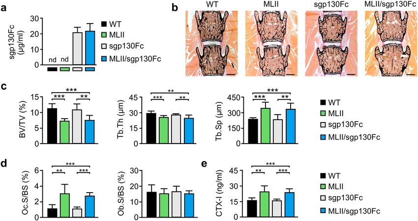
www.nature.com/scientificreports/
a number of other cytokines have been identified to induce non-canonical osteoclastogenesis with IL-6 being
one of them7–10. Indeed, increased production and/or action of IL-6 have been implicated in the pathogenesis
of disease states characterized by excessive bone resorption in adulthood such as postmenopausal osteoporosis
and Paget’s disease11–13.
Depending on the cell type, IL-6 signal transduction occurs via classic and trans-signaling14. In the classic
signaling pathway, IL-6 binds to the membrane-bound IL-6 receptor (IL-6R) and subsequently associates with
ubiquitously expressed membrane-bound, homodimeric gp130 co-receptors. In the trans-signaling pathway,
secreted IL-6 binds to the soluble form of IL-6R (sIL-6R) to further transmit the signal via binding to the gp130
co-receptor on the membrane surface. Thus, the trans-signaling allows stimulation of cells that do not express
membrane-bound IL-6R but the gp130 co-receptor15. Based on the finding that IL-6 promotes osteoclast forma-
tion only in the presence of sIL-6R10,16, IL-6 seems to stimulate osteoclastogenesis via the trans-signaling pathway.
On the other hand, osteoclast precursors and mature osteoclasts express both IL-6R and g p13017–20, suggesting
that osteoclasts may have the capacity to respond to IL-6 via the classic signaling pathway as well. In fact, stud-
ies in human osteoclast cultures support this idea8,9,21. In addition, it has been proposed that IL-6 modulates
osteoclastogenesis induced by RANKL-producing osteoblasts16,22,23. Thus, it remains controversial which target
cells are stimulated by IL-6 and which role IL-6 plays in basal and pathological bone resorption.
Besides the membrane-bound gp130, a soluble form of gp130 (sgp130) functions as a natural inhibitor of
IL-6 trans-signaling as it competes with the membrane-bound gp130 for the formation of the complex with IL-6/
sIL-6R14. Since sgp130 binds IL-6 only in the presence of sIL-6R, it specifically inhibits the trans-signaling, while
the classic signaling pathway remains u naffected24. Therefore, sgp130 represents a blocking agent to neutralize
25
circulating IL-6 and provides a basis for the treatment of osteoporosis and other skeletal diseases associated
with elevated IL-6 levels.
Severe skeletal pathologies and growth retardation are typical symptoms in patients with mucolipidosis
type II (MLII), a rare lysosomal storage disorder of c hildhood26. MLII is caused by mutations in the GNPTAB
gene encoding the catalytic α- and β-subunits of the GlcNAc-1-phosphotransferase, which generates mannose
6-phosphate residues on lysosomal enzymes for their efficient delivery to lysosomes27–29. In cells from patients
with MLII, missorting and hypersecretion of lysosomal enzymes lacking the mannose 6-phosphate targeting
signals results in accumulation of non-degraded material in lysosomes and subsequently impairs the function of
various cell types and tissues28. The systematic skeletal analysis of MLII mice, harboring an MLII patient muta-
tion, revealed that the progressive bone loss observed in these animals is caused by dysfunction of bone-forming
osteoblasts and increased number of bone-resorbing osteoclasts30. Importantly, since the expression of IL-6 was
strongly increased in primary cultured MLII o steoblasts29, we hypothesized that the osteoporotic phenotype in
MLII is mainly caused by excessive osteoclastogenesis, which is potentially induced by osteoblast-derived IL-6.
In this study we have focused on a potential treatment option to prevent the bone loss and skeletal deformi-
ties in MLII mice. More specifically, we addressed the clinically relevant question whether a blockade of IL-6
trans-signaling can reduce the excessive osteoclastogenesis in MLII mice and, subsequently normalize their
bone remodeling pathology.
Results
Increased expression of IL‑6 in terminally differentiated primary osteoblasts and chondro‑
cytes from MLII mice. Osteoblast differentiation requires the coordinated stepwise expression of multiple
genes. Previously we have shown that most of these genes are expressed at lower levels in MLII osteoblasts
during the early stage of osteoblastogenic differentiation30. Since lysosomal storage accumulation was more pro-
nounced in mature MLII o steoblasts30, we aimed to assess genome-wide transcriptional differences between
wild-type and MLII osteoblasts, which were cultured for 25 days to reach an osteocyte-like state of terminal
differentiation31. The transcriptome and gene ontology (GO) enrichment analysis revealed that dysregulated
genes related to the GO-category "Bone biological processes" were significantly enriched in MLII osteoblasts.
The increased or decreased mRNA expression pattern was associated with the GO-terms "Bone mineralization"
or "Osteoclast differentiation" (Fig. 1a, Supplementary Material, Table S1). More specifically, the transcription
of genes important for bone mineralization (e.g. Bglap, Ifitm5, Phex and Mepe) was strongly decreased in MLII
osteoblasts, which was confirmed by quantitative PCR analysis (Supplementary Material, Fig. S1). On the other
hand, osteoclastogenic factors such as the cytokines Il6 and Ccl516,32 were significantly induced in osteoblasts
from MLII mice (Fig. 1a, Supplementary Material, Fig. S1).
Next, we confirmed the elevated expression of Il6 transcripts in MLII osteoblasts on the protein level. For
this purpose, we performed IL-6 immunostaining of primary wild-type and MLII osteoblasts, which revealed
strongly increased levels of intracellular IL-6 in MLII cultures (Fig. 1b). Besides, MLII osteoblasts excessively
secreted IL-6 into the cell culture medium (Fig. 1c). A fivefold increased Il6 transcript level was also found in
primary chondrocytes of MLII mice, whereas the Il6 mRNA expression in primary MLII osteoclasts was not
affected (Fig. 1d). Interestingly, we found the basal mRNA level of Il6 to be very low in wild-type osteoblasts and
osteoclasts, whereas in chondrocytes of the cartilage the Il6 transcript concentration was approximately 50-fold
higher than in bone cells (Fig. 1d).
These in-vitro studies corroborated our previous findings in osteoblasts at the early stage of d
ifferentiation30
and suggested that the progressive bone loss and osteoporotic phenotype in MLII might be caused by osteoblast
dysfunction and an increased expression of the osteoclastogenic factor IL-6 derived from MLII osteoblasts and
chondrocytes.
In‑vitro differentiation of primary osteoclasts is enhanced via IL‑6 classic and trans‑signal‑
ing. To study the responsiveness of primary osteoclasts from wild-type and MLII mice to IL-6 we first deter-
Scientific Reports | (2021) 11:3556 | https://doi.org/10.1038/s41598-021-82802-3 2
Vol:.(1234567890)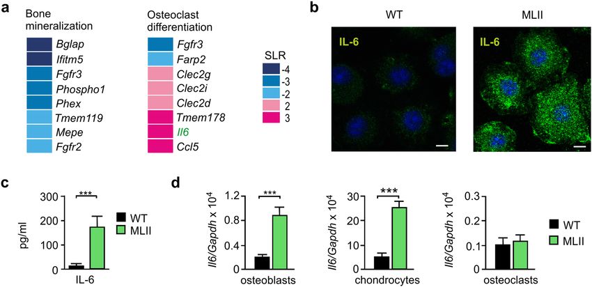
www.nature.com/scientificreports/
Figure 1. Increased expression of IL-6 in primary cultured osteoblasts and chondrocytes from MLII mice. (a)
Signal log ratio (SLR) of differentially expressed genes (SLR ≥ 2 or ≤ -2) in terminally differentiated osteoblasts
from wild-type (WT) and MLII mice related to the gene ontology (GO) "Bone biological processes": "Bone
mineralization" (GO 0030282) and "Osteoclast differentiation" (GO 0030316). (b) IL-6 immunostaining
(green) of WT and MLII osteoblasts. Nuclei were visualized by 4′,6-diamidino-2-phenylindole (DAPI) staining
(blue). Scale bar: 10 μm. (c) Concentration of IL-6 in conditioned media from WT and MLII osteoblasts
(n = 5, mean ± SD, ***p ≤ 0.001). (d) Expression levels of Il6 mRNA related to Gapdh in primary osteoblasts,
chondrocytes and osteoclasts from WT and MLII mice (n = 3, mean ± SD, ***p ≤ 0.001).
mined the mRNA levels of Il6ra (encoding IL-6R) and Il6st (encoding gp130) by quantitative PCR in bone
marrow cells incubated for 7 days in the presence of the osteoclastogenic factors 1,25-dihydroxyvitamin-D3,
M-CSF and RANKL. According to previous findings17–20, both receptors were found to be expressed in wild-
type osteoclasts, with the mRNA level of Il6st being 30-fold higher compared to Il6ra (Fig. 2a). Interestingly,
transcription of both genes was significantly increased in MLII osteoclasts (Fig. 2a).
The formation of multinucleated giant cells is an indicator for proper osteoclast differentiation. These multi-
nucleated cells (MNC) were visualized by staining with the osteoclast marker protein TRAP (lysosomal tar-
trate-resistant acid phosphatase) and were mainly detectable in RANKL-supplemented cultures (Supplementary
Material, Fig. S2a). When we differentiated wild-type bone marrow cells in the presence of recombinant IL-6, a
significant increase in the RANKL-dependent osteoclastic MNC formation was observed (Supplementary Mate-
rial, Fig. S2b). Due to lacking mannose 6-phosphate formation on lysosomal enzymes in MLII cells, TRAP is
less present intracellularly30 (Supplementary Material, Fig. S2c). The osteoclast marker Ctsk (encoding lysosomal
cathepsin K) is also transcriptionally activated during osteoclast differentiation of bone marrow cells33. However,
unlike TRAP, the newly synthesized Ctsk was not mistargeted to the extracellular space in MLII cells, and its
intracellular amounts were comparable to those in wild-type cells, which were demonstrated by the presence of
the lysosomal mature forms of the enzyme in both cell types (Fig. 2b). Therefore, we quantified and compared
the osteoclast formation of wild-type and MLII cells in response to IL-6 by measuring the mRNA expression of
Ctsk instead of TRAP activity staining. As a proof of principle, we thereby confirmed the previous finding30 that
the RANKL-dependent differentiation of MLII osteoclasts was comparable to wild-type cultures (Fig. 2c). In
addition, the osteoclast formation was enhanced in wild-type and MLII cultures in the presence of IL-6, which is
known to induce osteoclastogenesis via both classic and trans-signaling (Fig. 2d). A similar but less pronounced
effect was observed in the presence of hyper-IL-6 (Fig. 2d), an activator of trans-signaling which represents a
recombinant IL-6 fused to the sIL-6R via a flexible peptide l inker34,35. Importantly, both signaling pathways were
found to be significantly enhanced ~ 1.6-fold in MLII osteoclasts differentiated in the presence of either IL-6 or
hyper-IL-6, as compared to the respectively treated wild-type cultures (Fig. 2d). This was most likely caused by
the elevated expression of IL-6R and gp130 in MLII cells (Fig. 2a). Similar to wild-type osteoclasts, hyper-IL-6
enhanced osteoclastogenesis in MLII cultures less strongly than IL-6, suggesting the involvement of both classic
and trans-signaling in osteoclast formation (Fig. 2d).
Reduced bone mass and increased osteoclastogenesis in MLII mice is not prevented by IL‑6
trans‑signaling inhibition. The obtained in-vitro results led us to further expand our analysis by address-
ing the potential relevance of IL-6 in activated osteoclastogenesis of MLII mice in-vivo. Thereby we aimed to
inhibit the increased local bone-specific IL-6 production and to prevent bone loss in MLII. Since IL-6 mediates
the activation of anti-inflammatory pathways in the immune system14, the complete block of IL-6 signaling
might lead to serious side effects. Thus, we decided rather to reduce the potency of IL-6 in MLII mice by block-
ing specifically the IL-6 trans-signaling pathway by the use of soluble gp130Fc. Since the early development
Scientific Reports | (2021) 11:3556 | https://doi.org/10.1038/s41598-021-82802-3 3
Vol.:(0123456789)www.nature.com/scientificreports/
Figure 2. In-vitro osteoclastogenesis can be mediated by IL-6 classic and trans-signaling. (a) mRNA Expression
levels of Il6ra (encoding IL-6R) and Il6st (encoding gp130) related to Gapdh in primary osteoclasts from wild-
type (WT) and MLII mice (n = 3, mean ± SD, **p ≤ 0.01). (b) Representative western blot analysis of extracts
(25 µg protein) of WT and MLII osteoclasts using an antibody against cathepsin K (Ctsk). The positions of
precursor (open arrowhead) and mature form (black arrowhead) are indicated. Endogenous Gapdh was used
as loading control. (c) mRNA expression levels of Ctsk related to Gapdh in primary osteoclasts from WT
and MLII mice cultured in the presence or absence of 40 ng/ml RANKL (n = 3, mean ± SD, ***p ≤ 0.001). (d)
Relative mRNA expression levels of Ctsk related to Gapdh in primary osteoclast cultures from WT and MLII
mice (supplemented with 40 ng/ml RANKL) in the presence or absence of 100 ng/ml IL-6 and/or 100 ng/ml
hyper-IL-6 (Hy-IL-6) as indicated (n = 3, mean ± SD, *p ≤ 0.05, **p ≤ 0.005, ***p ≤ 0.001).
and prenatal or neonatal onset of MLII-associated skeletal alterations in humans and mice26,30, we decided for
a transgenic approach for constant sgp130Fc expression in the circulation from the birth instead of sgp130Fc
protein injection.
Therefore we crossed heterozygous MLII mice with transgenic mice expressing the human recombinant
fusion protein sgp130Fc, which comprises the soluble extracellular portion of gp130 with the constant portion
of the mouse IgG1 a ntibody36. The expression is driven by the liver-specific PEPCK promoter and results in
the concentration of sgp130Fc as high as 20–30 µg/ml in the circulation of transgenic m ice36. Accordingly, we
detected approximately 25 µg/ml sgp130Fc in the serum of sgp130Fc and MLII/sgp130Fc, which was not present
in wild-type and MLII mice, as expected (Fig. 3a). For skeletal analysis, we performed histology and quantitative
histomorphometry of undecalcified vertebra sections from 12-week-old wild-type, MLII, sgp130Fc and MLII/
sgp130Fc mice (Fig. 3b–d). Similar to our previous d ata30, we found by von Kossa/van Gieson staining of the
mineralized bone matrix that the trabecular bone volume was significantly reduced in MLII mice, which was
also reflected by decreased trabecular thickness and increased trabecular spacing (Fig. 3b,c). However, we did not
observe significant differences in the trabecular bone parameters between MLII and MLII/sgp130Fc mice. Fur-
thermore, we confirmed that the low trabecular bone mass in MLII mice results from a highly elevated number
of osteoclasts, while the osteoblast number was not affected in 12-week-old MLII mice (Fig. 3d). Likewise, the
quantification of the bone resorption biomarker CTX-I in the serum revealed that MLII mice display excessive
bone resorption (Fig. 3e). Again, we did not observe significant differences between MLII and MLII/sgp130Fc
mice, since excessive bone resorption was also detected in the latter. These analyses demonstrated that the block-
ade of the IL-6 trans-signaling pathway by sgp130Fc does not normalize increased osteoclast differentiation to
prevent the osteoporotic phenotype in MLII mice. Furthermore, and in contrast to a previous study reporting
that high doses of sgp130Fc result in impaired growth and low bone mass in mice37, we did not observe any
detrimental effect of sgp130Fc on the skeleton.
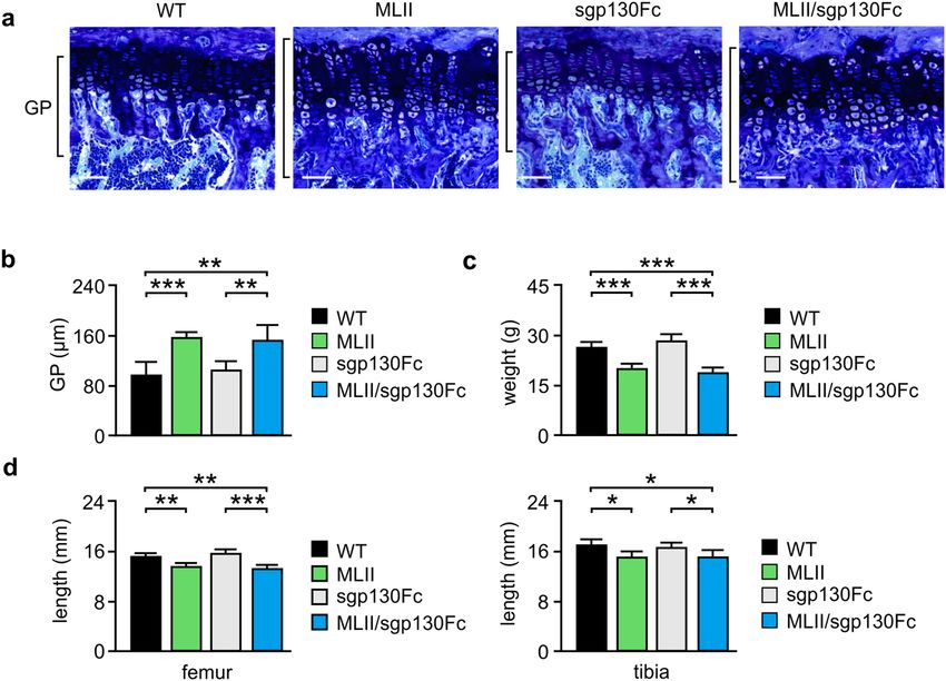
IL‑6 trans‑signaling inhibition does not prevent growth retardation in MLII mice. Skeletal
growth is primarily dependent on the coordinated differentiation of growth plate chondrocytes38. MLII mice
at the age of 4 and 12 weeks are characterized by growth retardation accompanied by growth plate widening30.
Similarly, transgenic IL-6 mice are also reduced in size and display defective growth plates39,40. Interestingly, Il6
Scientific Reports | (2021) 11:3556 | https://doi.org/10.1038/s41598-021-82802-3 4
Vol:.(1234567890)www.nature.com/scientificreports/
Figure 3. Reduced trabecular bone mass of female 12-week-old MLII mice is not prevented by sgp130Fc.
(a) Concentration of sgp130Fc protein in serum from wild-type (WT), MLII, sgp130Fc and MLII/sgp130Fc
mice (n ≥ 5, mean ± SD). nd: not detected. (b) Representative von Kossa/van Gieson staining of undecalcified
vertebra sections from WT, MLII, sgp130Fc and MLII/sgp130Fc mice (scale bars: 1 mm). (c) Quantification of
the vertebral trabecular bone volume per tissue volume (BV/TV), trabecular thickness (Tb.Th) and trabecular
spacing (Tb.Sp) from the same mice (n = 5, mean ± SD, **p ≤ 0.005, ***p ≤ 0.001). (d) Quantification of cellular
parameters in vertebra sections from the same mice: osteoclast surface per bone surface (Oc.S/BS) and
osteoblast surface per bone surface (Ob.S/BS) (n = 5, mean ± SD, **p ≤ 0.005, ***p ≤ 0.001). (e) Concentration
of C-terminal telopeptides of type I collagen (CTX-I) in serum from WT, sgp130Fc, MLII and MLII/sgp130Fc
mice (n ≥ 7, mean ± SD, **p ≤ 0.005, ***p ≤ 0.001).
mRNA was found to be much higher expressed in chondrocytes compared to bone cells (Fig. 1d). These findings
indicate an important role of IL-6 in skeletal development.
We therefore performed toluidine-blue staining of the growth plates in undecalcified tibia sections from
12-week-old mice and found that the growth plate width was significantly increased in MLII and MLII/sgp130
mice compared to wild-type and sgp130Fc mice (Fig. 4a,b). This was accompanied by reduced body weight as
well as decreased femur and tibia length of MLII and MLII/sgp130 mice in comparison to the respective controls
(Fig. 4c,d). Similar results were obtained in 4- and 50-week-old animals (Supplementary Material, Fig. S3a). Of
note, the dramatic body weight loss in old MLII and MLII/sgp130 mice is additionally enhanced by the general
pathological constitution including severe neurodegeneration in MLII m ice41. In addition, similar to 12-week-old
mice, the severe bone remodeling phenotype of 50-week-old MLII mice was not corrected by sgp130Fc (Sup-
plementary Material, Fig. S3b). Taken together, transgenic inhibition of IL-6 trans-signaling could not normalize
the skeletal abnormalities of MLII mice.
Discussion
Osteoporosis, representing the most common bone disorder and one of the most prevalent diseases in the aged
population, is primarily caused by increased bone resorption mediated by o steoclasts42. It is therefore of high
clinical relevance that there is accumulating evidence indicating that increased IL-6 production in pathological
conditions, such as inflammation, induces o steoclastogenesis23. More specifically, enhanced IL-6 production has
been reported to be associated with bone loss in postmenopausal w omen12. Similarly, despite showing no skeletal
-/-
abnormalities in the absence of specific challenges, IL-6 mice were reported to be protected from bone loss
induced by ovariectomy43. Likewise, IL-6 transgenic mice with high circulating IL-6 levels are characterized by
an increased number of osteoclasts, causing bone loss and alterations of the skeletal m icrostructure40. Further-
more, bone loss induced by spaceflight was found to be associated with increased IL-6 expression in rodents44,45.
Importantly however, the precise role of IL-6 in physiological bone remodeling and skeletal maintenance is not
fully understood so far, especially regarding the responsible signaling pathways.
MLII is a severe multi-systemic disease resulting in premature death during early childhood. To date, there
is no established treatment to cure MLII. Although the skeletal alterations associated with MLII, such as short
stature, bowed limbs, progressive joint stiffness, hip and knee contractures, thoracal asymmetry and kyphosco-
liosis, do not contribute to premature mortality, they lead to chronic pain, progressive decline of mobility and
social stigmatization of the affected c hildren26. Since patients with MLII are also characterized by progressive
bone loss, it was important that our previous analysis of a corresponding mouse model revealed that these ani-
mals display a remarkable increase of osteoclastogenesis. Consistently, the osteoporotic phenotype of MLII mice
was prevented by administration of the established anti-resorptive bisphosphonate a lendronate30. However, as
Scientific Reports | (2021) 11:3556 | https://doi.org/10.1038/s41598-021-82802-3 5
Vol.:(0123456789)www.nature.com/scientificreports/
Figure 4. Retarded growth of female 12-week-old MLII mice is not prevented by sgp130Fc. (a) Representative
toluidine blue staining of the growth plate in undecalcified tibia sections from wild-type (WT), MLII, sgp130Fc
and MLII/sgp130Fc mice. Scale bars: 1 mm. (b) Quantification of the growth plate width in tibia of the same
mice (n ≥ 4, mean ± SD, **p ≤ 0.005, ***p ≤ 0.001). (c) Body weight of WT, MLII, sgp130Fc and MLII/sgp130Fc
mice (n ≥ 7, mean ± SD, ***p ≤ 0.001). (d) Femur and tibia length of WT, MLII, sgp130Fc and MLII/sgp130Fc
mice (n ≥ 5, mean ± SD, *p ≤ 0.05, **p ≤ 0.005, ***p ≤ 0.001).
long-term bisphosphonate administration, due to its interference with physiological bone r emodeling42,46, is not
a recommended treatment for children, it is relevant to establish alternative therapeutic options to prevent bone
loss and skeletal deformities in MLII.
Our previous analyses of MLII mice clearly demonstrated that these animals recapitulate the hallmark clini-
cal symptoms observed in MLII patients, thereby underscoring their value as a disease model. Importantly,
the skeletal phenotype of MLII mice, i.e. low bone mass due to increased osteoclastogenesis, was associated
with increased local expression of IL-6 in the bone microenvironment. In the present manuscript we studied
the relevance of this previous observation to address two major questions. Is the unexpected induction of IL-6
expression in skeletal cell types of MLII mice causing their osteoporotic phenotype? Is blockade of IL-6 trans-
signaling a potential therapeutic option to prevent bone loss in MLII?
Our present study demonstrated that IL-6 is released from osteoblasts to stimulate RANKL-dependent osteo-
clast formation. Noteworthy, the osteoclastogenesis appeared to be mediated via IL-6 classic and trans-signaling,
both pathways being enhanced in osteoclasts from MLII mice. This observation prompted us to reduce the
potency of IL-6 in MLII mice via blocking selectively the IL-6 trans-signaling pathway by recombinant sgp130Fc,
which represents a novel therapeutic agent for the treatment of chronic inflammatory diseases47. Importantly,
since spg130Fc has been suggested to prevent side effects triggered by a global IL-6 signaling blockade, it is
principally an available treatment option for patients with MLII. However, deep skeletal phenotyping of MLII
and MLII/sgp130Fc mice revealed that bone loss in MLII mice cannot be prevented by blockade of IL-6 trans-
signaling with sgp130Fc. It is also important to state that we did not observe an impact of high circulating
sgp130Fc levels on bone mass and skeletal remodeling on a wild-type genetic background. Thus, although IL-6
signaling represents a potential anti-resorptive therapeutic target, the specific role of classic and trans-IL-6
signaling is highly demanded to treat bone diseases associated with elevated IL-6 levels.
Methods
Mice. Generation and genotyping of MLII mice and sgp130Fc transgenic mice was described elsewhere36,41.
Heterozygous MLII mice were crossed with sgp130Fc transgenic mice to generate homozygous MLII-sgp130Fc
animals. Wild-type (WT), sgp130Fc and MLII littermates were used as controls. All mice were kept in a path-
ogen-free environment with a 12-h light/dark cycle, 45% to 65% relative humidity and 20 °C to 24 °C ambient
temperature in open or individually ventilated cages with wood shavings bedding and nesting material in groups
not surpassing 6 animals. The mice had access to tap water and standard rodent chow ad libitum. All experi-
Scientific Reports | (2021) 11:3556 | https://doi.org/10.1038/s41598-021-82802-3 6
Vol:.(1234567890)www.nature.com/scientificreports/
mental procedures were performed according to the institutional guidelines and approved by the “Behörde für
Gesundheit und Verbraucherschutz”. The study was carried out in compliance with the ARRIVE guidelines
(http://www.nc3rs.org.uk/page.asp?id=1357).
Primary osteoblasts. For transcriptome analysis, osteoblast progenitors were isolated from individual cal-
variae of 8 to 10 mice at the age of 3 to 5 days. Cells were released by collagenase/dispase digestion and plated in
α-MEM containing 10% FBS (α-MEM/FBS) at an initial density of 10,000 cells per cm2. Osteoblast differentia-
tion was induced at 80% confluency by the addition of 50 µg/ml ascorbic acid and 10 mM β-glycerophosphate
(both from Sigma-Aldrich) followed by culture for 25 days to induce an osteocyte-like state of terminal differ-
entiation.
For all other experiments, bone marrow was flushed out of the femora from 6 to 8 mice at the age of 12 weeks
with α-MEM/FBS. Cells were then plated at a density of 5 × 10⁶ cells per ml, and after 24 h the adherent cells
were cultured for 25 days in α-MEM/FBS containing 50 µg/ml ascorbic acid and 10 mM β-glycerophosphate.
Primary osteoclasts. For osteoclast differentiation, bone marrow was flushed out of the femora from 6
to 8 mice at the age of 12 weeks with α-MEM/FBS. Cells were then plated at a density of 5 × 10⁶ cells per ml,
and after 24 h the adherent cells were cultured in α-MEM/FBS containing 10 nM 1,25-dihydroxyvitamin-D3
(Sigma-Aldrich). Beginning at day 4 after seeding M-CSF and RANKL (both from Peprotech) were added to
a final concentration of 20 ng/ml and 40 ng/ml, respectively, and the cells were cultured for 7 days to generate
osteoclasts. For the stimulation experiments, 100 ng/ml IL-647 and 100 ng/ml hyper-IL-634,48 were added to the
culture medium during the whole period of osteoclast differentiation.
Formation of multinuclear cells was assessed by tartrate-resistant acid phosphatase (TRAP) activity staining
as described previously49. In brief, after removal of the medium and two washing steps with phosphate-buffered
saline (PBS), cells were fixed with cold methanol for 5 min. After washing and drying, cells were stained with
Naphthol AS-MX-Phosphate (Sigma-Aldrich) for 30 min before the number of TRAP-positive multinuclear
cells per well was counted.
Primary chondrocytes. Chondrocyte progenitor cells were isolated from a single sternum of 8 to 10 wild-
type and Gnptgko mice at the age of 10 days. Cells were separated by digesting the tissue initially in 0.1% col-
lagenase Ia solution followed by 0.2% collagenase Ia solution and cultured in DMEM/Ham’s F‐12 (1:1) medium
supplemented with 10% FCS. At a total cell confluence of 80%, chondrocyte differentiation was induced by the
addition of ascorbic acid (50 µg/ml) and culture for 10 days.
Transcriptome analysis. Total RNAs from calvarial osteoblasts of each four WT and MLII mice were
isolated with the PEQ Gold Total RNA Isolation Kit (VWR) according to manufacturer’s instructions. The
genome-wide gene expression analysis on ArrayXS Agilent microarrays was performed by OakLabs (Hen-
ningsdorf, Germany) using the mouse GE 8 × 60 k v2 Affymetrix microarray. Quality control was performed
using arrayQualityMetrics and quantile normalization of the expression data was performed with limma50,51.
Probes for the same gene were summarized to the median intensity. Surrogate variable analysis was performed
to correct for batch effects and the resulting co-variants were incorporated into limma to identify differentially
expressed genes52. Benjamini–Hochberg correction was performed to correct for multiple testing. For gene
ontology enrichment analysis, differentially expressed genes (absolute 1.5 SLR and adjusted p value ≤ 0.05) were
used to find enriched gene ontology biological processes using G Oseq53. Gene ontology terms with an adjusted
p value ≤ 0.05 were regarded as statistically significant.
For quantitative mRNA expression analysis, RNA isolation from cultured cells, cDNA synthesis and quan-
titative PCR using pre-designed Taqman-Assays (Thermo Fisher Scientific) were performed as previously
described54. The relative mRNA expression levels of analyzed genes were normalized to the level of Gapdh
mRNA in the same cDNA using the comparative CT method (2–ΔΔCT).
Protein analysis. For western blot analysis, cells were lysed in PBS containing 0.5% Triton X-100 and pro-
tease inhibitors for 30 min at 4 °C. After centrifugation at 16,000g supernatants were used for measurement of
the protein content. Cell extracts (25 µg protein) were processed for SDS-PAGE and western blot analysis using
the mouse anti-Ctsk or rabbit anti-Gapdh antibodies (both from Santa Cruz Biotechnology), the latter serving
as loading control, as previously described54.
Concentration of IL-6 in cell culture supernatants of primary osteoblasts was quantified by mouse IL-6 ELISA
(KMC0061; Thermo Fisher Scientific) according to the manufacturers’ instructions. The activity of TRAP in cell
extracts was determined as described p reviously55. Serum concentration of bone-specific collagen degradation
products (C-terminal telopeptides of type I collagen, CTX-I) was determined by ELISA (Immunodiagnostic
Systems). Serum concentrations of human sg130Fc protein in 4- and 12-week-old transgenic mice was deter-
mined by the DuoSet human gp130 ELISA Kit (R&D Systems) according to the manufacturers’ instructions.
Immunofluorescence microscopy. Primary cultured osteoblasts grown on cover slips were fixed with
4% paraformaldehyde in PBS for 30 min. After washing with 50 mM ammonium chloride, cells were permeabi-
lized with 0.1% saponine in PBS for 10 min and blocked in PBS containing 0.1% saponine and 3% bovine serum
albumin for 30 min. Subsequently, cells were incubated with rabbit anti-IL-6 antibody (MAB406; R&D Systems)
for 2 h. After washing with 0.1% saponine in PBS, cells were incubated with corresponding secondary antibodies
conjugated to Alexa Fluor 488 (Thermo Fisher Scientific) and 4′,6-diamidino-2-phenylindole (DAPI) for 1 h and
Scientific Reports | (2021) 11:3556 | https://doi.org/10.1038/s41598-021-82802-3 7
Vol.:(0123456789)www.nature.com/scientificreports/
embedded in Aqua-Poly/Mount. Fluorescence was detected and images were obtained using an Olympus digital
scanning confocal microscope (FluoView F1000) and Adobe Photoshop and Image J software.
Skeletal analysis. Dissected skeletons were fixed in 3.7% PBS-buffered formaldehyde for 18 h at 4 °C and
stored in 80% ethanol. All skeletons were first analyzed by contact radiography (Faxitron Xray) to measure
the length of femora and tibia. For non-decalcified bone histology, the lumbar vertebral bodies L1 to L4 of
each mouse were dehydrated in ascending alcohol concentrations and then embedded in methylmetacrylate as
described previously49. Sections of 4 μm thickness were cut sagittally on a Microtec rotation microtome (Techno-
Med GmbH) and stained by von Kossa/van Gieson procedures. Histomorphometry was performed according to
the ASBMR g uidelines56 using the OsteoMeasure system (Osteometrics).
Statistical analysis. Results were expressed as means ± standard deviations (SD) and the values were com-
pared using the unpaired Student’s t-test. Value differences with p < 0.05, p < 0.005 and p values < 0.001 were
considered statistically significant, whereas *, ** or *** indicate the significance levels.
Received: 25 September 2020; Accepted: 25 January 2021
References
1. Ducy, P., Schinke, T. & Karsenty, G. The osteoblast: a sophisticated fibroblast under central surveillance. Science 289, 1501–1504
(2000).
2. Cappariello, A., Maurizi, A., Veeriah, V. & Teti, A. The Great Beauty of the osteoclast. Arch. Biochem. Biophys. 558, 70–78 (2014).
3. Dallas, S. L., Prideaux, M. & Bonewald, L. F. The osteocyte: an endocrine cell … and more. Endocr Rev 34, 658–690 (2013).
4. Yasuda, H. et al. Osteoclast differentiation factor is a ligand for osteoprotegerin/osteoclastogenesis-inhibitory factor and is identical
to TRANCE/RANKL. Proc. Natl. Acad. Sci. USA 95, 3597–3602 (1998).
5. Boyle, W. J., Simonet, W. S. & Lacey, D. L. Osteoclast differentiation and activation. Nature 423, 337–342 (2003).
6. Blair, H. C., Robinson, L. J. & Zaidi, M. Osteoclast signalling pathways. Biochem. Biophys. Res. Commun. 328, 728–738 (2005).
7. Kotake, S. et al. Interleukin-6 and soluble interleukin-6 receptors in the synovial fluids from rheumatoid arthritis patients are
responsible for osteoclast-like cell formation. J. Bone Miner. Res. 11, 88–95 (1996).
8. Kurihara, N., Bertolini, D., Suda, T., Akiyama, Y. & Roodman, G. D. IL-6 stimulates osteoclast-like multinucleated cell formation
in long term human marrow cultures by inducing IL-1 release. J. Immunol. 144, 4226–4230 (1990).
9. Kudo, O. et al. Interleukin-6 and interleukin-11 support human osteoclast formation by a RANKL-independent mechanism. Bone
32, 1–7 (2003).
10. Feng, W. et al. Combination of IL-6 and sIL-6R differentially regulate varying levels of RANKL-induced osteoclastogenesis through
NF-κB, ERK and JNK signaling pathways. Sci. Rep. 7, 41411 (2017).
11. Clowes, J. A., Riggs, B. L. & Khosla, S. The role of the immune system in the pathophysiology of osteoporosis. Immunol. Rev. 208,
207–227 (2005).
12. Scheidt-Nave, C. et al. Serum interleukin 6 is a major predictor of bone loss in women specific to the first decade past menopause.
J. Clin. Endocrinol. Metab. 86, 2032–2042 (2001).
13. Teramachi, J. et al. Increased IL-6 expression in osteoclasts is necessary but not sufficient for the development of Paget’s disease
of bone. J. Bone Miner. Res. 29, 1456–1465 (2014).
14. Scheller, J., Garbers, C. & Rose-John, S. Interleukin-6: from basic biology to selective blockade of pro-inflammatory activities.
Semin. Immunol. 26, 2–12 (2014).
15. Schaper, F. & Rose-John, S. Interleukin-6: Biology, signaling and strategies of blockade. Cytokine Growth Factor Rev. 26, 475–487
(2015).
16. Tamura, T. et al. Soluble interleukin-6 receptor triggers osteoclast formation by interleukin 6. Proc. Natl. Acad. Sci. USA 90,
11924–11928 (1993).
17. Romas, E. et al. The role of gp130-mediated signals in osteoclast development: regulation of interleukin 11 production by osteoblasts
and distribution of its receptor in bone marrow cultures. J. Exp. Med. 183, 2581–2591 (1996).
18. Gao, Y. et al. Expression of IL-6 receptor and GP130 in mouse bone marrow cells during osteoclast differentiation. Bone 22, 487–493
(1998).
19. Adebanjo, O. A. et al. Mode of action of interleukin-6 on mature osteoclasts. Novel interactions with extracellular C a2+ sensing in
the regulation of osteoclastic bone resorption. J. Cell Biol. 142, 1347–1356 (1998).
20. Langub, M. C. Jr. et al. Bone resorption and mRNA expression of IL-6 and IL-6 receptor in patients with renal osteodystrophy.
Kidney Int. 50, 515–520 (1996).
21. Danks, L., Workman, S., Webster, D. & Horwood, N. J. Elevated cytokine production restores bone resorption by human Btk-
deficient osteoclasts. J. Bone Miner. Res. 26, 182–192 (2011).
22. Udagawa, N. et al. Interleukin (IL)-6 induction of osteoclast differentiation depends on IL-6 receptors expressed on osteoblastic
cells but not on osteoclast progenitors. J. Exp. Med. 182, 1461–1468 (1995).
23. Sims, N. A. Cell-specific paracrine actions of IL-6 family cytokines from bone, marrow and muscle that control bone formation
and resorption. Int. J. Biochem. Cell Biol. 79, 14–23 (2016).
24. Rose-John, S. IL-6 trans-signaling via the soluble IL-6 receptor: importance for the pro-inflammatory activities of IL-6. Int. J. Biol.
Sci. 8, 1237–1247 (2012).
25. Liu, X., Jones, G. W., Choy, E. H. & Jones, S. A. The biology behind interleukin-6 targeted interventions. Curr. Opin. Rheumatol.
28, 152–160 (2016).
26. Velho, R. V. et al. The lysosomal storage disorders mucolipidosis type II, type III alpha/beta and type III gamma: update on
GNPTAB and GNPTG mutations. Hum. Mutat. 40, 842–864 (2019).
27. Marschner, K., Kollmann, K., Schweizer, M., Braulke, T. & Pohl, S. A key enzyme in the biogenesis of lysosomes is a protease that
regulates cholesterol metabolism. Science 333, 87–90 (2011).
28. Kollmann, K. et al. Mannose phosphorylation in health and disease. Eur. J. Cell Biol. 89, 117–123 (2010).
29. Tiede, S. et al. Mucolipidosis II is caused by mutations in GNPTA encoding the alpha/beta GlcNAc-1-phosphotransferase. Nat.
Med. 11, 1109–1112 (2005).
30. Kollmann, K. et al. Decreased bone formation and increased osteoclastogenesis cause bone loss in mucolipidosis II. EMBO Mol.
Med. 5, 1871–1886 (2013).
Scientific Reports | (2021) 11:3556 | https://doi.org/10.1038/s41598-021-82802-3 8
Vol:.(1234567890)www.nature.com/scientificreports/
31. Schinke, T. et al. The protein tyrosine phosphatase Rptpzeta is expressed in differentiated osteoblasts and affects bone formation
in mice. Bone 42, 524–534 (2008).
32. Brylka, L. J. & Schinke, T. Chemokines in physiological and pathological bone remodeling. Front. Immunol. 10, 2182 (2019).
33. Pohl, S. et al. The lysosomal protein arylsulfatase B is a key enzyme involved in skeletal turnover. J. Bone Miner. Res. 33, 2186–2201
(2018).
34. Fischer, M. et al. I. A bioactive designer cytokine for human hematopoietic progenitor cell expansion. Nat Biotechnol 15, 142–145
(1997).
35. Schmidt-Arras, D. & Rose-John, S. IL-6 pathway in the liver: from physiopathology to therapy. J. Hepatol. 64, 1403–1415 (2016).
36. Rabe, B. et al. Transgenic blockade of interleukin 6 transsignaling abrogates inflammation. Blood 111, 1021–1028 (2008).
37. McGregor, N. E. et al. IL-6 exhibits both cis- and trans-signaling in osteocytes and osteoblasts, but only trans-signaling promotes
bone formation and osteoclastogenesis. J. Biol. Chem. 294, 7850–7863 (2019).
38. Karsenty, G. Transcriptional control of skeletogenesis. Annu. Rev. Genom. Hum. Genet. 9, 183–196 (2008).
39. De Benedetti, F. The impact of chronic inflammation on the growing skeleton: lessons from interleukin-6 transgenic mice. Horm.
Res. 72(Suppl 1), 26–29 (2009).
40. De Benedetti, F. et al. Impaired skeletal development in interleukin-6-transgenic mice: a model for the impact of chronic inflam-
mation on the growing skeletal system. Arthritis Rheumatol. 54, 3551–3563 (2006).
41. Kollmann, K. et al. Lysosomal dysfunction causes neurodegeneration in mucolipidosis II “knock-in” mice. Brain 135, 2661–2675
(2012).
42. Rachner, T. D., Khosla, S. & Hofbauer, L. C. Osteoporosis: now and the future. Lancet 377, 1276–1287 (2011).
43. Poli, V. et al. Interleukin-6 deficient mice are protected from bone loss caused by estrogen depletion. EMBO J. 13, 1189–1196
(1994).
44. Grano, M. et al. Rat hindlimb unloading by tail suspension reduces osteoblast differentiation, induces IL-6 secretion, and increases
bone resorption in ex vivo cultures. Calcif. Tissue Int. 70, 176–185 (2002).
45. He, B. et al. Blockade of IL-6 alleviates bone loss induced by modeled microgravity in mice. Can. J. Physiol. Pharmacol. 98, 1–6
(2020).
46. Teufel, S. et al. Inhibition of bone remodeling in young mice by bisphosphonate displaces the plasma cell niche into the spleen. J.
Immunol. 193, 223–233 (2014).
47. Mackiewicz, A., Schooltink, H., Heinrich, P. C. & Rose-John, S. Complex of soluble human IL-6-receptor/IL-6 up-regulates expres-
sion of acute-phase proteins. J. Immunol. 149, 2021–2027 (1992).
48. Schroers, A. et al. Dynamics of the gp130 cytokine complex: a model for assembly on the cellular membrane. Protein Sci. 14,
783–790 (2005).
49. Schulze, J. et al. Interleukin-33 is expressed in differentiated osteoblasts and blocks osteoclast formation from bone marrow pre-
cursor cells. J. Bone Miner. Res. 26, 704–717 (2011).
50. Kauffmann, A., Gentleman, R. & Huber, W. arrayQualityMetrics—a bioconductor package for quality assessment of microarray
data. Bioinformatics 25, 415–416 (2009).
51. Smyth, G. K. Linear models and empirical bayes methods for assessing differential expression in microarray experiments. Stat.
Appl. Genet. Mol. Biol. 3, Article3 (2004).
52. Leek, J. T. & Storey, J. D. Capturing heterogeneity in gene expression studies by surrogate variable analysis. PLoS Genet. 3, 1724–1735
(2007).
53. Young, M. D., Wakefield, M. J., Smyth, G. K. & Oshlack, A. Gene ontology analysis for RNA-seq: accounting for selection bias.
Genome Biol. 11, R14 (2010).
54. Di Lorenzo, G. et al. Lysosomal proteome and secretome analysis identifies missorted enzymes and their non-degraded substrates
in mucolipidosis III mouse cells. Mol. Cell. Proteom. 17, 1612–1626 (2018).
55. Saftig, P. et al. Mice deficient in lysosomal acid phosphatase develop lysosomal storage in the kidney and central nervous system.
J. Biol. Chem. 272, 18628–18635 (1997).
56. Parfitt, A. M. et al. Bone histomorphometry: standardization of nomenclature, symbols, and units. Report of the ASBMR Histo-
morphometry Nomenclature Committee. J. Bone Miner. Res. 2, 595–610 (1987).
Acknowledgements
This work was funded by the Deutsche Forschungsgemeinschaft (DFG, German Research Foundation, SFB877,
Projects A1, A10, A14 and B3; PO 1539/1-1), the International Advocate for Glycoprotein Storage Diseases
(ISMRD), the National MPS Society, the Austrian MPS Society, the Wagner Foundation, the Spanish MPS Society,
the Australian MPS Society, the Irish MPS Society and the European Community’s Seventh Framework Pro-
gramme under Grant Agreement n°602300 (SYBIL). We thank the UKE Research Animal Facility for the support
as well as Olga Winter, Lukas Sandoval Flores, Mona Neven und Johannes Brand for excellent technical assistance.
Author contributions
S.R.J., C.G. and S.P. conceived the study. T.S. and S.P. coordinated and supervised the project. L.M.W., A.B.,
G.D.L., T.D., J.S., J.M.S. and G.H. performed the experiments and/or analyzed the data. S.P. and T.D. wrote the
manuscript. S.P. prepared the figures. M.A., S.R.J., C.G. and T.S. provided materials and/or edited the manuscript.
All authors reviewed the manuscript.
Funding
Open Access funding enabled and organized by Projekt DEAL.
Competing interests
S.R.J. is an inventor on patents describing the function of sgp130Fc. He is also a shareholder of the CONARIS
Research Institute (Kiel, Germany), which is commercially developing sgp130Fc proteins as therapeutics for
inflammatory diseases. All other authors declare no competing financial interests.
Additional information
Supplementary Information The online version contains supplementary material available at https://doi.
org/10.1038/s41598-021-82802-3.
Correspondence and requests for materials should be addressed to S.P.
Scientific Reports | (2021) 11:3556 | https://doi.org/10.1038/s41598-021-82802-3 9
Vol.:(0123456789)www.nature.com/scientificreports/
Reprints and permissions information is available at www.nature.com/reprints.
Publisher’s note Springer Nature remains neutral with regard to jurisdictional claims in published maps and
institutional affiliations.
Open Access This article is licensed under a Creative Commons Attribution 4.0 International
License, which permits use, sharing, adaptation, distribution and reproduction in any medium or
format, as long as you give appropriate credit to the original author(s) and the source, provide a link to the
Creative Commons licence, and indicate if changes were made. The images or other third party material in this
article are included in the article’s Creative Commons licence, unless indicated otherwise in a credit line to the
material. If material is not included in the article’s Creative Commons licence and your intended use is not
permitted by statutory regulation or exceeds the permitted use, you will need to obtain permission directly from
the copyright holder. To view a copy of this licence, visit http://creativecommons.org/licenses/by/4.0/.
© The Author(s) 2021
Scientific Reports | (2021) 11:3556 | https://doi.org/10.1038/s41598-021-82802-3 10
Vol:.(1234567890)You can also read