The phosphorylated retinoid X receptor-α promotes diethylnitrosamine-induced hepatocarcinogenesis in mice through the activation of β-catenin ...
←
→
Page content transcription
If your browser does not render page correctly, please read the page content below
Carcinogenesis, 2021, 1–9

                                                                                  https://doi.org/10.1093/carcin/bgab099
                                                                                  Advance Access publication October 20, 2021
                                                                                  Original Article

                                                                                                                                                         Downloaded from https://academic.oup.com/carcin/advance-article/doi/10.1093/carcin/bgab099/6404558 by guest on 14 November 2021
Original Article
The phosphorylated retinoid X receptor-α promotes
diethylnitrosamine-induced hepatocarcinogenesis
in mice through the activation of β-catenin
signaling pathway
Hiroyasu Sakai1,*, , Yasuhiro Yamada2, Masaya Kubota1, Kenji Imai1,
Yohei Shirakami1, Hiroyuki Tomita3, Akira Hara3 and Masahito Shimizu1
1
 Department of Gastroenterology, Gifu University Graduate School of Medicine, Gifu, Japan, 2Division of Stem Cell Pathology,
Center for Experimental Medicine and Systems Biology, Institute of Medical Science, University of Tokyo, Tokyo, Japan and
3
 Department of Tumor Pathology, Gifu University Graduate School of Medicine, Gifu, Japan
*To whom correspondence should be addressed. Tel: +81 58 230 6308; Fax: +81 58 230 6310; Email: sakaih03@gifu-u.ac.jp

Abstract
Previous studies have shown that phosphorylation of the retinoid X receptor-α (RXRα) is associated with the development
of hepatocellular carcinoma (HCC). However, these findings were revealed using HCC cell lines that express phosphorylated-
RXRα (p-RXRα) proteins; therefore, it remains unclear whether p-RXRα affects hepatocarcinogenesis in vivo. Therefore,
to investigate the biological function of p-RXRα in vivo, we developed a doxycycline-inducible ES cell line and transgenic
mouse, both of which overexpress the phosphomimetic mutant form of RXRα, T82D/S260D, in a doxycycline-dependent
manner. We found that the development of liver tumors, especially high-grade adenoma and HCC, was enhanced in
diethylnitrosamine (DEN)-treated T82D/S260D-inducible mice. Moreover, the increased incidence of liver tumors in the
transgenic mice was attributable to the promotion of cell cycle progression. Interestingly, the expression of β-catenin
protein and its target gene cyclin D1 was elevated in the liver tumors of DEN-treated T82D/S260D-inducible mice, concurrent
with increased cytoplasmic and nuclear β-catenin protein expression, indicating its stabilization and transcriptional
activation. These results indicate that p-RXRα promotes DEN-induced hepatocarcinogenesis in mice through the activation
of the β-catenin signaling pathway, suggesting that p-RXRα may serve as a possible therapeutic target for HCC.

Introduction
Hepatocellular carcinoma (HCC) is one of the most common ma-                    and regorafenib has been reported to improve the survival of
lignancies worldwide; more than half a million people are diag-                 patients with advanced HCC (4–6). Despite the progress in diag-
nosed with HCC annually (1). The development of HCC is generally                nosis and treatment strategies, the prognosis for patients with
associated with chronic liver inflammation and subsequent cir-                  HCC remains poor owing to its high recurrence rate (70% of
rhosis. Cirrhosis is a well-established major risk factor for HCC,              cases at 5 years), which is attributed to either intrahepatic me-
irrespective of the underlying liver disease (1). Surveillance for              tastasis or the development of de novo tumors (2). Moreover,
patients at high risk of HCC has increased the possibility of early             there are currently no established agents for the curative treat-
diagnosis and curative treatments, including surgical resection                 ment of this malignancy (2). Therefore, to improve the prognosis
and percutaneous ablation, thereby providing long-term control                  of patients with HCC, the underlying molecular mechanisms of
for patients with early-stage HCC (2,3). In addition, treatment                 hepatocarcinogenesis need to be clarified, and more effective
with molecularly targeted agents such as sorafenib, lenvatinib                  chemopreventive and chemotherapeutic strategies are needed.

Received: June 15, 2021; Revised: October 5, 2021; Accepted: October 18, 2021
© The Author(s) 2021. Published by Oxford University Press.
This is an Open Access article distributed under the terms of the Creative Commons Attribution-NonCommercial License (https://creativecommons.org/
licenses/by-nc/4.0/), which permits non-commercial re-use, distribution, and reproduction in any medium, provided the original work is properly cited.
For commercial re-use, please contact journals.permissions@oup.com                                                                                1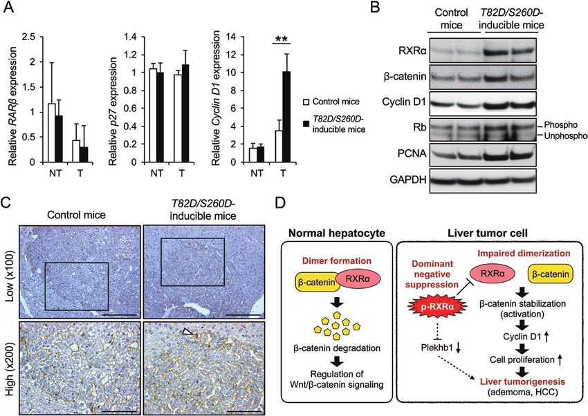
2   |   Carcinogenesis, 2021, Vol. XX, No. XX

Abbreviations                                                                was generated as described previously (14), and then cloned into pcr2.1-
                                                                             TOPO. Sequence-verified T82D/S260D cDNA was subcloned into a unique
APC                           adenomatous polyposis coli
                                                                             EcoR1 site of the pBS31 prime vector (20,21). KH2 ES cells obtained from
DEN                           diethylnitrosamine
                                                                             Open Biosystems, Huntsville, AL were used to insert a single copy of T82D/
DOX                           doxycycline                                    S260D by flippase recombination into the Col1A1 locus under the control
HCC                           hepatocellular carcinoma                       of a minimal CMV tetracycline-inducible promoter using a previously de-
LEF                           lymphoid enhancer factor                       scribed method (20), and ES cells were selected for hygromycin resistance.
PCNA                          proliferating cell nuclear antigen
qRT-PCR                       quantitative real-time reverse                 Mouse generation
                              transcription-polymerase chain                 Fertilized zygotes were isolated from the oviducts of day-0.5 pregnant

                                                                                                                                                            Downloaded from https://academic.oup.com/carcin/advance-article/doi/10.1093/carcin/bgab099/6404558 by guest on 14 November 2021
                              reaction                                       B6D2F1 females and allowed to develop to the blastocyst stage in culture.
RARs                          retinoic acid receptors                        Subsequently, 7-12 ES cells were injected into the blastocysts, which were
RXRα                          retinoid X receptor-α                          then transferred into day-2.5 pseudo-pregnant females.
TCF                           T-cell factor
TUNEL                         terminal
                                                                             Doxycycline treatment
                              deoxynucleotidyltransferase-                   Mice were administered 2 mg/ml (wt/vol) DOX (D9891; Sigma–Aldrich, St.
                              mediated dUTP-biotin nick end                  Louis, MO) in their drinking water, which was supplemented with 10 mg/
                                                                             ml sucrose. For culture cells, DOX was used at a concentration of 2 μg/
                              labeling.
                                                                             ml (w/v).

    Retinoids, a derivative of vitamin A, are physiological
                                                                             RNA preparation and quantitative real-time reverse
signaling molecules that regulate cell proliferation, tissue dif-
                                                                             transcription-polymerase chain reaction
ferentiation and organism development primarily through two
                                                                             Total RNA was extracted using the RNAqueous-4PCR kit (Ambion, Carlsbad,
distinct nuclear receptors, retinoic acid receptors (RARs) and ret-
                                                                             CA) according to the manufacturer’s instructions. Thereafter, cDNA was
inoid X receptors (RXRs), both of which are composed of three
                                                                             synthesized using the SuperScript III First-Strand Synthesis System
subtypes (α, β and γ) (7). Notably, abnormalities in the expression          (Invitrogen Life Technologies, Carlsbad, CA). Quantitative real-time reverse
and function of these receptors are associated with the develop-             transcription-polymerase chain reaction (qRT-PCR) analysis using the
ment of various malignancies, including HCC (8). RXRα can bind               fluorescent SYBR green method was performed according to previously
to the enhancer element of HBV (9). In addition, the RARα gene               described protocols (22). The expression of each gene was normalized to
is located near one of the integration sites of HBV and its ex-              that of β-actin using the standard curve method. Primer sequences are
pression is induced in HBV-related HCC (10). In addition, genetic            shown in Supplementary Table S1, available at Carcinogenesis Online.
variants of RXR have been associated with the risk of hepatitis
C virus (HCV) infection chronicity among the Chinese popula-                 Western blotting analysis
tion with a high risk of HCC (11). Thus, abnormal expression and             Western blotting analysis was performed as described previously (22).
mutations of RXR and RAR genes have been identified in HBV-                  The following primary antibodies were used: anti-RXRα (rabbit IgG, 1:500
                                                                             dilution, sc-553; Santa Cruz Biotechnology, Inc., Santa Cruz, CA), anti-β-
or HCV-related HCC. Moreover, altered expression of RARs has
                                                                             catenin (mouse IgG, 1:8000 dilution, #610154; BD Transduction, San Jose,
been reported to be associated with malignant transformation
                                                                             CA), anti-Rb (mouse IgG, 1:500 dilution, #554136; BD Pharmingen, San
of animal tissues or cultured cells (12,13). Our previous studies
                                                                             Jose, CA), anti-cyclin D1 (rabbit IgG, 1:1000 dilution, sc-753; Santa Cruz
have reported that a malfunction of RXRα due to its aberrant                 Biotechnology), anti-proliferating cell nuclear antigen (PCNA) (mouse IgG,
phosphorylation plays a role in the development of HCC (14,15).              1:2000 dilution, #2586; Cell Signaling Technology, Danvers, MA) and anti-
In HCC cell lines, RXRα is constitutively phosphorylated at both             GAPDH (rabbit IgG, 1:2000 dilution, #2118; Cell Signaling Technology).
threonine 82 and serine 260 owing to the activation of Ras/
MAPK signaling (14), and it accumulates by avoiding ubiquitin-               Animals and experimental procedures
ation and proteasome-mediated degradation (15). The accumu-                  Heterozygous Rosa26::M2rtTA mice with heterozygous tetO-T82D/
lation of phosphorylated-RXRα (p-RXRα) protein interferes with               S260D allele (Rosa/+; Rxr/+) were generated by breeding homozygous
the functioning of the remaining normal RXRα in a dominant-                  Rosa26::M2rtTA male mice with homozygous tetO-T82D/S260D allele (Rosa/
negative manner, thereby promoting the development of HCC                    Rosa; Rxr/Rxr) to C3H/HeN females without the Rosa26::M2rtTA allele or
cells (14). In contrast, acyclic retinoid, a synthetic retinoid that         tetO-T82D/S260D allele (SLC Japan, Shizuoka, Japan). In contrast, hetero-
                                                                             zygous Rosa26::M2rtTA mice (Rosa/+), which served as a control for Rosa/+;
can inhibit the phosphorylation of RXRα and restore its receptor
                                                                             Rxr/+ mice, were generated by breeding homozygous Rosa26::M2rtTA
function (16), has been reported to inhibit the growth of human
                                                                             (Rosa/Rosa) males to C3H/HeN females without Rosa26::M2rtTA or tetO-
hepatoma cells (16–19). Thus, our previous in vitro studies have             T82D/S260D allele (SLC Japan) (Supplementary Figure S1A, available at
revealed the important role of p-RXRα in hepatocarcinogenesis.               Carcinogenesis Online). First, to investigate the spontaneous development
    Although our previous studies showed that p-RXRα is as-                  of liver tumors in vivo, T82D/S260D-inducible male mice (Rosa/+; Rxr/+) and
sociated with the development of HCC cell lines, the effects                 control male mice (Rosa/+) were administered tap water containing 2 mg/
of p-RXRα expression on hepatocarcinogenesis in vivo remain                  ml (wt/vol) DOX (D9891; Sigma–Aldrich) starting at 4 weeks of age and
unknown. Therefore, in the current study, we established a                   killed at either 6 (Supplementary Figure S1B, available at Carcinogenesis
phosphomimetic mutant form of human RXRα, T82D/S260D,                        Online) or 8 (Supplementary Figure S1C, available at Carcinogenesis Online)
                                                                             months of age for both macroscopic inspection and histological analysis.
transgenic mice using a doxycycline (DOX)-dependent expres-
                                                                             Next, to induce liver tumors, the liver carcinogen diethylnitrosamine
sion system to investigate the role of T82D/S260D expression in
                                                                             (DEN) (442687; Sigma–Aldrich) dissolved in 0.9% saline was administered
hepatocarcinogenesis in mice.
                                                                             intraperitoneally (25 mg/kg of body weight) to 15-day-old male pups,
                                                                             which were obtained from each breeding pair described above. The DEN-
                                                                             treated T82D/S260D-inducible male mice (Rosa/+; Rxr/+) and DEN-treated
Materials and methods                                                        control male mice (Rosa/+) were given tap water containing 2 mg/ml (wt/
                                                                             vol) DOX from 4 weeks to 6 months of age. At 6 months of age, both geno-
Molecular cloning and gene targeting in ES cells                             types of mice were killed for both macroscopic inspection and histological
A phosphomimetic mutant form of human RXRα cDNA, T82D/S260D, in              analysis (Supplementary Figure S1D, available at Carcinogenesis Online). All
which threonine 82 and serine 260 were mutated to aspartate, respectively,   mice received humane care and were housed at the Gifu University Life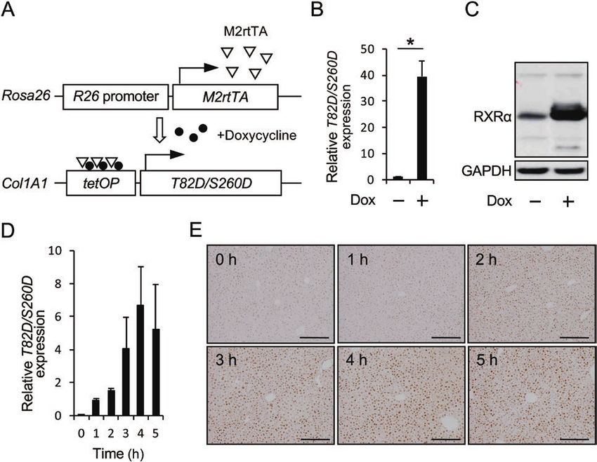
H.Sakai et al. | 3
Science Research Center in accordance with the Institutional Animal Care a time-dependent increase in T82D/S260D mRNA and protein
Guidelines. All animal experiments were approved by the Institutional expression in the liver, indicating that the phosphomimetic
Committee on Animal Experiments of Gifu University. mutant form of human RXRα was effectively induced in these
transgenic mice by the administration of DOX.
Macroscopic inspection, histopathologic analysis,
immunohistochemical analysis and terminal
T82D/S260D-inducible mice are highly susceptible to
deoxynucleotidyltransferase-mediated dUTP-biotin
the development of DEN-induced liver tumors
nick end labeling assay
We investigated the effects of a phosphomimetic mutant form
After killing the mice, the livers were immediately removed and weighed,
of human RXRα, T82D/S260D, on the spontaneous development
Downloaded from https://academic.oup.com/carcin/advance-article/doi/10.1093/carcin/bgab099/6404558 by guest on 14 November 2021
and the number of hepatic tumors on the surface of the left lobe with
diameter ≥1 mm was macroscopically counted. Maximum sagittal
of liver tumors in vivo. Initially, both T82D/S260D-inducible male
sections of three sublobes (left lateral, medial and right medial lobes) were mice (Rosa/+; Rxr/+) and control male mice (Rosa/+) were adminis-
histopathologically examined. Four-micrometer-thick sections of 10% buf- tered DOX starting at 4 weeks of age and were killed at 6 months
fered formalin-fixed, paraffin-embedded liver sections were stained with of age (Supplementary Figure S1B, available at Carcinogenesis
hematoxylin and eosin for conventional histopathology. The numbers and Online). Macroscopic inspection and microscopic analysis re-
the histological grades of liver tumors were evaluated microscopically ac- vealed no development of liver tumors in both genotypes of
cording to previously described criteria by which liver tumors were clas- mice, and histological findings of the liver were comparable be-
sified as Grade 1, 2 or 3 adenomas and HCC depending on the atypical tween both genotypes (data not shown). We then extended DOX
degree (23). Per these criteria (23), liver tumors were defined as Grade 1:
treatment up to 8 months of age (Supplementary Figure S1C,
the lesion showed a focal trabecular pattern with single cell plates; Grade
available at Carcinogenesis Online); however, the development of
2: the lesion showed a prominent trabecular pattern with plates of two
cell layers, slight cellular pleomorphism and increased cell size; Grade 3:
liver tumors was not observed in either group (data not shown).
the lesion was composed of prominent abnormal trabeculation with more To induce liver tumors, we administered the liver carcinogen
than two cell layers, increased N/C ratio, high mitotic rates and marked DEN (25 mg/kg of body weight) intraperitoneally to 15-day-old
cellular pleomorphism; and HCC: the lesion showed features similar to male pups of each group and started DOX treatment at 4 weeks
that of Grade 3, but necrosis was present with no visible remnant of ad- of age, and killed the mice at 6 months of age (Supplementary
enoma (Supplementary Figure S2A, available at Carcinogenesis Online). In Figure S1D, available at Carcinogenesis Online). As shown in Table
addition, foci of cellular alteration (FCA), which are hepatic preneoplastic 1 and Figure 2, the number and maximum size of macroscopic
lesions with a basophilic cytoplasm and hyperchromatic nuclei (24), were
liver tumors were significantly increased in DEN-treated T82D/
also evaluated (Supplementary Figure S2B, available at Carcinogenesis
S260D-inducible mice (Rosa/+; Rxr/+) compared with those in
Online). Immunohistochemistry was performed using an avidin–biotin
DEN-treated control mice (Rosa/+). In addition, microscopic
immunoperoxidase assay, according to a previously described protocol (22).
The following primary antibodies were used: anti-PCNA (rabbit IgG, 1:100 analysis revealed that the number of high-grade liver tumors,
dilution, ab2426; Abcam, Cambridge, MA), anti-cleaved caspase 3 (rabbit including grade 3 adenoma and HCC, was significantly increased
IgG, 1:100 dilution, #9661; Cell Signaling Technology) and anti-β-catenin in DEN-treated T82D/S260D-inducible mice (Rosa/+; Rxr/+) (Table
(mouse IgG, 1:500 dilution, #610154; BD Transduction). Apoptotic cells in 2). No signs of liver cirrhosis, such as pseudolobule formation
the liver were evaluated using the ApopTag peroxidase in situ apoptosis and liver fibrosis, were observed in either mice genotype (data
detection kit (#S7100; Millipore, Billerica, MA), which labels DNA strand not shown). Thus, these results indicate that a phosphomimetic
breaks by the terminal deoxynucleotidyltransferase-mediated dUTP- mutant form of human RXRα, T82D/S260D, by itself, does not
biotin nick end labeling (TUNEL) method, according to the manufacturer’s
initiate liver tumorigenesis, but promotes hepatocarcinogenesis
instructions.
following the administration of DEN.
Statistical analysis
Statistical analysis was performed using GraphPad Prism 4 software Development of liver tumors in DEN-treated T82D/
(GraphPad Software, Inc., San Diego, CA). The mean ± standard deviation S260D-inducible mice is primarily attributed to the
(SD) was calculated for all parameters determined. Statistical significance promotion of cell proliferation
was evaluated using either Student’s t-test or Welch’s t-test for paired
The retinoid receptor RXRα plays a role in maintaining homeo-
samples. Statistical significance was set at P < 0.05.
stasis by regulating fundamental cell activities, including cell
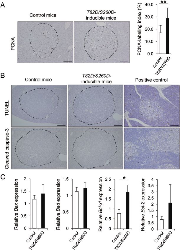
proliferation and apoptosis (7,25). Previous studies have shown
Results that RXRα loses its receptor function by undergoing phospho-
modifications, which are associated with hepatocarcinogenesis
Inducible expression of a phosphomimetic mutant in vitro (14,17–19,26,27). We then investigated the levels of cell
form of the human RXRα (T82D/S260D) in ES cells proliferation and apoptosis in liver tumors developed in either
and mice DEN-treated T82D/S260D-inducible mice (Rosa/+; Rxr/+) or DEN-
We generated DOX-inducible T82D/S260D ES cells, in which a treated control mice (Rosa/+). Immunohistochemistry for PCNA
phosphomimetic mutant form of the human RXRα gene, T82D/ showed that the percentage of PCNA-positive cells per liver
S260D, can be induced under the control of a tetracycline- tumor was significantly increased in DEN-treated T82D/S260D-
responsive regulatory element (Figure 1A). Upon treatment of inducible mice (Rosa/+; Rxr/+) compared with that in DEN-
these ES cells with DOX, the expression of T82D/S260D mRNA treated control mice (Rosa/+) (Figure 3A). In contrast, the levels
was significantly increased compared with that in ES cells of apoptosis, as ascertained by immunohistochemical staining
without DOX treatment (Figure 1B). Increased expression of of tumor sections with TUNEL and cleaved caspase 3, were iden-
T82D/S260D protein, which was evaluated using an anti-RXRα tical in both genotypes of mice (Figure 3B). The expression of
antibody, was also confirmed in DOX-treated ES cells (Figure the anti-apoptotic Bcl-2 and Bcl-xl genes tended to increase in
1C). Next, we administered DOX to T82D/S260D-inducible mice the liver tumors of DEN-treated T82D/S260D-inducible mice
(Rosa/+; Rxr/+) to confirm the expression of the phosphomimetic (Rosa/+; Rxr/+), but no difference was observed in the gene ex-
mutant form of human RXRα, T82D/S260D, in the liver. As pression levels of pro-apoptotic Bax and Bad genes between the
shown in Figure 1D and E, the administration of DOX induced two genotypes (Figure 3C). Thus, our findings suggest that the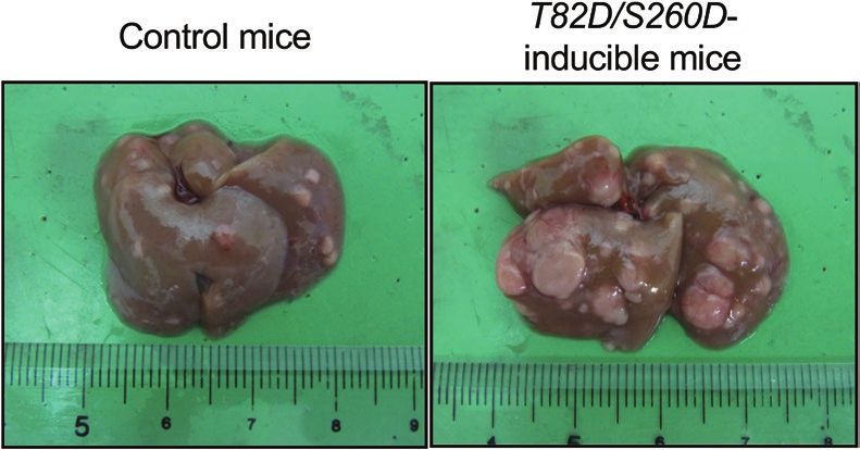
4   |   Carcinogenesis, 2021, Vol. XX, No. XX

                                                                                                                                                                               Downloaded from https://academic.oup.com/carcin/advance-article/doi/10.1093/carcin/bgab099/6404558 by guest on 14 November 2021
Figure 1. Inducible expression of T82D/S260D. (A) Schematic of the doxycycline-inducible T82D/S260D alleles. (B) The expression of T82D/S260D mRNA in T82D/S260D-
inducible ES cells was detected by qRT-PCR using specific primers. The administration of doxycycline (2 μg/ml in culture medium) for 12 h significantly induced T82D/
S260D mRNA expression in the ES cells. Transcript levels were normalized to that of β-actin. Data are presented as mean ± SD (n = 3). *P < 0.05, Student’s t-test. (C) T82D/
S260D protein expression in T82D/S260D-inducible ES cells was evaluated by western blotting analysis using an anti-RXRα antibody, as there is no specific antibody
for the T82D/S260D protein. The administration of doxycycline (2 μg/ml in culture medium) for 24 h induced RXRα protein expression in the ES cells. GAPDH served as
the loading control. Representative images of three independent experiments are shown. (D) The expression of T82D/S260D mRNA in the liver of T82D/S260D-inducible
mice (Rosa/+; Rxr/+) was detected by qRT-PCR using specific primers. The administration of doxycycline (2 mg/ml in drinking water) induced T82D/S260D mRNA expres-
sion in the liver in a time-dependent manner after starting the treatment. Transcript levels were normalized to that of β-actin. Data are presented as mean ± SD (n = 3).
(E) The expression and localization of T82D/S260D protein in the liver of T82D/S260D-inducible mice (Rosa/+; Rxr/+) were analysed by immunohistochemical analysis
using an anti-RXRα antibody. The administration of doxycycline (2 mg/ml in drinking water) induced T82D/S260D protein expression in a time-dependent manner, and
the protein was primarily localized in the nucleus of liver cells. Representative images of RXRα-stained liver sections are shown. Scale bar, 200 μm.

Table 1. Experimental data of macroscopic liver tumors observed in each genotype of mice

Genotype                                                    No. of mice           Incidence (%)              Multiplicitya         Max. size (mm)             LW/BW (%)

Control mice (Rosa/+)                                       21                    21/21 (100)                 8.1 ± 7.7b           2.9 ± 1.2                  5.0 ± 0.8
T82D/S260D-inducible mice (Rosa/+; Rxr/+)                   19                    19/19 (100)                33.6 ± 12.0c          5.4 ± 2.5c                 6.9 ± 1.3c

BW, body weight; LW, liver weight; Max., maximum; No., number.
a
  Number of tumors per mouse.
b
  Mean ± SD.
c
 Significantly different from control mice (Rosa/+) by unpaired t-test with Welch’s correction (P < 0.01).

increased liver tumors observed in DEN-treated T82D/S260D-                               on the mRNA levels of these molecules. As shown in Figure
inducible mice are primarily attributed to the promotion of cell                         4A, the expression levels of RARβ and p27 mRNA in liver tu-
proliferation.                                                                           mors and non-tumorous liver tissues were comparable be-
                                                                                         tween DEN-treated T82D/S260D-inducible mice (Rosa/+; Rxr/+)
The β-catenin signaling pathway is activated                                             and DEN-treated control mice (Rosa/+). In contrast, the ex-
in the liver tumors of DEN-treated T82D/                                                 pression levels of cyclin D1 mRNA were significantly increased
S260D-inducible mice                                                                     in the liver tumors of DEN-treated T82D/S260D-inducible mice
We previously reported that an impaired receptor function                                (Rosa/+; Rxr/+). Cyclin D1 is known as a downstream target
of RXRα due to its phosphorylation is associated with the                                of β-catenin/Tcf transcription (28), and the activation of the
growth of HCC cells, which was attributed to either a de-                                Wnt/β-catenin pathway promotes the development of several
crease in RARβ and p27 or an increase in cyclin D1 (17–19).                              types of cancer, including HCC (29–33). As shown in Figure
Therefore, we examined the effect of T82D/S260D expression                               4B, the levels of β-catenin and cyclin D1 proteins in the liver
H.Sakai et al. | 5
Downloaded from https://academic.oup.com/carcin/advance-article/doi/10.1093/carcin/bgab099/6404558 by guest on 14 November 2021
Figure 2. Representative macroscopic images of the livers from each genotype of mice.
Table 2. The number of FCA, adenoma and HCC observed microscopically in each genotype of mice
Multiplicitya
Genotype No. of mice FCA G1 G2 G3 HCC Total
Control mice (Rosa/+) 21 1.0 ± 0.8 b
1.1 ± 0.9 3.0 ± 1.4 13.4 ± 3.9 1.6 ± 1.0 20.2 ± 5.3
T82D/S260D-inducible mice (Rosa/+; Rxr/+) 19 1.2 ± 0.9 1.2 ± 0.8 4.8 ± 2.1 21.5 ± 5.5c 13.5 ± 4.6d 42.1 ± 11.1d
FCA, foci of cellular alteration; G1, grade 1 adenoma; G2, grade 2 adenoma; G3, grade 3 adenoma; HCC, hepatocellular carcinoma; No., number.
a
Number of tumors per mouse.
b
Mean ± SD.
c
Significantly different from control mice (Rosa/+) by unpaired Student’s t-test (P < 0.05).
d
Significantly different from control mice (Rosa/+) by unpaired t-test with Welch’s correction (P < 0.05).
tumors of DEN-treated T82D/S260D-inducible mice (Rosa/+; Reduced expression of Pleckstrin homology
Rxr/+) were higher than in the liver tumors of DEN-treated domain-containing family B member 1 (Plekhb1)
control mice (Rosa/+). In addition, the level of phosphoryl- mRNA may be associated with DEN-induced liver
ated Rb protein, which induces G1-S checkpoint transition tumorigenesis in T82D/S260D-inducible mice
of the cell cycle under the control of cyclin D1 (34,35), was
We also investigated other molecular targets that can be regu-
also increased in the liver tumors of DEN-treated T82D/S260D-
lated by a phosphomimetic mutant form of human RXRα.
inducible mice (Rosa/+; Rxr/+). Consistent with these results,
Previously, Plekhb1 was shown to be downregulated in human
the protein levels of PCNA, which assists in DNA replication
HCC (38). In addition, the Plekhb1 gene was identified to have
and is a well-known marker of cell proliferation (36), were
a canonical retinoic acid response element (RARE) in the pro-
also increased in the liver tumors of DEN-treated T82D/S260D-
moter regions, and the levels of Plekhb1 expression in germ
inducible mice (Rosa/+; Rxr/+).
cells were downregulated in RARα conditional knockout mice
To histologically evaluate the activation of the β-catenin
(39). Given that the overexpression of T82D/S260D inhibited
signaling pathway, it is important to determine the subcellular
transactivation through RARE in normal human hepatocytes
and nuclear localization of β-catenin, because signal transduc-
(14), we estimated the association between the expression of
tion via this protein involves its post-transcriptional stabil-
T82D/S260D and Plekhb1 mRNA. We then focused on validating
ization and translocation into the nucleus (37). Therefore, we
gene expression in the liver of DOX-treated T82D/S260D-
compared the localization of β-catenin protein in precancerous
inducible mice (Rosa/+; Rxr/+) using qRT-PCR. In contrast with
Grade 3 adenomas, defined according to previous criteria (23),
the significant increase in T82D/S260D mRNA levels after DOX
between DEN-treated T82D/S260D-inducible mice (Rosa/+; Rxr/+)
treatment, the mRNA expression of Plekhb1 was significantly
and DEN-treated control mice (Rosa/+). As shown in Figure 4C,
reduced, suggesting a negative correlation between the ex-
the expression of β-catenin protein in DEN-treated control mice
pression of each gene (Supplementary Figure S3A, available at
(Rosa/+) was primarily localized in the membrane of tumor
Carcinogenesis Online). Meanwhile, in DEN-treated T82D/S260D-
cells. In contrast, relatively strong cytoplasmic expression of
inducible mice (Rosa/+; Rxr/+), the expression of Plekhb1 mRNA
β-catenin protein in tumor cells was observed in DEN-treated
was significantly reduced in liver tumors compared with non-
T82D/S260D-inducible mice (Rosa/+; Rxr/+). β-catenin protein
tumorous liver tissues (Supplementary Figure S3B, available at
was localized in the nucleus in some tumor cells of DEN-treated
Carcinogenesis Online). Notably, compared with DEN-treated con-
T82D/S260D-inducible mice (Rosa/+; Rxr/+). Overall, these find-
trol mice (Rosa/+), the mRNA level of Plekhb1 in the liver tumors
ings suggest that in DEN-treated T82D/S260D-inducible mice
of DEN-treated T82D/S260D-inducible mice (Rosa/+; Rxr/+) was
(Rosa/+; Rxr/+), β-catenin signaling pathway is activated even in
significantly reduced (Supplementary Figure S3C, available at
precancerous lesions of HCC and may play a role in promoting
Carcinogenesis Online). In contrast, increased T82D/S260D mRNA
hepatocarcinogenesis.6 | Carcinogenesis, 2021, Vol. XX, No. XX
Downloaded from https://academic.oup.com/carcin/advance-article/doi/10.1093/carcin/bgab099/6404558 by guest on 14 November 2021
Figure 3. Effects of T82D/S260D expression on cellular proliferation and apoptosis in DEN-induced liver tumors. (A) Liver sections from DEN-treated control mice
(Rosa/+) and DEN-treated T82D/S260D-inducible mice (Rosa/+; Rxr/+) were stained with anti-PCNA antibody. Representative images from each group are shown in the
left panels. PCNA-positive cells were counted and expressed as a percentage of the total number of cells per liver tumor. The positive cell indices are shown in the right
panels. The dotted lines indicate the margin of liver tumors. Scale bar, 200 μm. (B) TUNEL and cleaved caspase 3-positive cells in liver tumors were evaluated using an
apoptosis detection kit and immunohistochemical analysis, respectively. Representative images from each group are shown in the left panels. Sections of rat thymus
were used as the positive control in each experiment. The dotted lines indicate the margin of liver tumors. Scale bar, 200 μm. (C) The mRNA expression levels of Bax,
Bad, Bcl-xl and Bcl-2 in liver tumors were detected by qRT-PCR using specific primers. Transcript levels were normalized to that of β-actin. Data are presented as mean
± SD (n = 3). *P < 0.05, Student’s t-test.
expression was observed in liver tumors of DEN-treated T82D/ suggest that the expression of Plekhb1 mRNA may be negatively
S260D-inducible mice (Rosa/+; Rxr/+) (Supplementary Figure S3C, regulated by T82D/S260D expression, and the reduced expres-
available at Carcinogenesis Online). Collectively, these results sion of the Plekhb1 gene may be associated with DEN-inducedH.Sakai et al. | 7
Downloaded from https://academic.oup.com/carcin/advance-article/doi/10.1093/carcin/bgab099/6404558 by guest on 14 November 2021
Figure 4. The β-catenin signaling pathway is activated in the liver tumors of DEN-treated T82D/S260D-inducible mice (Rosa/+; Rxr/+). (A) The mRNA expression levels
of RARβ, p27 and cyclin D1 in the liver tumors (T) and adjacent non-tumor tissues (NT) of either DEN-treated control mice (Rosa/+) or DEN-treated T82D/S260D-inducible
mice (Rosa/+; Rxr/+) were detected by qRT-PCR using specific primers. Transcript levels were normalized to that of β-actin. Data are presented as mean ± SD (n = 3).
**P < 0.01, Student’s t-test. (B) Total proteins were extracted from DEN-induced liver tumors of each genotype of mice, and the protein expression of RXRα, β-catenin,
cyclin D1, Rb and PCNA were examined by western blotting analysis using specific antibodies. GAPDH served as the loading control. Representative images of three
independent experiments are shown. (C) Liver sections from each genotype of mice were stained with anti-β-catenin antibody. Representative images from each group
are shown. Lower panels indicate the enlarged images of the regions enclosed within the solid lines in the respective upper panels. Arrowhead indicates a tumor cell in
which β-catenin is localized in the nucleus. Scale bar, 200 μm (upper panels), 100 μm (lower panels). (D) Proposed molecular mechanisms underlying the role of p-RXRα
in promoting DEN-induced hepatocarcinogenesis in mice.
liver tumorigenesis in T82D/S260D-inducible mice (Rosa/+; Rxr/+) Nuclear and cytoplasmic β-catenin expression has been
(Figure 4D). shown to play an important role in tumor progression (30,42).
The stabilized cytoplasmic β-catenin enters the nucleus by
binding to the T-cell factor (TCF) and lymphoid enhancer factor
Discussion (LEF) family of proteins, and induces the transcription of target
In the present study, our findings demonstrate that the genes, including cyclin D1. Cyclin D1 promotes the transition
in vivo expression of T82D/S260D promotes DEN-induced between the G1-S checkpoint of the cell cycle by influencing the
hepatocarcinogenesis in mice through the activation of the activity of Rb protein and induces cell proliferation in the cell
β-catenin signaling pathway. cycle (34,35). Indeed, cytoplasmic and nuclear accumulation of
β-Catenin is a structural protein in the cadherin-mediated β-catenin in HCC tissues has been reported in previous clinical
cell–cell adhesive system which plays a role in the differentiation and experimental studies (13,29–31,43,44), and the amplification
and repair of normal tissues (40). It is also known to act as a medi- of cyclin D1 genes and its overexpression have been shown to be
ator in the canonical Wnt signaling pathway; inappropriate acti- associated with aggressive forms of liver tumors, including HCC
vation of this pathway has been implicated in the development of (45,46). In this study, β-catenin protein was localized in the cyto-
several types of malignancies, including HCC (30–33). A previous plasm and the nucleus of liver tumor cells in DEN-treated T82D/
study showed that overexpression of β-catenin accelerates liver S260D-inducible mice, and the downstream proliferative signals,
tumorigenesis and progression to HCC following DEN exposure such as cyclin D1 and phosphorylated Rb proteins, were elevated
(41). In this study, a high prevalence of high-grade liver tumors and in these liver tumors (Figure 4B and C), indicating the activation
increased expression of β-catenin protein in tumor cells was ob- of the β-catenin signaling pathway in these mouse models. Our
served in DEN-treated T82D/S260D-inducible mice (Table 2, Figure findings suggest that the acceleration of hepatocarcinogenesis
4B and C), suggesting the involvement of the β-catenin signaling observed in DEN-treated T82D/S260D-inducible mice is attrib-
pathway in hepatocarcinogenesis in this experimental model. utable to the activation of the β-catenin signaling pathway.8 | Carcinogenesis, 2021, Vol. XX, No. XX
In addition, as previously indicated (45,46), increased cyclin Supplementary material
D1 expression may be associated with the high prevalence of
Supplementary data are available at Carcinogenesis online.
high-grade liver tumors observed in this mouse model.
Previous studies have reported that retinoid receptors,
including RARα and RXRα, directly interact with β-catenin and Funding
regulate the Wnt/β-catenin signaling pathway. Han et al. revealed This work was supported by the Japan Society for the Promotion
that RXRα overexpression directly inhibited both β-catenin/TCF/ of Science KAKENHI (grant number 15K19320).
LEF transcriptional activity and β-catenin protein levels in colo-
rectal cancer cells, whereas downregulation of RXRα by small
Acknowledgements
Downloaded from https://academic.oup.com/carcin/advance-article/doi/10.1093/carcin/bgab099/6404558 by guest on 14 November 2021
interfering RNA abolished these inhibitory effects and elevated
both β-catenin protein levels and β-catenin/TCF/LEF transcrip- The authors are grateful to Chiyoko Sano and Hitomi Fujisawa
tional activity (47). In addition, a previous study using transgenic (Department of Gastroenterology, Gifu University Graduate
mice expressing the RARα-dominant negative form in hepato- School of Medicine) for their secretarial assistance.
cytes showed that the reduction of the RARα/β-catenin complex Conflict of Interest Statement: None declared.
caused an increase in the β-catenin/TCF complex, thus inducing
the expression of cyclin D1 and leading to hepatocarcinogenesis
Authors’ contributions
(13). Thus, RXRα and RARα regulate free β-catenin protein levels
by directly forming a complex with β-catenin, thereby inhibiting H.S., Y.Y. and M.S. collaborated with the conception and de-
the Wnt/β-catenin/TCF function. Notably, the direct regulation sign of the experiment. H.S., M.K. and Y.S. performed the ex-
of β-catenin by these retinoid receptors is perturbed by not only periments and acquired the data and images. H.S., Y.Y., K.I. and
the reduced expression of retinoid receptors, but also their mal- H.T. analyzed the data. H.S. wrote the main manuscript text and
function (13,29,43,47). T82D/S260D, the phosphomimetic mutant prepared all the figures and tables. A.H., Y.Y. and M.S. super-
form of human RXRα used in this study, has been reported to vised the manuscript preparation. All authors reviewed the
interfere with the function of the remaining normal RXRα in manuscript.
a dominant-negative manner (14). Previous studies combined
with our present findings may lead to the hypothesis that the
References
in vivo expression of T82D/S260D may inhibit complex forma-
tion between normal RXRα and β-catenin, thereby causing an 1. El-Serag, H.B. (2011) Hepatocellular carcinoma. N. Engl. J. Med., 365,
increase in the free levels of β-catenin, activating the Wnt/β- 1118–1127.
2. European Association for the Study of the Liver. (2018) EASL clin-
catenin signaling pathway, inducing overexpression of cyclin
ical practice guidelines: management of hepatocellular carcinoma. J.
D1 and thereby contributing to hepatocarcinogenesis in DEN-
Hepatol., 69, 182–236.
treated T82D/S260D-inducible mice (Figure 4D). 3. Njei, B. et al. (2015) Emerging trends in hepatocellular carcinoma inci-
The levels of free β-catenin are regulated by two adenomatous dence and mortality. Hepatology, 61, 191–199.
polyposis coli (APC)-dependent proteasomal degradation path- 4. Llovet, J.M. et al.; SHARP Investigators Study Group. (2008) Sorafenib in
ways: glycogen synthesis kinase-3β-regulated pathway involving advanced hepatocellular carcinoma. N. Engl. J. Med., 359, 378–390.
the APC/Axin complex and a p53-inducible pathway involving 5. Bruix, J. et al.; RESORCE Investigators. (2017) Regorafenib for patients
Siah-1 (48,49). Mutations in either the key components of the with hepatocellular carcinoma who progressed on sorafenib treatment
above two pathways, such as APC, Axin and p53, or β-catenin itself, (RESORCE): a randomised, double-blind, placebo-controlled, phase 3
trial. Lancet, 389, 56–66.
have been reported to lead to dysregulation of β-catenin turnover
6. Kudo, M. et al. (2018) Lenvatinib versus sorafenib in first-line treatment
in several malignancies, including HCC, subsequently resulting in
of patients with unresectable hepatocellular carcinoma: a randomised
its cytoplasmic and nuclear accumulation and abnormal activa-
phase 3 non-inferiority trial. Lancet, 391, 1163–1173.
tion of TCF/LEF-regulated genes that are involved in oncogenesis 7. Chambon, P. (1996) A decade of molecular biology of retinoic acid re-
(43,50). To date, there have been no reports of an association be- ceptors. FASEB J., 10, 940–954.
tween retinoid receptor malfunction and the above-mentioned 8. Sun, S.Y. et al. (2002) Retinoids and their receptors in cancer develop-
mutations. However, given that the β-catenin signaling pathway ment and chemoprevention. Crit. Rev. Oncol. Hematol., 41, 41–55.
was activated in the liver tumors of DEN-treated T82D/S260D- 9. Huan, B.et al.(1992) Retinoid X receptor RXR alpha binds to and trans-activates
inducible mice, the possibility that the in vivo expression of T82D/ the hepatitis B virus enhancer. Proc. Natl Acad. Sci. USA, 89, 9059–9063.
S260D may be associated with these mutations cannot be ruled 10. Benbrook, D. et al. (1988) A new retinoic acid receptor identified from a
hepatocellular carcinoma. Nature, 333, 669–672.
out. Further experiments are required to address this issue.
11. Zhang, R. et al. (2021) Genetic variant of RXR involved in the vitamin
The levels of the Plekhb1 gene were significantly reduced in
D metabolic pathway was linked to HCV infection outcomes among a
liver tumors of DEN-treated T82D/S260D-inducible mice com- high-risk Chinese population. Infect. Genet. Evol., 87, 104641.
pared with those of DEN-treated control mice (Supplementary 12. Bushue, N. et al. (2010) Retinoid pathway and cancer therapeutics. Adv.
Figure S3C, available at Carcinogenesis Online). Given that the Drug Deliv. Rev., 62, 1285–1298.
gene expression has been revealed to be downregulated in 13. Yanagitani, A. et al. (2004) Retinoic acid receptor alpha dominant nega-
human HCC (38), it has been suggested that the reduced ex- tive form causes steatohepatitis and liver tumors in transgenic mice.
pression of Plekhb1 gene may be associated with DEN-induced Hepatology, 40, 366–375.
liver tumorigenesis in T82D/S260D-inducible mice (Figure 4D). 14. Matsushima-Nishiwaki, R. et al. (2001) Phosphorylation of retinoid X
receptor alpha at serine 260 impairs its metabolism and function in
Further experiments are necessary to elucidate the underlying
human hepatocellular carcinoma. Cancer Res., 61, 7675–7682.
molecular mechanisms.
15. Adachi, S. et al. (2002) Phosphorylation of retinoid X receptor suppresses
In conclusion, our findings demonstrate that p-RXRα plays
its ubiquitination in human hepatocellular carcinoma. Hepatology, 35,
a role in chemically induced hepatocarcinogenesis in mice. The 332–340.
fact that the abnormal phosphorylation of RXRα is involved in 16. Matsushima-Nishiwaki, R. et al. (2003) Molecular mechanism for
liver carcinogenesis in vivo suggests that p-RXRα may serve as a growth suppression of human hepatocellular carcinoma cells by
possible therapeutic target for HCC. acyclic retinoid. Carcinogenesis, 24, 1353–1359.H.Sakai et al. | 9
17. Shimizu, M. et al. (2004) Synergistic effects of acyclic retinoid and OSI- 34. Edamoto, Y. et al. (2003) Alterations of RB1, p53 and Wnt pathways in
461 on growth inhibition and gene expression in human hepatoma hepatocellular carcinomas associated with hepatitis C, hepatitis B and
cells. Clin. Cancer Res., 10, 6710–6721. alcoholic liver cirrhosis. Int. J. Cancer, 106, 334–341.
18. Suzui, M. et al. (2002) Growth inhibition of human hepatoma cells by 35. Lévy, L. et al. (2002) Genetic alterations and oncogenic pathways in
acyclic retinoid is associated with induction of p21(CIP1) and inhib- hepatocellular carcinoma. Ann. N. Y. Acad. Sci., 963, 21–36.
ition of expression of cyclin D1. Cancer Res., 62, 3997–4006. 36. Kelman, Z. et al. (1995) Structural and functional similarities of pro-
19. Suzui, M. et al. (2004) Acyclic retinoid activates retinoic acid receptor karyotic and eukaryotic DNA polymerase sliding clamps. Nucleic Acids
beta and induces transcriptional activation of p21(CIP1) in HepG2 Res., 23, 3613–3620.
human hepatoma cells. Mol. Cancer Ther., 3, 309–316. 37. Rubinfeld, B. et al. (1996) Binding of GSK3beta to the APC-beta-catenin
20. Beard, C. et al. (2006) Efficient method to generate single-copy trans- complex and regulation of complex assembly. Science, 272, 1023–1026.
Downloaded from https://academic.oup.com/carcin/advance-article/doi/10.1093/carcin/bgab099/6404558 by guest on 14 November 2021
genic mice by site-specific integration in embryonic stem cells. Genesis, 38. Ho, D.W. et al. (2015) TCGA whole-transcriptome sequencing data re-
44, 23–28. veals significantly dysregulated genes and signaling pathways in
21. Hochedlinger, K. et al. (2005) Ectopic expression of Oct-4 blocks hepatocellular carcinoma. Front. Med., 9, 322–330.
progenitor-cell differentiation and causes dysplasia in epithelial tis- 39. Law, S.M. (2013) Retinoic Acid Receptor Alpha in Germ Cells Is Important for
sues. Cell, 121, 465–477. Mitosis of Spermatogonia, Spermatogonial Differentiation and Meiosis. School of
22. Sakai, H. et al. (2010) Genetic ablation of Tnfalpha demonstrates no Molecular Biosciences. ProQuest LLC: Washington State University, p. 291.
detectable suppressive effect on inflammation-related mouse colon 40. Wei, Y. et al. (2002) Altered expression of E-cadherin in hepatocellular
tumorigenesis. Chem. Biol. Interact., 184, 423–430. carcinoma: correlations with genetic alterations, beta-catenin expres-
23. Jang, J.J. et al. (1992) Progressive atypia in spontaneous and sion, and clinical features. Hepatology, 36, 692–701.
N-nitrosodiethylamine-induced hepatocellular adenomas of C3H/ 41. Nejak-Bowen, K.N. et al. (2010) Accelerated liver regeneration and
HeNCr mice. Carcinogenesis, 13, 1541–1547. hepatocarcinogenesis in mice overexpressing serine-45 mutant beta-
24. Uehara, T. et al. (2021) The DEN and CCl4-induced mouse model of fi- catenin. Hepatology, 51, 1603–1613.
brosis and inflammation-associated hepatocellular carcinoma. Curr. 42. Tien, L.T. et al. (2005) Expression of beta-catenin in hepatocellular car-
Protoc., 1, e211. cinoma. World J. Gastroenterol., 11, 2398–2401.
25. Germain, P. et al. (2006) International Union of Pharmacology. LXIII. 43. Xiao, J.H. et al. (2003) Adenomatous polyposis coli (APC)-independent
Retinoid X receptors. Pharmacol. Rev., 58, 760–772. regulation of beta-catenin degradation via a retinoid X receptor-
26. Shimizu, M. et al. (2009) Strategy and mechanism for the prevention of mediated pathway. J. Biol. Chem., 278, 29954–29962.
hepatocellular carcinoma: phosphorylated retinoid X receptor alpha is 44. Ando, N. et al. (2007) Expression of retinoid X receptor alpha is decreased
a critical target for hepatocellular carcinoma chemoprevention. Cancer in 3’-methyl-4-dimethylaminoazobenzene-induced hepatocellular
Sci., 100, 369–374. carcinoma in rats. Oncol. Rep., 18, 879–884.
27. Yoshimura, K. et al. (2007) Phosphorylated retinoid X receptor alpha 45. Nishida, N. et al. (1994) Amplification and overexpression of the cyclin
loses its heterodimeric activity with retinoic acid receptor beta. Cancer D1 gene in aggressive human hepatocellular carcinoma. Cancer Res., 54,
Sci., 98, 1868–1874. 3107–3110.
28. Tetsu, O. et al. (1999) Beta-catenin regulates expression of cyclin D1 in 46. Ito, Y. et al. (1999) Expression and prognostic roles of the G1-S modu-
colon carcinoma cells. Nature, 398, 422–426. lators in hepatocellular carcinoma: p27 independently predicts the re-
29. Easwaran, V. et al. (1999) Cross-regulation of beta-catenin-LEF/TCF and currence. Hepatology, 30, 90–99.
retinoid signaling pathways. Curr. Biol., 9, 1415–1418. 47. Han, A. et al. (2008) A direct protein-protein interaction is involved in
30. Lustig, B. et al. (2003) The Wnt signaling pathway and its role in tumor the suppression of beta-catenin transcription by retinoid X receptor
development. J. Cancer Res. Clin. Oncol., 129, 199–221. alpha in colorectal cancer cells. Cancer Biol. Ther., 7, 454–459.
31. Asaoka, Y. et al. (2020) Clinical implications of WNT/β-catenin signaling 48. Ha, N.C. et al. (2004) Mechanism of phosphorylation-dependent binding
for hepatocellular carcinoma. Glob. Health Med., 2, 269–272. of APC to beta-catenin and its role in beta-catenin degradation. Mol.
32. Chen, J. et al. (2016) The microtubule-associated protein PRC1 promotes Cell, 15, 511–521.
early recurrence of hepatocellular carcinoma in association with the 49. Liu, J. et al. (2001) Siah-1 mediates a novel beta-catenin degradation
Wnt/β-catenin signalling pathway. Gut, 65, 1522–1534. pathway linking p53 to the adenomatous polyposis coli protein. Mol.
33. Liu, L.J. et al. (2016) Aberrant regulation of Wnt signaling in Cell, 7, 927–936.
hepatocellular carcinoma. World J. Gastroenterol., 22, 7486–7499. 50. Polakis, P. (2000) Wnt signaling and cancer. Genes Dev., 14, 1837–1851.You can also read