Cuscuta chinensis Lam. Protects Against Light-Induced Retinal Degeneration: Therapeutic Implications for Photoreceptor Degenerative Disorders ...
←
→
Page content transcription
If your browser does not render page correctly, please read the page content below
ORIGINAL RESEARCH
                                                                                                                                                 published: 08 June 2022
                                                                                                                                         doi: 10.3389/fphar.2022.904849

                                            Cuscuta chinensis Lam. Protects
                                            Against Light-Induced Retinal
                                            Degeneration: Therapeutic
                                            Implications for Photoreceptor
                                            Degenerative Disorders
                                            Hanhan Wu 1†, Beijing Zhu 2†, Daijin Li 1, Jing Xu 1,3, Jie Chang 1, Xiaoye Du 1,3, Jingang Cui 1,3,
                                            Ning Zhang 4, Teng Zhang 1,3 and Yu Chen 1,3*
                                            1
                                              Yueyang Hospital of Integrated Traditional Chinese and Western Medicine, Shanghai University of Traditional Chinese Medicine,
                                            Shanghai, China, 2Baoshan Hospital of Integrated Traditional Chinese and Western Medicine, Shanghai University of Traditional
                                            Chinese Medicine, Shanghai, China, 3Clinical Research Institute of Integrative Medicine, Shanghai Academy of Traditional
                                            Chinese Medicine, Shanghai, China, 4Science and Technology Laboratory Center, Shanghai University of Traditional Chinese
                                            Medicine, Shanghai, China
                          Edited by:
                          Youhua Xu,
     Macau University of Science and        Cuscuta chinensis Lam. (CCL) is a medicinal herb widely used in traditional Chinese
                 Technology, China          medicine for the treatment of ophthalmic diseases, including age-dependent vision-
                      Reviewed by:          threatening retinal degenerative disorders that involve irreversible loss of the first-order
                           Silvia Bisti,
                                            retinal neurons, photoreceptors. However, evidence is lacking if CCL is pharmacologically
          University of L’Aquila, Italy
                     Juan J. Salazar,       active at protecting against loss of photoreceptors and photoreceptor degeneration-
   Complutense University of Madrid,        associated retinal structural and functional impairment. The current study thus evaluates
                                 Spain
                                            the potential photoreceptor protective effects of CCL to better support its clinical
                   *Correspondence:
                            Yu Chen         applications in the prevention and treatment of photoreceptor degenerative diseases.
                chenyu@shutcm.edu.cn        Non-invasive full-retinal optical coherence tomography, electroretinography, histological
     †
         These authors have contributed     examination, immunohistochemistry and real-time qPCR analysis were performed to
                    equally to this work
                                            assess the retinal protective effects of CCL in light-exposed BALB/c mice
                      Specialty section:
                                            characterized by photooxidative stress-mediated photoreceptor loss and associated
           This article was submitted to    retinal morphological and functional impairment. The results showed that CCL
                    Ethnopharmacology,
                                            treatment protected against light-induced degeneration of the photoreceptor structure
                 a section of the journal
              Frontiers in Pharmacology     and deterioration of the retinal function. Furthermore, CCL treatment increased the retinal
             Received: 26 March 2022        expression of rhodopsin, S-opsin and M-opsin, supporting the protective effects of CCL in
              Accepted: 16 May 2022         both rod and cone photoreceptors. CCL treatment suppressed photoreceptor cell death
             Published: 08 June 2022
                                            in the light-exposed retinas. The morphological integrity of the second-order retinal
                             Citation:
Wu H, Zhu B, Li D, Xu J, Chang J, Du X,
                                            neurons was also preserved as a result of CCL treatment. In addition, CCL treatment
Cui J, Zhang N, Zhang T and Chen Y          attenuated light-induced reactive müller gliosis, microglial activation and inflammation in
       (2022) Cuscuta chinensis Lam.
                                            the retina. In conclusion, the current work demonstrates for the first time that CCL protects
Protects Against Light-Induced Retinal
            Degeneration: Therapeutic       against photooxidative stress-mediated degeneration of photoreceptors and associated
        Implications for Photoreceptor      disturbance of structural, functional and immune homeostasis of the retina. The findings
               Degenerative Disorders.
        Front. Pharmacol. 13:904849.
                                            here thus provide novel experimental evidence supporting the clinical application of CCL in
     doi: 10.3389/fphar.2022.904849         the prevention and treatment photoreceptor degenerative diseases.

Frontiers in Pharmacology | www.frontiersin.org                                     1                                             June 2022 | Volume 13 | Article 904849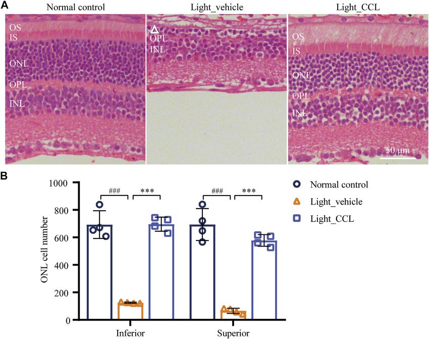
Wu et al.                                                                                                   Cuscuta Chinensis Lam. Protects Photoreceptors

                                           Keywords: Cuscuta chinensis lam., retina, photoreceptor degeneration, cell death, müller gliosis, microglial
                                           activation, retinal inflammation

INTRODUCTION                                                                     MATERIALS AND METHODS
Photoreceptors are the first-order retinal neurons that execute the               HPLC Analysis
function of light absorption and phototransduction, playing an                   CCL seeds (Lot No. 190208) were purchased from Shanghai
essential role in the formation of vision. Photoreceptor                         Kangqiao Chinese Medicine Tablet Co., Ltd. (Shanghai,
degeneration constitutes the central pathology that accounts                     China). For the quality control analysis, ground CCL powder
for vision impairment or in the worst case scenario, blindness                   was accurately weighed, dissolved in 80% methanol and extracted
in patients with retinal degenerative diseases, for instance, age-               by sonication (Power 300 W, Frequency 40 kHz) for 60 min. CCL
related macular degeneration (AMD), Stargardt disease, and                       decoction (2.4 g/ml) was diluted with 80% methanol to a final
retinitis pigmentosa (RP) (Curcio et al., 1996; Wenzel et al.,                   concentration of 0.48 g/ml. The solution was filtrated through a
2005). Currently, effective photoreceptor protective therapies are               0.22-µm filter. 2 μl of solution was injected for HPLC analysis.
still lacking in Western medicine.                                               The stock solutions of the standard compounds neochlorogenic
    Oxidative stress directly leads to photoreceptor cell death,                 acid (Lot No. 19081403, Chengdu Pufei De Biotech Co., Ltd.,
causing degeneration of the photoreceptor structure and                          China), chlorogenic acid (Lot No. Y20A11K111541, Shanghai
impairment of the retinal function (Carmody et al., 1999;                        Yuanye Bio-Technology Co., Ltd., China), cryptochlorogenic acid
Krishnamoorthy et al., 1999; Carmody and Cotter, 2000; Ryter                     (Lot No. 20111206, Chengdu Pufei De Biotech Co., Ltd., China),
et al., 2007). Exaggerated inflammatory responses resulting from                  caffeic acid (Lot No. 20110501, Shanghai Yuanye Bio-Technology
the initial degenerative changes in the retina further exacerbates               Co., Ltd., China), p-coumaric acid (Lot No. 18011605, Chengdu
photoreceptor death, playing an important role in the progression                Pufei De Biotech Co., Ltd., China), hyperoside (Lot No.
and deterioration of photoreceptor degenerative disorders                        P14A11F121347, Shanghai Yuanye Bio-Technology Co., Ltd.,
(Telander, 2011; Kohno et al., 2013; Chen and Xu, 2015).                         China), Astragaline (Lot No. 20062173, Shanghai Shifeng
Cuscuta chinensis Lam (CCL) has a long history of the clinical                   Biological Technology Co., Ltd., China), quercetin (Lot No.
usage as a component of traditional Chinese medicine (TCM)                       20071651, Shanghai Shifeng Biological Technology Co., Ltd.,
formulas for the treatment of ophthalmic disorders manifesting                   China) and kaempferol (Lot No. 20061127, Shanghai Shifeng
impaired vision encountered in patients with various ophthalmic                  Biological Technology Co., Ltd., China) were prepared in 80%
diseases including photoreceptor degenerative diseases (Zheng                    methanol and further diluted using methanol to a final
HZD and She, 1998; Zhu et al., 2020). However, TCM-guided                        concentration of 14.5 μg/ml, 55 μg/ml, 18.5 μg/ml, 13 μg/ml,
clinical application of CCL in the treatment of photoreceptor                    18.5 μg/ml, 83.4 μg/ml, 28.4 μg/ml, 9.04 μg/ml, and 9.9 μg/ml,
degenerative disorders is to a large extent empirical, which is                  respectively. All of the solutions were stored at 4°C until
primarily based on the symptoms (e.g. dim or blurred vision)                     analysis. The content of the major phenolic acids including
instead of pathological mechanisms or disease entities.                          neochlorogenic acid, chlorogenic acid, cryptochlorogenic acid,
Meanwhile, pharmacological studies have demonstrated the                         caffeic acid, p-coumaric acid and major flavonoids such as
anti-apoptotic, anti-oxidant and anti-inflammatory activities of                  hyperoside, astragaline, quercetin and kaempferol in the
CCL in various pathophysiological processes (Donnapee et al.,                    ground CCL powder and CCL decoction was analyzed using
2014), while the pharmacological implications of CCL in                          an Agilent HPLC 1200 system (Agilent Technologies,
photoreceptor degenerative diseases remain unexplored. It is                     United States). Chromatographic separation was conducted on
unknown whether CCL is pharmacologically effective at                            Symmetry Shield RP18 column (4.6 × 250 mm, 5 μm) at the
protecting against the retinal structural and functional                         column temperature of 30°C. The mobile phase for the developed
impairment caused by photoreceptor degeneration. Clarifying                      method consisted of 0.1% phosphoric acid in water (solvent A)
the photoreceptor protective properties of CCL helps to better                   and acetonitrile (solvent B). The method involved a stepwise
orient the clinical application of CCL in the prevention and                     linear gradient as follows: 8% solvent B at 0–8 min, 8–14% solvent
treatment of photoreceptor degenerative diseases.                                B at 8–50 min, 14–20% solvent B at 50–65 min, 20–30% solvent B
    Bright light-induced retinal degeneration in BALB/c mice is                  at 65–80 min, 30–50% solvent B at 80–90 min, 50–95% solvent B
characterized        by      photooxidative       stress-mediated                at 90–95 min, and then 95% solvent B at 95–105 min. In addition,
photoreceptor degeneration and ensuing second-order                              the velocity of flow was 1 ml/min. The detection wavelength was
neuronal impairment, reactive müller gliosis, microglial                         set at 328 nm. Chemstation software (Agilent Technology) was
activation and retinal inflammation, recapitulating the                           used for peak detection and peak area calculation.
hallmark pathologies in patients with photoreceptor
degenerative disorders such as AMD, Stargardt disease and                        Animals and Treatments
RP (Young, 1988; Organisciak et al., 1998). Thus, the                            Four to five-week-old BALB/c mice (20 ± 1.1 g body weight, bw)
protective effects of CCL against photoreceptor                                  (Shanghai Laboratory Animal Research Center, China) were
degeneration were evaluated in experimental light-exposed                        maintained under a 12/12 h light/dark cycle with temperature
BALB/c mice in the current study.                                                set at 24 ± 2°C. The mean luminance of the animal research core

Frontiers in Pharmacology | www.frontiersin.org                              2                                      June 2022 | Volume 13 | Article 904849
Wu et al.                                                                                          Cuscuta Chinensis Lam. Protects Photoreceptors

facility is kept at 15–20 lux. CCL decoction at the concentration of       processing using a Benchtop Tissue Processor (Leica
2.4 g/ml and 0.6 g/ml was prepared by boiling and concentrating            TP1020, Germany). Tissue blocks were then embedded
CCL seeds in water. The mice were dark-adapted for 24 h prior to           using a paraffin dispenser in conjunction with a cold plate
white light exposure (Compact Fluorescence Lamp, 45 W,                     (Leica EG1150H&C Embedding Center, Germany). Paraffin
Chaoya Lighting, Shanghai, China) delivered at 15,000 lux for              sections in the thickness of 4 μm were cut using a rotary
30 min. The mice unexposed to the experimental light were                  microtome (Leica RM2235, Germany). For the making of
treated with sterile water to serve as the normal controls.                cryosections, the cornea and lens were removed from the
Light-exposed mice were intraperitoneally injected with sterile            enucleated eyes and the remaining eye cups were fixed in
water or a single dose of CCL at 3 g/kg bw (designated as low-dose         4% paraformaldehyde. After washing the fixed eye cups in
CCL, CCL-L) and 12 g/kg bw (designated as high-dose CCL,                   PBS, manual dehydration was performed using 5, 10, 15 and
CCL-H or CCL for the indicated analyses) in a controlled volume            30% sucrose solutions, followed by embedding in optimal
of 100 μL per mouse 30 min prior to light exposure following the           cutting temperature compound (Tissue-Tek, Sakura Finetek,
treatment protocols as described in our previous studies (Bian             United States). Cryosections in the thickness of 12 μm were cut
et al., 2016; Chen et al., 2016). All protocols were reviewed and          using a cryostat (Leica CM3050S, Germany). Gross retinal
approved by the Institutional Animal Care and Use Committee of             histology was examined by hematoxylin and eosin (H&E)
Yueyang Hospital of Integrated Traditional Chinese Medicine,               staining of the paraffin sections and observed using a light
Shanghai University of TCM (YYLAC-2019-067) and carried out                microscope (DM 2000, Leica, Germany). The number of the
in adherence to the Association for Research in Vision and                 photoreceptor cell nuclei was counted using ImageJ/FIJI
Ophthalmology Statement for the Use of Animals in                          (https://fiji.sc/, NIH, United States) as previously described
Ophthalmic and Vision Research.                                            (Byun et al., 2006). Paraffin sections were also examined for the
                                                                           expression of rhodopsin and short wavelength-sensitive cone
Optical Coherence Tomography                                               opsin (S-opsin) and middle wavelength-sensitive opsin
Image-guided OCT (OCT 2 with Micron IV, Phoenix Research                   (M-opsin) in the retina. Cryosections were examined for the
Labs, United States) was performed to obtain full-retinal scans in         retinal expression of glial fibrillary acid protein (GFAP),
the live animals as previously described (Wu et al., 2020). Briefly,        PKCα, Calbindin D and Iba-1. Briefly, deparaffinized
in preparation for the OCT imaging, anesthesia was induced by              sections or cryosections were incubated with primary
intraperitoneal injection of ketamine hydrochloride (82.5 mg/kg            antibodies including mouse anti-rhodopsin antibody (1:
bw) and xylazine (8.25 mg/kg bw), which was followed by pupil              1,000, Novus Bio, United States), rabbit anti-opsin S
dilation using 1% tropicamide (Santen Pharmaceutical, Japan).              antibody (1:100, Millipore, United States), rabbit anti-opsin
OCT scans acquired and averaged using Phoenix Reveal OCT                   M antibody (1:100, Millipore, United States), rabbit anti-
Software (Phoenix Research Labs, United States) were then                  protein kinase C α (PKCα) antibody (1:5,000, Sigma,
subjected to thickness measurement of the outer nuclear layer              United States), rabbit anti-calbindin D antibody (1:1,000,
(ONL) using Insight Image Segmentation Software for the                    Abcam, United States), goat anti-GFAP antibody (1:500,
Phoenix OCT and Retinal Imaging System (Version 2.0.5490,                  Dako, United States), or rabbit anti-Iba1 antibody (1:500,
Voxeleron LLC, United States).                                             Wako, Japan), which was followed by incubation of
                                                                           secondary antibodies including Cy3-conjugated sheep anti-
Electroretinography                                                        mouse or sheep anti-rabbit secondary antibodies (1:1,000,
The scotopic ERG responses were measured using Ganzfeld                    Sigma, United States). Counterstaining of 4-6-diamidino-2-
(ERG 2, Phoenix Research Labs, United States) and analyzed                 phenylindole (DAPI) was performed to visualize the nuclei.
by LabScribe software (Phoenix Research Labs, United States)               The immunoreactivity was observed by a fluorescent
as previously described (Wu et al., 2020). In brief, dark-                 microscope (DM6000B, Leica, Germany). Image processing
adapted mice were anesthetized by ketamine hydrochloride                   was performed using ImageJ/FIJI in reference to the previously
and xylazine cocktail as indicated above, followed by pupil                published methods (Carvalho et al., 2018; Akiba et al., 2019).
dilation and ERG recording under safe-light conditions (5
lux). For ERG recording, flashes of green light (504 nm) were
delivered at the intensity of −2 (0.5 msec duration and 5 s                TdT-Mediated dUTP Nick-End Labeling
inter-stimulus-interval), −0.8 (1 msec duration and 5 s inter-             (TUNEL) Assay
stimulus-interval), 0.4 (1 msec duration and 10 s inter-                   Cryosections were used for the detection of cell death by a
stimulus-interval), 1.6 (1 msec duration and 20 s inter-
stimulus-interval) and 3.1 (1 msec duration and 60 s inter-
                                                                           TUNEL assay (DeadEnd       ™ Fluorometric TUNEL system,
                                                                           Promega, United States) following the manufacturer’s
stimulus-interval) log cd·s·m−2.                                           instructions. Briefly, sections were fixed in 4%
                                                                           paraformaldehyde and permeabilized with 20 μg/ml
                                                                           proteinase K solution. Sections were then incubated with
Histological and Immunohistochemical                                       equilibration buffer at room temperature for 10 min, which
Examination                                                                was followed by incubation with TdT reaction buffer at 37°C
For the making of paraffin sections, eyes were enucleated and               for 45 min in a humidified chamber. Images were observed by a
fixed in 4% paraformaldehyde and subject to further                         fluorescent microscope (DM6000B, Leica, Germany).

Frontiers in Pharmacology | www.frontiersin.org                        3                                   June 2022 | Volume 13 | Article 904849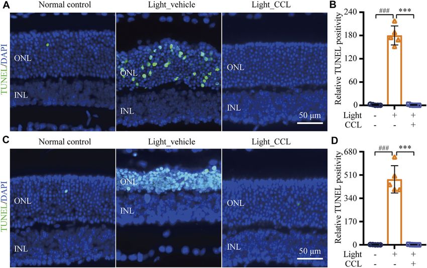
Wu et al. Cuscuta Chinensis Lam. Protects Photoreceptors
TABLE 1 | The primer sequences. STATISTICAL ANALYSIS
Gene name Primer sequences (5939)
All results were expressed as mean ± standard deviation (SD).
Ccl2 Forward primer AGCTGTAGTTTTTGTCACCAAGC Statistical analyses were performed using independent-samples
Reverse primer GTGCTGAAGACCTTAGGGCA
t-test (GraphPad Prism 8, United States). Differences were
Gfap Forward primer CCGAGTACTGAAGCCAAGGG
Reverse primer GCAGTTTGTAACCCCTCCCA considered statistically significant if p < 0.05.
Il1b Forward primer TGCCACCTTTTGACAGTGATG
Reverse primer AAGGTCCACGGGAAAGACAC
Il6 Forward primer CCAAGAACGATAGTCAATTCCAGAA RESULTS
Reverse primer AAGAAGGCAACTGGATGGAAGT
Tnf Forward primer ACGTCGTAGCAAACCACCAA
Reverse primer GCAGCCTTGTCCCTTGAAGA CCL Treatment Protects Photoreceptors
18S rRNA Forward primer GAGGTTCGAAGACGATCAGA From Developing Light-Induced Structural
Reverse primer TCGCTCCACCAACTAAGAAC
Impairment
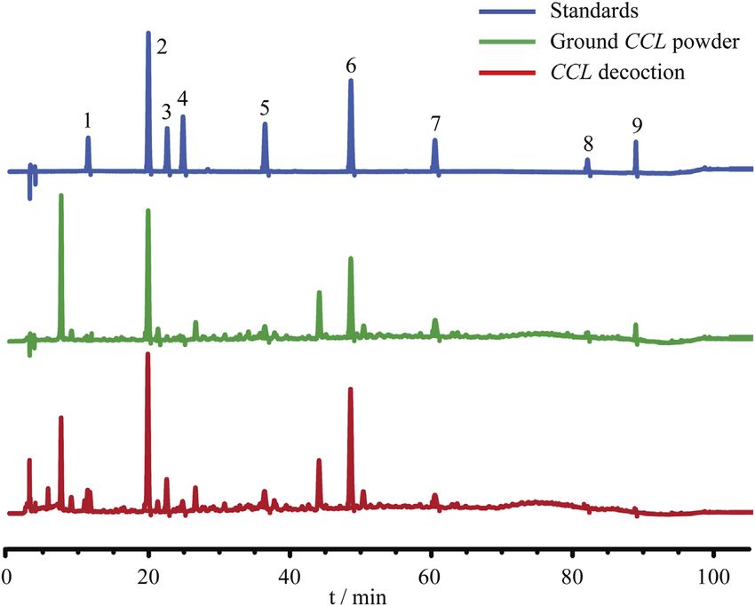
Prior to the indicated analysis, HPLC was performed to obtain the
chemical profile of the major components in CCL for the quality
Real-Time Quantitative Polymerase Chain control purpose. The results showed that phenolic acids including
Reaction (Real-Time qPCR) neochlorogenic acid, chlorogenic acid, cryptochlorogenic acid,
The retinas were collected after the indicated treatment. Total caffeic acid, p-coumaric acid and flavonoids including
RNA was then isolated usingTRIzol (TaKaRa, Japan) reagent, hyperoside, astragaloside, quercetin and kaempferol could be
followed by reverse transcription using the PrimeScriptTM RT detected in the ground CCL powder and CCL decoction
reagent Kit (TaKaRa, Japan). Real-time qPCR was subsequently (Figure 1). According to Pharmacopoeia of the People’s
performed with the SYBR Green I Master (Roche, United States) Republic of China (2020 Edition), the content of hyperoside,
on a Roche Light Cycler 480 II (Roche, United States). The the quality control compound for CCL, should be no lower than
sequences of primers used for real-time qPCR analysis were 0.01% in the dried CCL. The results from the HPLC analysis
listed in Table 1. 18S ribosomal RNA (rRNA) was amplified as showed that the amount of hyperoside in the ground CCL powder
an internal control. The fold change in the gene expression was was estimated to be approximately 0.1296%, validating the quality
calculated based on 2−[Ct (specific gene)−Ct (18s rRNA)]. of CCL used in the current study. Meanwhile, HPLC analysis also
FIGURE 1 | Quality control analysis of CCL. For the quality control purpose, HPLC analysis was performed to estimate the content of neochlorogenic acid,
chlorogenic acid, cryptochlorogenic acid, caffeic acid, p-coumaric acid, hyperoside, astragaline, quercetin and kaempferol in ground CCL powder and CCL decoction.
The detection wavelength was set at 328 nm. Peak detection and peak area calculation was performed using Chemstation software.
Frontiers in Pharmacology | www.frontiersin.org 4 June 2022 | Volume 13 | Article 904849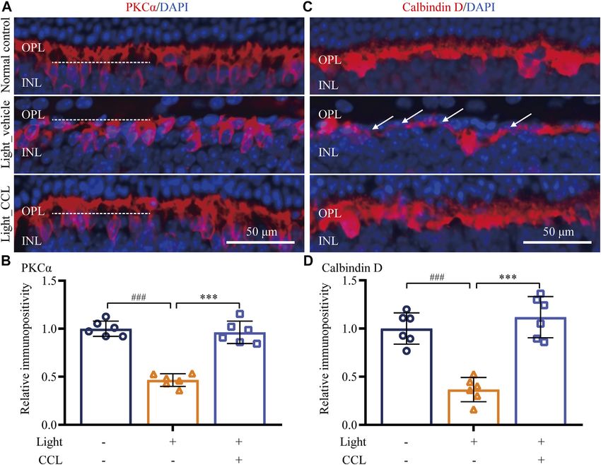
Wu et al.                                                                                                                  Cuscuta Chinensis Lam. Protects Photoreceptors

TABLE 2 | Estimated content of the major chemical components in CCL.                           Next, the pharmacological potentials of CCL in protecting
Compound                     Estimated content (%)a       Estimated content (%)b           against photoreceptor degeneration were assessed by multiple
                                                                                           experimental approaches in the light-exposed BALB/c mice
Neochlorogenic acid                   0.0069                        0.0029                 (Figure 2A). Bright light-induced photoreceptor degeneration is
Chlorogenic acid                      0.1653                        0.0149
                                                                                           characterized by a gradual loss of photoreceptor structure that
Cryptochlorogenic acid                0.0123                        0.0031
Caffeic acid                          0.0047                        0.0009                 physically stabilizes 7 days after illumination (Wenzel et al., 2005).
p-Coumaric acid                       0.0221                        0.0023                 Meanwhile, impairment of the second-order retinal neurons such
Hyperoside                            0.1296                        0.0145                 as bipolar cells and horizontal cells as well as reactive gliosis in
Astragaline                           0.0350                        0.0022                 müller cells are hallmark pathologies secondary to the degenerative
Quercetin                             0.0053                        0.0002
Kaempferol                            0.0104                        0.0001
                                                                                           changes in photoreceptors, which persist into the end-stage
a
                                                                                           photoreceptor degeneration and collectively contribute to
    Ground CCL powder was analyzed.
b
    CCL decoction was analyzed.
                                                                                           irreversible deterioration of the retinal structure and function
                                                                                           (Chen et al., 2016; Pfeiffer et al., 2020). Thus, the impact of
                                                                                           CCL treatment on light-induced structural, functional and
revealed that the major phenolic acids and flavonoids were                                  morphological impairment of photoreceptors and the
present in the CCL decoction used for the following                                        pathological alterations in bipolar cells, horizontal cells and
assessment (Table 2).                                                                      müller cells were evaluated by OCT, ERG, H&E staining and

    FIGURE 2 | CCL protects against light-induced impairment of the photoreceptor structure. (A) The schematic drawing of the experimental design. BALB/c mice
    were pretreated with CCL or water. Thirty min later, the mice were exposed to white light at 15,000 lux for 30 min, followed by the indicated assessments. (B) OCT
    imaging was carried out 7 days after light exposure to obtain the full-retinal structural images. (C) ONL thickness was measured at 125, 250, 500 and 1,000 μm off ONH
    in the inferior and superior retinas. The white triangle signifies the damaged ONL. INL, inner nuclear layer; ONH, optic nerve head; ONL, outer nuclear layer. Scale
    bar, 100 μm. Data were expressed as mean ± SD (n = 5 per group). ### Compared to Normal control, p < 0.001; * compared to Light_vehicle, p < 0.05; ** compared to
    Light_vehicle, p < 0.01; *** compared to Light_vehicle, p < 0.001.

Frontiers in Pharmacology | www.frontiersin.org                                        5                                            June 2022 | Volume 13 | Article 904849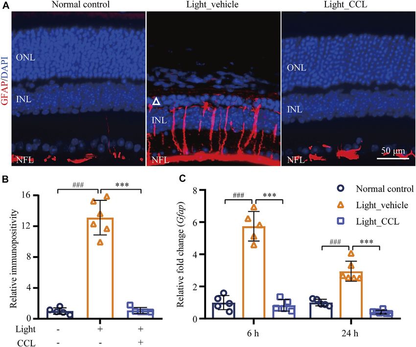
Wu et al. Cuscuta Chinensis Lam. Protects Photoreceptors
IHC assessments 7 days after the light exposure. In addition, our significantly decreased a wave and b wave amplitudes in the
previous studies have demonstrated that microglial activation is light-exposed vehicle-treated retinas compared to the vehicle-
most notable prior to the eventual loss of photoreceptor structure treated normal controls (a wave: p < 0.001; b wave: p < 0.01 or
(Bian et al., 2016; Wu et al., 2020). IHC examination of microglial 0.001). Both low-dose and high-dose CCL treatment resulted in
activation was thus assessed 24 h and 3 days after light exposure. increased a wave and b wave amplitudes compared to that from
Furthermore, findings from our previous work indicate that the light-exposed vehicle-treated mice (a wave: p < 0.05, 0.01 or
photoreceptor cell death becomes evident 24 h after light 0.001; b wave: p < 0.05, 0.01 or 0.001), with better effects observed
exposure and peaks around 3 days after illumination (Bian in the light-exposed high-dose CCL-treated mice (Figure 3B).
et al., 2016). Therefore, TUNEL assay was performed 3 days These results indicate that CCL treatment protects against light-
after the light exposure to assess the effect of CCL treatment on induced impairment of the retinal function.
photoreceptor cell death. Lastly, it has been shown in our previous
studies that the retinal expression of genes involved in CCL Confers Protection to Rod and Cone
inflammatory responses and reactive gliosis was altered at the Photoreceptors in the Light-Exposed
early stage of photoreceptor degeneration (Bian et al., 2016; Wu
Retinas
et al., 2020), real-time qPCR was thus performed 6 h and/or 24 h As demonstrated above, high-dose CCL treatment resulted in
after light exposure to evaluate the impact of CCL treatment on the nearly complete protection of the photoreceptor structure and
retinal expression of genes implicated in these processes. retinal function in the light-exposed mice. Therefore, detailed
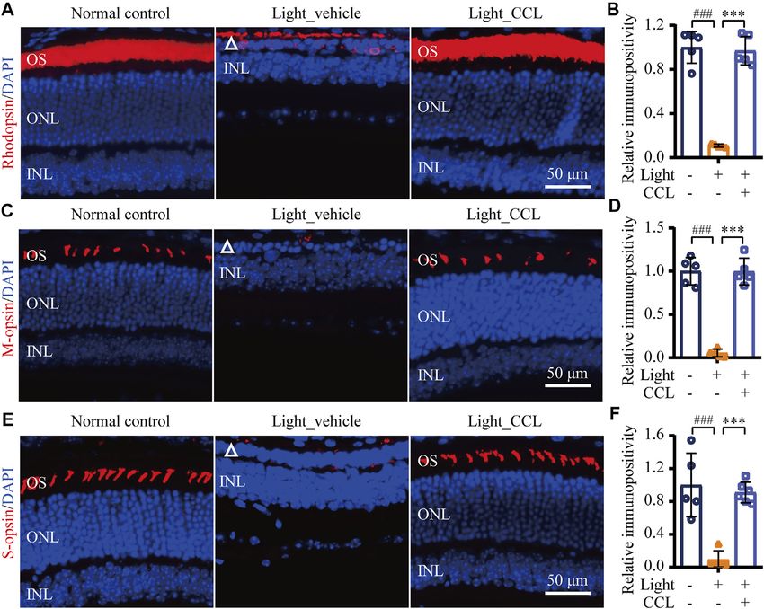
To evaluate the photoreceptor protective effects of CCL, histological and immunohistochemical examinations were
BALB/c mice were exposed to experimental white light at the carried out to further characterize the protective effective of
intensity of 15,000 lux for 30 min and treated by vehicle or CCL at high-dose CCL (marked as CCL in the following experiments)
3 g/kg (low-dose CCL) and 12 g/kg bw (high-dose CCL). Non- on the light-exposed retinas. First, H&E staining was performed
invasive full-retinal OCT imaging was carried out 7 days after to gain a better visualization of the gross retinal morphology. As
light exposure to obtain an objective and comprehensive view of shown in Figure 4A, compared to the vehicle-treated normal
the retinal structure. As shown in Figure 2B, compared to the controls, photoreceptor outer segment (OS) and inner segment
vehicle-treated normal controls, the ONL consisting of the cell (IS) were nearly diminished and the ONL was severely damaged
bodies of rod and cone photoreceptors was evidently diminished in the vehicle-treated light-exposed mice. However, the
in the light-exposed vehicle-treated mice. However, the ONL morphology of OS, IS and ONL was preserved in the light-
structure was partial preserved in the light-exposed mice treated exposed mice treated with CCL. Measurement of the number of
with low-dose CCL, whereas nearly complete preservation of the nuclei in ONL demonstrated a significant reduction in the
ONL structure was observed in the light-exposed mice treated number of photoreceptor cell bodies in both inferior and
with high-dose CCL. Measurement of the ONL thickness revealed superior retinas from the light-exposed vehicle-treated mice
that the ONL thickness was significantly decreased across the compared to the vehicle-treated normal controls (p < 0.001 in
retina in the light-exposed vehicle-treated mice compared to that both the inferior and superior retinas), whereas the number of
from the vehicle-treated normal controls (p < 0.001). Significantly nuclei in ONL in the inferior and superior retinas was increased
increased ONL thickness was observed as a result of the low-dose in the light-exposed CCL-treated mice compared to that from the
CCL (p < 0.05 or 0.01) and high-dose CCL treatment (p < 0.01 or light-exposed vehicle-treated mice (p < 0.001 in both the inferior
0.001) compared to that from the light-exposed vehicle-treated and superior retinas) (Figure 4B). Next, to better visualize the
mice, with greater effect observed in the light-exposed high-dose protection of rod and cone photoreceptors conferred by CCL
CCL-treated mice (Figure 2C). The results from OCT imaging treatment, IHC was performed to specifically label rods, short
indicate that CCL protects photoreceptor against light-induced wavelength-sensitive cones and mid wavelength-sensitive cones
structural impairment. in the retina. As shown in Figures 5A,B, compared to the
abundantly expressed rod-specific rhodopsin in the retinas
from the vehicle-treated normal controls, only residual
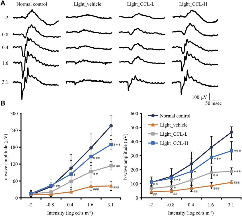
CCL Protects Against Light-Induced expression of rhodopsin was detected in the retinas from the
Functional Deterioration in the Retina light-exposed vehicle-treated mice (p < 0.001). In contrast, the
Next, full-retinal ERG recording was performed to evaluate the expression of rhodopsin was well preserved in the light-exposed
effect of CCL treatment on the scotopic a wave and b wave, which CCL-treated retinas compared to that from the light-exposed
reflect the retinal function conducted by rod photoreceptors and vehicle-treated mice (p < 0.001). Similarly, the retinal expression
second-order neurons, respectively. As shown in Figure 3A, of short wavelength-sensitive cone-specific S-opsin and mid
compared to light-evoked responses in a wave and b wave in wavelength-sensitive cone-specific M-opsin was found to be
the vehicle-treated normal controls, depressed a wave and b wave nearly diminished in the light-exposed vehicle-treated retinas
responses to light stimuli were readily observed in the light- (p < 0.001), whereas the expression of both S-opsin (Figures
exposed vehicle-treated mice. In contrast, a wave and b wave 5C,D) and M-opsin (Figures 5E,F) was preserved in the light-
responses to the light stimuli were improved in the light-exposed exposed CCL-treated retinas (p < 0.001). These results collectively
mice treated with both low-dose and high-dose CCL. demonstrate that CCL treatment protects rod and cone
Quantification of a wave and b wave amplitudes revealed photoreceptors against light-induced morphological damage.
Frontiers in Pharmacology | www.frontiersin.org 6 June 2022 | Volume 13 | Article 904849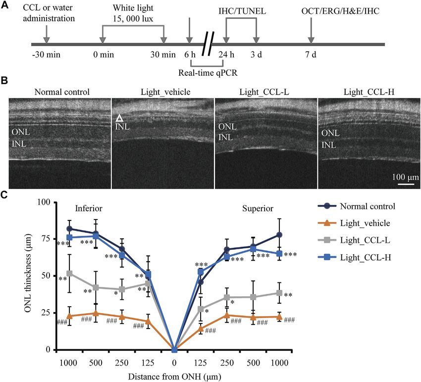
Wu et al.                                                                                                              Cuscuta Chinensis Lam. Protects Photoreceptors

 FIGURE 3 | CCL maintains the retinal function in the light-exposed mice. Scotopic ERG was performed 7 days after light exposure to assess the retinal function. (A)
 Representative electroretinograms from the indicated experimental groups. (B) Amplitudes of a wave and b wave were plotted. Data were expressed as mean ± SD (n =
 four to five per group). # Compared to NC, p < 0.05; ## Compared to Normal control, p < 0.01; ### compared to NC, p < 0.001; * compared to Light_vehicle, p < 0.05; **
 compared to Light_vehicle, p < 0.01; *** compared to Light_vehicle, p < 0.001.

CCL Maintains the Integrity of the                                                     CCL Attenuates Photoreceptor Cell Death
Second-Order Neurons in the                                                            in the Light-Exposed Retinas
Light-Exposed Retinas                                                                  Cell death is the central cellular mechanism that directly leads
Photoreceptor degeneration often leads to impairment of second-                        to loss of the photoreceptor structure and function. Our
order retinal neurons, which contributes to the functional                             previous study has demonstrated a progressive increase in
deterioration of the visual processing pathway. Thus, the effect of                    photoreceptor cell death prior to the eventual loss of
CCL treatment on the integrity of the second-order retinal neurons,                    photoreceptor structure (Bian et al., 2016). To further
bipolar cells and horizontal cells, were further examined. As shown in                 understand the impact of CCL treatment on photoreceptor
Figures 6A,B, PKCα labeling revealed that the dendrites of rod                         cell death in the light-exposed retinas, TUNEL assay was
bipolar cells were impaired in the retinas from the light-exposed                      performed. As shown in Figure 7, TUNEL positive cells
vehicle-treated mice (p < 0.001). However, the dendritic morphology                    were rare in the retinas from the vehicle-treated normal
of the bipolar cells was preserved in the retinas from the light-exposed               controls. However, a visible increase in TUNEL positivity
CCL-treated mice (p < 0.001). Moreover, calbindin D labeling                           was readily observed in the ONL from the light-exposed
showed that compared to the vehicle-treated normal controls, the                       vehicle-treated mice 1 day (Figures 7A,B) and 3 days
dendrites of horizontal cells were evidently damaged in the light-                     (Figures 7C,D) after the light exposure (p < 0.001). In
exposed vehicle-treated mice (p < 0.001). In sharp contrast, the                       distinct contrast, TUNEL positivity was barely observed in
morphological integrity of horizontal cell dendrites was preserved in                  the retinas from the light-exposed CCL-treated mice at both
the light-exposed CCL-treated mice (p < 0.001) (Figures 6C,D).                         time points (p < 0.001). These results indicate that CCL
These results demonstrate that CCL confers remarkable protection to                    treatment suppresses photoreceptor cell death in the light-
the second-order neurons in the light-exposed retinas.                                 exposed retinas.

Frontiers in Pharmacology | www.frontiersin.org                                    7                                             June 2022 | Volume 13 | Article 904849
Wu et al.                                                                                                            Cuscuta Chinensis Lam. Protects Photoreceptors

 FIGURE 4 | CCL protects against light-induced morphological damage to the photoreceptors. Histological examination was performed by H&E staining. (A)
 Representative micrographs from the indicated experimental groups. (B) The number of photoreceptors was counted from ONH to 400 μm off ONH in the inferior and
 superior retinas. White triangle signifies the damage ONL. INL, inner nuclear layer; IS, inner segment; ONH, optic nerve head; ONL, outer nuclear layer; OPL, outer
 plexiform layer; OS, outer segment. Scale bar, 50 μm. The data were expressed as mean ± SD (n = 4 per group). ### Compared to Normal control, p < 0.001; ***
 compared to Light_vehicle, p < 0.001.

CCL Alleviates Müller Cell Gliosis in the                                             compared to that from the vehicle-treated normal controls
Light-Exposed Retinas                                                                 (p < 0.001). Compared to that from the light-exposed vehicle-
Photoreceptor degeneration is often accompanied by excessive                          treated retinas, GFAP immunopositivity was markedly reduced
müller cell gliosis. The müller cell gliosis is not only a sensitive                  as a result of the CCL treatment (Figure 8B) (p < 0.001). In
pathological alteration signifying even subtle disturbance of the                     addition, real-time qPCR was performed to analyze the mRNA
retinal homeostasis, it may further aggravates retinal                                level of GFAP in the retina. As shown in Figure 8C, significantly
degeneration (Dyer and Cepko, 2000). The retinal expression                           increased mRNA expression of GFAP was detected in the light-
of GFAP was thus evaluated to assess the effect of CCL on the                         exposed vehicle-treated retinas as early as 6 h (p < 0.001) and 24 h
reactive changes in the müller cells in the light-exposed retinas.                    (p < 0.001) after the light exposure. In sharp contrast, the mRNA
As shown in Figure 8A, GFAP positivity was primarily restricted                       level of GFAP was significantly decreased in the light-exposed
to the nerve fiber layer (NFL) in the retinas from the vehicle-                        CCL-treated retinas compared to that from the light-exposed
treated normal controls. In distinct contrast, the GFAP positivity                    vehicle-treated retinas (p < 0.001 at both 6 and 24 h). These
was readily detected not only in the NFL, but also in the inner                       results demonstrate that CCL treatment suppresses light-induced
plexiform layer (IPL), inner nuclear layer (INL) and ONL in the                       reactive müller gliosis in the retinas.
light-exposed vehicle-treated retinas 7 days after the experimental
light exposure, indicative of müller gliosis accompanying                             CCL Suppresses Microglial Activation and
photoreceptor degeneration. However, this aberrancy in the
                                                                                      Inflammatory Responses in the
GFAP expression pattern was attenuated in the light-exposed
CCL-treated retinas. Quantification of the GFAP positivity                             Light-Exposed Retinas
consistently     revealed    significantly       increased    GFAP                     Following photoreceptor degeneration, resident microglial cells
immunopositivity in the light-exposed vehicle-treated retinas                         are activated to clear the damaged retinal neurons. However,

Frontiers in Pharmacology | www.frontiersin.org                                   8                                           June 2022 | Volume 13 | Article 904849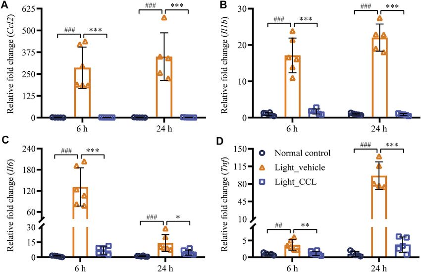
Wu et al.                                                                                                                Cuscuta Chinensis Lam. Protects Photoreceptors

 FIGURE 5 | CCL protects against light-induced ablation of rod and cone photoreceptors. IHC was performed to examine the expression of rhodopsin, M-opsin and
 S-opsin in the retina (in red). DAPI counterstaining (in blue) was performed to visualize the nuclei. The immunopositivity of rhodopsin, S-opsin and M-opsin in
 photoreceptor OS was quantified using ImageJ. (A) Representative micrographs showing the expression of rhodopsin in the retina. (B) The relative fold change in
 rhodopsin immunopositivity was plotted against that from Normal control. (C) Representative micrographs showing the expression of S-opsin in the retina. (D) The
 relative fold change in the immunopositivity of S-opsin was plotted against that from Normal control. (E) Representative micrographs showing the expression of M-opsin
 in the retina. (F) The relative fold change in the immunopositivity of M-opsin was plotted against that from Normal control. White triangle points to damaged ONL. INL,
 inner nuclear layer; ONL, outer nuclear layer; OS, outer segment. Scale bar, 50 μm. The data were expressed as mean ± SD (n = 5 per group). ### Compared to Normal
 control, p < 0.001; *** compared to Light_vehicle, p < 0.001.

uncontrolled microglial activation exacerbates neuronal cell                            observed in the light-exposed vehicle-treated mice 6 h after the
death in the damaged retina through excessive inflammatory                               light exposure. In distinct contrast, significantly decreased
responses (Xu et al., 2009). To evaluate the effect of CCL                              expression of Ccl2 (p < 0.001), Il1b (p < 0.001), Il6 (p <
treatment      on     photoreceptor      degeneration-associated                        0.001) and Tnf (p < 0.01) was noted in the light-exposed
microglial activation in the retina, the retinal expression of                          CCL-treated retinas 6 h after the light exposure. Similar
the microglial marker Iba-1 was examined. The results showed                            results were observed 24 h after light exposure. Compared to
that Iba-1 positivity was increased in the light-exposed vehicle-                       the vehicle-treated normal controls, increased retinal
treated retinas 1 day (p < 0.001) (Figures 9A,B) and 3 days (p <                        expression of Ccl2 (p < 0.001), Il1b (p < 0.001), Il6 (p <
0.001) (Figures 9C,D) after the light exposure, whereas much                            0.001), and Tnf (p < 0.001) was observed in the light-
less Iba-1 positivity was observed in the light-exposed CCL-                            exposed vehicle-treated mice 24 h after the light exposure.
treated retinas at both time points (p < 0.001). Furthermore, the                       However, significantly decreased expression of Ccl2 (p <
retinal expression of genes encoding inflammation mediators                              0.001), Il1b (p < 0.001), Il6 (p < 0.05), and Tnf (p < 0.001)
including Ccl2, Il1b, Il6, and Tnf was further analyzed. As                             was noted in the light-exposed CCL-treated retinas. These
shown in Figure 10, compared to the vehicle-treated normal                              results collectively indicate that CCL treatment alleviates
controls, significantly increased retinal expression of Ccl2 (p <                        photoreceptor degeneration-associated microglial activation
0.001), Il1b (p < 0.001), Il6 (p < 0.001) and Tnf (p < 0.01) was                        and inflammatory responses in the retina.

Frontiers in Pharmacology | www.frontiersin.org                                     9                                             June 2022 | Volume 13 | Article 904849
Wu et al.                                                                                                                 Cuscuta Chinensis Lam. Protects Photoreceptors

 FIGURE 6 | CCL attenuates the morphological impairment of bipolar and horizontal cells in the light-exposed retinas. IHC was performed to examine the expression
 of PKCα (in red) and calbindin D (in red) in the retina. Counterstaining of DAPI (in blue) was performed. The immunopositivity of PKCα and calbindin D in OPL was
 quantified by ImageJ. (A) Representative micrographs showing the expression of PKCα in the retina. (B) The relative fold change in the immunopositivity of PKCα was
 plotted against that from Normal control. (C) Representative micrographs showing the expression of calbindin D in the retina. (D) The relative fold change in the
 immunopositivity of calbindin D was plotted against that from Normal control. INL, inner nuclear layer; OPL, outer plexiform layer. Scale bar, 50 μm. Data were expressed
 as mean ± SD (n = 6 per group). ### Compared to Normal control, p < 0.001; *** compared to Light_vehicle, p < 0.001.

DISCUSSION                                                                                Chinese pharmacopoeia, Shennong’s Herbal as well as the classic
                                                                                          work of Chinese materia medica, Compendium of Materia
The current study demonstrates that CCL provides structural,                              Medica. In terms of the ophthalmic diseases, CCL has been
morphological and functional protection against light-induced                             included as one of the major components in various TCM
photooxidative stress-mediated photoreceptor degeneration.                                formulas to treat not only degenerative retinal diseases such as
These photoreceptor protective effects of CCL are in part                                 dry AMD, RP, glaucoma and ischemic optic neuropathy, but also
attributable to inhibition of photoreceptor cell death,                                   neovascular retinal disorders, for instance, wet AMD and diabetic
preservation of the morphological integrity of the second-                                retinopathy. In addition, CCL-containing TCM formulas have
order retinal neurons as well as mitigation of müller gliosis,                            also been widely used to treat cataract and dry eye syndrome,
microglial activation and retinal inflammation. Thus, the                                  disease entities that affect the conjunctiva and lens (Zhu et al.,
findings here provide direct evidence supporting the protective                            2020). The reproductive effects, hepatoprotective, renoprotective,
effects of CCL on photoreceptors and the retinal                                          cardioprotective, anti-aging, neuroprotective and anti-diabetic
microenvironment under photooxidative stress conditions.                                  activities of CCL have been supported by multiple lines of
    The major findings from the current work relate to                                     pharmacological studies (Donnapee et al., 2014). Our previous
photoreceptor protection conferred by CCL. CCL has been                                   study has also demonstrated that a CCL-containing TCM
used for the treatment of a broad range of disorders including                            formula, Shihu Yeguang Pill, protects against light-induced
reproductive disorders, hepatic diseases, renal diseases, aging and                       photoreceptor degeneration (Wu et al., 2020). However, to our
ophthalmic diseases affecting vision (Zheng HZD and She, 1998;                            best knowledge, the pharmacological implications of CCL per se
Donnapee et al., 2014; Zhu et al., 2020). The traditional views on                        in photoreceptor degenerative diseases have not been addressed.
the vision-enhancing effects of CCL can be found in the earliest                          Here, by demonstrating that CCL counteracts light-induced

Frontiers in Pharmacology | www.frontiersin.org                                      10                                             June 2022 | Volume 13 | Article 904849
Wu et al. Cuscuta Chinensis Lam. Protects Photoreceptors FIGURE 7 | CCL suppresses photoreceptor cell death in the light-exposed retinas. TUNEL assay was performed to assess photoreceptor cell death. Nuclei were stained by DAPI (in blue). TUNEL positivity (in green) in ONL was quantified by ImageJ. (A) Representative micrographs demonstrating TUNEL positivity from the eyes enucleated 24 h after light exposure. (B) The relative fold change in the TUNEL positivity from (A) was plotted against that from Normal control. (C) Representative micrographs demonstrating TUNEL positivity from the eyes enucleated 3 days after light exposure. (D) The relative fold change in the TUNEL positivity from (C) was plotted against that from Normal control. INL, inner nuclear layer, ONL, outer nuclear layer. Scale bar, 50 μm. Data were expressed as mean ± SD (n = 5 per group). ### Compared to Normal control, p < 0.001; *** compared to Light_vehicle, p < 0.001. impairment of the photoreceptor structure and associated retinal conditions such as AMD and RP (Hoon et al., 2014). The function, our study provides direct in vivo experimental evidence photoreceptors are exposed to high concentration of oxygen that supports the photoreceptor protective properties of CCL, from choriocapillaries (Wangsa-Wirawan and Linsenmeier, thereby justifying the clinical application of CCL in the 2003). Photoreceptor OS is rich in lipids and is vulnerable prevention and treatment of photoreceptor degenerative to oxidation. Light exposure, oxidization of polyunsaturated diseases such as dry AMD and RP. fatty acids, and phagocytosis of photoreceptor OS routinely As an integral part of the central nervous system, the light- produce high levels of free radicals (Fliesler and Bretillon, sensing retina conducts the essential function of vision 2010). Therefore, even under physiological conditions, formation (Demb and Singer, 2015). It contains millions of photoreceptors are under constant oxidative stress. Under light-sensitive first-order neurons, namely, photoreceptors as pathological conditions, whether caused by genetic well as the second-order and the third-order neurons. mutation/deficiency or environmental insults, oxidative Photoreceptors, the first-order retinal neurons, are the stress is regarded as the major mechanism mediating primary sensory neurons in the retina. They capture and multiple biochemical and molecular changes that eventually convert the incoming light into an electrical signal that is lead to the death and degeneration of the stressed further processed through the second-order and the third- photoreceptors (Domènech and Marfany, 2020). Bright light order retinal neurons. The electrical visual signal initially exposure creates excessive photo-oxidative stress to the retina generated in the photoreceptors is eventually carried to the and thus has been extensively used to recapitulate oxidative brain to create conscious vision. There are two types of stress-induced photoreceptor degeneration (Organisciak et al., photoreceptors, namely rods and cones. Rods are extremely 1998; Wenzel et al., 2005). Our work here demonstrates that sensitive and carry out specialized function in mediating low- CCL attenuates bright light-induced photoreceptor cell death light vision. Cones are responsible for the generation of and preserves the structure and function integrity of the retina daylight and color vision. Like other neurons in the central in the bright light-exposed mice (Figures 2–7), supporting the nervous system, photoreceptors are postmitotic and the loss of notion that CCL attenuates oxidative stress-mediated photoreceptors is irreversible, which directly leads to vision photoreceptor degeneration. Future studies are worth impairment in the patients with related retinal degenerative pursuing to further elucidate the molecular mechanisms Frontiers in Pharmacology | www.frontiersin.org 11 June 2022 | Volume 13 | Article 904849
Wu et al.                                                                                                                Cuscuta Chinensis Lam. Protects Photoreceptors

 FIGURE 8 | CCL attenuates reactive müller gliosis in the light-exposed retinas. IHC was performed to examine the expression of GFAP in the retina (in red).
 Counterstaining of DAPI (in blue) was performed. The immunopositivity of GFAP in the retina was quantified by ImageJ. (A) Representative micrographs showing the
 expression of GFAP in the retina. (B) The relative immunopositivity of GFAP in the indicated experimental groups was plotted against that from Normal control. (C) Total
 RNA was isolated from the retinas collected from the indicated experimental groups 6 and 24 h after light exposure. Real-time qPCR was then performed to analyze
 the retinal expression of GFAP. 18S rRNA was amplified as an internal control. The relative fold change in the expression of GFAP was plotted against that from Normal
 control. INL, inner nuclear layer; NFL, nerve fiber layer; ONL, outer nuclear layer. Scale bar, 50 μm. Data were expressed as mean ± SD (n = 5-6 per group). ### Compared
 to Normal control, p < 0.001; *** compared to Light_vehicle, p < 0.001.

underlying the anti-oxidant effects of CCL in the pathological                           generated in photoreceptors to the third-order retinal
context of photoreceptor degeneration.                                                   ganglion cells, the specialized retinal neurons that send out
   Secondly, our work here demonstrates that other than                                  the vision information to the brain. Given that bipolar cells are
significant protection over photoreceptors, CCL treatment                                 in charge of relaying the information for vision formation, they
maintains the morphological integrity of the second-order                                constitute the elementary building blocks of vision. Horizontal
bipolar and horizontal cells under photooxidative stress                                 cells are the interneurons in the retina, forming triad synapses
conditions (Figure 6). Bipolar cells and horizontal cells are                            with photoreceptors and bipolar cells. Horizontal cells
the essential components of the retinal neuronal network                                 function      to    modulate     signal    transmission     from
(Euler et al., 2014). Operating in the similar fashion as that                           photoreceptors to bipolar cells (Thoreson and Mangel,
of the brain, the function of the neuronal network of the retina                         2012). Ablation of horizontal cells leads to morphological
is also dependent on the spatially ordered organization of                               degeneration of rod photoreceptors and impaired retinal
projection neurons and interneurons as well as their                                     function (Sonntag et al., 2012). Therefore, maintaining the
synaptic connections. Photoreceptors form synaptic                                       morphological integrity of bipolar cells and horizontal cells
connections with bipolar cells and horizontal cells,                                     contribute to the protective effects on the retinal structure and
constituting the first information relay point in the visual                              function as a result of CCL treatment. However, whether CCL
pathway (Burger et al., 2021). Bipolar cells are the                                     provides direct protection to bipolar and/or horizontal cells
projection neurons in the retina, relaying the information                               requires further investigation in the future studies.

Frontiers in Pharmacology | www.frontiersin.org                                     12                                             June 2022 | Volume 13 | Article 904849
Wu et al.                                                                                                                  Cuscuta Chinensis Lam. Protects Photoreceptors

 FIGURE 9 | CCL mitigates microglial activation in the light-exposed retinas. IHC was performed to examine the expression of Iba-1 in the retina (in red).
 Counterstaining of DAPI (in blue) was performed. The immunopositivity of Iba-1 in the outer retina including ONL and the subretinal space was quantified by ImageJ. (A)
 Representative micrographs demonstrating Iba-1 positivity from the eyes enucleated 24 h after light exposure. (B) The relative fold change in the immunopositivity of Iba-
 1 from (A) was plotted against that from Normal control. (C) Representative micrographs demonstrating Iba-1 positivity from the eyes enucleated 3 days after light
 exposure. (D) The relative fold change in the immunopositivity of Iba-1 from (C) was plotted against that from Normal control. INL, inner nuclear layer; ONL, outer nuclear
 layer. Scale bar, 50 μm. Data were expressed as mean ± SD (n = 5 per group). ### Compared to Normal control, p < 0.001; *** compared to Light_vehicle, p < 0.001.

    In addition, CCL treatment results in a remarkable attenuation of                      expression in the light-exposed retinas. It is a well-established
the gliotic changes in the müller cells in the light-exposed retinas                       notion that enhanced inflammatory response exacerbates retinal
(Figure 8). Müller cells are macroglial cells in the retina that                           cell death (Xu et al., 2009; Anderson et al., 2002; Hollborn et al.,
structurally support the retinal neurons. Meanwhile, they are held                         2008). The activated microglial cells promote inflammatory
responsible for maintaining the homeostasis of the neuronal                                responses and aggravate cell death in the neuronal tissues.
microenvironment in the retina (Bringmann et al., 2006).                                   Accumulated evidence has revealed that microglial activation
However, under pathological conditions, instead of being                                   is not merely a common pathology associated with the
neuronal supportive, müller cells experience reactive gliotic                              degenerated retinas, it is also neurotoxic and aggravates
changes and develop gliotic scars that may detrimentally disturb                           photoreceptor loss. Suppressing microglial activation has been
the retinal homeostasis and further exacerbate neuronal cell death.                        demonstrated to be neuroprotective in light-challenged retinas
Thus, reactive müller gliosis is not only one of the most sensitive                        (Kohno et al., 2013; Scholz et al., 2015). Therefore, microglial
hallmark pathologies associated with nearly all known retinal                              activation has emerged as a new target for photoreceptor
disorders, it also promotes the progression of neuronal                                    protective treatment. Our work here demonstrates that CCL
degeneration by enhancing the susceptibility of retinal neurons to                         treatment suppresses microglial activation as well as the
stress stimuli (Bringmann et al., 2006). Considering the important                         expression of proinflammatory genes in the light-exposed
implications of müller cells in the retinal homeostasis, suppressed                        retinas (Figures 9, 10), which may in part contribute to its
müller gliosis may in part contribute to the photoreceptor protection                      protective effect against light-induced photoreceptor
observed in the light-exposed CCL-treated mice. The direct impact                          degeneration. Meanwhile, oxidative stress not only directly
of CCL treatment on reactive müller gliosis, however, requires                             leads to cell death, but also triggers inflammatory responses
further delineation.                                                                       (Ozawa, 2020). Therefore, it is also possible that attenuated
    Lastly, our work here demonstrates that CCL treatment                                  microglial activation occurs as a result of CCL-conferred
attenuates microglial activation and inflammatory gene                                      suppression of the photooxidative stress in the retina. Further

Frontiers in Pharmacology | www.frontiersin.org                                       13                                             June 2022 | Volume 13 | Article 904849
Wu et al. Cuscuta Chinensis Lam. Protects Photoreceptors FIGURE 10 | CCL suppresses the expression of proinflammatory genes in the light-exposed retinas. Total RNA was isolated from the retinas collected from the indicated experimental groups 6 and 24 h after light exposure. Real-time qPCR analyses were performed to examine the expression of Ccl2 (A), Il1b (B), Il6 (C), and Tnf (D) in the retina. Relative fold change was normalized against that from Normal control. The data were expressed as the mean ± SD (n = 5-6 per group). ## Compared to Normal control, p < 0.01; ### Compared to Normal control, p < 0.001; * compared to Light_vehicle, p < 0.05; ** compared to Light_vehicle, p < 0.01; *** compared to Light_vehicle, p < 0.001. studies are thus necessary to clarify whether CCL plays any direct CCL preparation in analyzing its photoreceptor protective roles in suppressing microglial inflammatory activation. activities. It is also worth noting that quercetin, a type of flavonoids In conclusion, our current work presents novel experimental found in CCL, has been shown to mitigate light-induced retinal evidence demonstrating that CCL protects against light-induced degeneration in rats (Koyama et al., 2019). The pharmacological oxidative stress-mediated photoreceptor cell death, second-order potential of quercetin in attenuating neuroinflammation retinal neuron impairment, reactive müller gliosis, microglial accompanying photoreceptor degeneration has also been activation and retinal inflammation, which may collectively demonstrated in a mouse model of RP (Ortega and contribute to CCL-conferred preservation of the structural and Jastrzebska, 2021). In addition, it has been reported that functional integrity of the retina. The findings here thus provide kaempferol, another flavonoid component of CCL, protects direct pharmacological evidence supporting TCM-guided clinical against oxidizing agent sodium iodate-induced photoreceptor application of CCL in the prevention and treatment of vision- degeneration (Du et al., 2018). These findings provide threatening photoreceptor degenerative disorders. pharmacological hypotheses in understanding the chemical basis of the retinal protective effects of CCL as revealed by the current work. However, future studies are required to DATA AVAILABILITY STATEMENT elucidate to what extent quercetin and kaempferol contribute to the effects of CCL in protecting the retina from developing The original contributions presented in the study are included in photooxidative stress-mediated photoreceptor degeneration the article/Supplementary Material, further inquiries can be and associated retinal inflammation. Additionally, given that directed to the corresponding author. CCL decoction instead of the individual bioactive compound in CCL was analyzed in the current study, our findings here only relate to the photoreceptor protective effects of the total ETHICS STATEMENT composition of CCL with indicated chemical profiles shown in Figure 1. A synergistic effect of different bioactive The animal study was reviewed and approved by the Institutional compounds in CCL decoction is likely at play in protecting Animal Care and Use Committee of Yueyang Hospital of the retina from developing bright light-induced degenerative Integrated Traditional Chinese Medicine, Shanghai University pathologies. This notion also stresses the standardization of of TCM. Frontiers in Pharmacology | www.frontiersin.org 14 June 2022 | Volume 13 | Article 904849
Wu et al.                                                                                                                        Cuscuta Chinensis Lam. Protects Photoreceptors

AUTHOR CONTRIBUTIONS                                                                            of   Shanghai    Academic/Technology       Research   Leader
                                                                                                (19XD1403700, YC) and Science Foundation of Yueyang
YC designed and supervised the study. HW, BZ, DL, JX, JC, JgC,                                  Hospital, Shanghai University of Traditional Chinese Medicine
and XD performed the experiments. HW, BZ and DL analyzed                                        (2019YYZ02, YC).
the data. HW and DL drafted part of the manuscript. NZ and TZ
supervised part of the study. YC wrote the manuscript.
                                                                                                ACKNOWLEDGMENTS
FUNDING                                                                                         We thank the research staff at the Clinical Research Institute
                                                                                                of Integrative Medicine, Shanghai Academy of Traditional
This work was supported by the National Natural Science                                         Chinese Medicine for their helpful discussion and technical
Foundation of China (81673790 and 81473732, YC), Program                                        support.

                                                                                                Du, W., An, Y., He, X., Zhang, D., and He, W. (2018). Protection of Kaempferol on
REFERENCES                                                                                         Oxidative Stress-Induced Retinal Pigment Epithelial Cell Damage. Oxid. Med.
                                                                                                   Cell Longev. 2018, 1610751. doi:10.1155/2018/1610751
Akiba, R., Matsuyama, T., Tu, H. Y., Hashiguchi, T., Sho, J., Yamamoto, S., et al.              Dyer, M. A., and Cepko, C. L. (2000). Control of Müller Glial Cell Proliferation and
   (2019). Quantitative and Qualitative Evaluation of Photoreceptor Synapses in                    Activation Following Retinal Injury. Nat. Neurosci. 3 (9), 873–880. doi:10.1038/
   Developing, Degenerating and Regenerating Retinas. Front. Cell Neurosci. 13,                    78774
   16. doi:10.3389/fncel.2019.00016                                                             Euler, T., Haverkamp, S., Schubert, T., and Baden, T. (2014). Retinal Bipolar Cells:
Anderson, D. H., Mullins, R. F., Hageman, G. S., and Johnson, L. V. (2002). A Role                 Elementary Building Blocks of Vision. Nat. Rev. Neurosci. 15 (8), 507–519.
   for Local Inflammation in the Formation of Drusen in the Aging Eye. Am.                          doi:10.1038/nrn3783
   J. Ophthalmol. 134 (3), 411–431. doi:10.1016/s0002-9394(02)01624-0                           Fliesler, S. J., and Bretillon, L. (2010). The Ins and Outs of Cholesterol in the
Bian, M., Du, X., Cui, J., Wang, P., Wang, W., Zhu, W., et al. (2016). Celastrol                   Vertebrate Retina. J. Lipid Res. 51 (12), 3399–3413. doi:10.1194/jlr.
   Protects Mouse Retinas from Bright Light-Induced Degeneration through                           R010538
   Inhibition of Oxidative Stress and Inflammation. J. Neuroinflammation 13,                      Hollborn, M., Francke, M., Iandiev, I., Bühner, E., Foja, C., Kohen, L., et al. (2008).
   50. doi:10.1186/s12974-016-0516-8                                                               Early Activation of Inflammation- and Immune Response-Related Genes after
Bringmann, A., Pannicke, T., Grosche, J., Francke, M., Wiedemann, P., Skatchkov,                   Experimental Detachment of the Porcine Retina. Invest. Ophthalmol. Vis. Sci.
   S. N., et al. (2006). Müller Cells in the Healthy and Diseased Retina. Prog. Retin              49 (3), 1262–1273. doi:10.1167/iovs.07-0879
   Eye Res. 25 (4), 397–424. doi:10.1016/j.preteyeres.2006.05.003                               Hoon, M., Okawa, H., Della Santina, L., and Wong, R. O. (2014). Functional
Burger, C. A., Jiang, D., Mackin, R. D., and Samuel, M. A. (2021). Development and                 Architecture of the Retina: Development and Disease. Prog. Retin Eye Res. 42,
   Maintenance of Vision’s First Synapse. Dev. Biol. 476, 218–239. doi:10.1016/j.                  44–84. doi:10.1016/j.preteyeres.2014.06.003
   ydbio.2021.04.001                                                                            Kohno, H., Chen, Y., Kevany, B. M., Pearlman, E., Miyagi, M., Maeda, T., et al.
Byun, J., Verardo, M. R., Sumengen, B., Lewis, G. P., Manjunath, B. S., and Fisher, S.             (2013). Photoreceptor Proteins Initiate Microglial Activation via Toll-like
   K. (2006). Automated Tool for the Detection of Cell Nuclei in Digital                           Receptor 4 in Retinal Degeneration Mediated by All-Trans-Retinal. J. Biol.
   Microscopic Images: Application to Retinal Images. Mol. Vis. 12, 949–960.                       Chem. 288 (21), 15326–15341. doi:10.1074/jbc.M112.448712
Carmody, R. J., and Cotter, T. G. (2000). Oxidative Stress Induces Caspase-                     Koyama, Y., Kaidzu, S., Kim, Y. C., Matsuoka, Y., Ishihara, T., Ohira, A., et al.
   independent Retinal Apoptosis In Vitro. Cell Death Differ. 7 (3), 282–291.                      (2019). Suppression of Light-Induced Retinal Degeneration by Quercetin via
   doi:10.1038/sj.cdd.4400646                                                                      the AP-1 Pathway in Rats. Antioxidants (Basel) 8 (4), 79. doi:10.3390/
Carmody, R. J., McGowan, A. J., and Cotter, T. G. (1999). Reactive Oxygen Species                  antiox8040079
   as Mediators of Photoreceptor Apoptosis In Vitro. Exp. Cell Res. 248 (2),                    Krishnamoorthy, R. R., Crawford, M. J., Chaturvedi, M. M., Jain, S. K., Aggarwal, B.
   520–530. doi:10.1006/excr.1998.4421                                                             B., Al-Ubaidi, M. R., et al. (1999). Photo-oxidative Stress Down-Modulates the
Carvalho, L. S., Xiao, R., Wassmer, S. J., Langsdorf, A., Zinn, E., Pacouret, S., et al.           Activity of Nuclear Factor-kappaB via Involvement of Caspase-1, Leading to
   (2018). Synthetic Adeno-Associated Viral Vector Efficiently Targets Mouse and                    Apoptosis of Photoreceptor Cells. J. Biol. Chem. 274 (6), 3734–3743. doi:10.
   Nonhuman Primate Retina In Vivo. Hum. Gene Ther. 29 (7), 771–784. doi:10.                       1074/jbc.274.6.3734
   1089/hum.2017.154                                                                            Organisciak, D. T., Darrow, R. M., Barsalou, L., Darrow, R. A., Kutty, R. K., Kutty,
Chen, M., and Xu, H. (2015). Parainflammation, Chronic Inflammation, and Age-                        G., et al. (1998). Light History and Age-Related Changes in Retinal Light
   Related Macular Degeneration. J. Leukoc. Biol. 98, 713–725. doi:10.1189/jlb.                    Damage. Invest. Ophthalmol. Vis. Sci. 39 (7), 1107–1116.
   3RI0615-239R                                                                                 Ortega, J. T., and Jastrzebska, B. (2021). Neuroinflammation as a Therapeutic
Chen, Y., Palczewska, G., Masuho, I., Gao, S., Jin, H., Dong, Z., et al. (2016).                   Target in Retinitis Pigmentosa and Quercetin as its Potential Modulator.
   Synergistically Acting Agonists and Antagonists of G Protein-Coupled                            Pharmaceutics 13 (11), 1935. doi:10.3390/pharmaceutics13111935
   Receptors Prevent Photoreceptor Cell Degeneration. Sci. Signal 9 (438),                      Ozawa, Y. (2020). Oxidative Stress in the Light-Exposed Retina and its Implication
   ra74. doi:10.1126/scisignal.aag0245                                                             in Age-Related Macular Degeneration. Redox Biol. 37, 101779. doi:10.1016/j.
Curcio, C. A., Medeiros, N. E., and Millican, C. L. (1996). Photoreceptor Loss in                  redox.2020.101779
   Age-Related Macular Degeneration. Invest. Ophthalmol. Vis. Sci. 37 (7),                      Pfeiffer, R. L., Marc, R. E., and Jones, B. W. (2020). Persistent Remodeling and
   1236–1249.                                                                                      Neurodegeneration in Late-Stage Retinal Degeneration. Prog. Retin Eye Res. 74,
Demb, J. B., and Singer, J. H. (2015). Functional Circuitry of the Retina. Annu. Rev.              100771. doi:10.1016/j.preteyeres.2019.07.004
   Vis. Sci. 1, 263–289. doi:10.1146/annurev-vision-082114-035334                               Ryter, S. W., Kim, H. P., Hoetzel, A., Park, J. W., Nakahira, K., Wang, X., et al.
Domènech, E. B., and Marfany, G. (2020). The Relevance of Oxidative Stress in the                  (2007). Mechanisms of Cell Death in Oxidative Stress. Antioxid. Redox Signal 9
   Pathogenesis and Therapy of Retinal Dystrophies. Antioxidants (Basel) 9 (4), 347.               (1), 49–89. doi:10.1089/ars.2007.9.49
Donnapee, S., Li, J., Yang, X., Ge, A. H., Donkor, P. O., Gao, X. M., et al. (2014).            Scholz, R., Sobotka, M., Caramoy, A., Stempfl, T., Moehle, C., and Langmann, T.
   Cuscuta Chinensis Lam.: A Systematic Review on Ethnopharmacology,                               (2015). Minocycline Counter-regulates Pro-inflammatory Microglia Responses
   Phytochemistry and Pharmacology of an Important Traditional Herbal                              in the Retina and Protects from Degeneration. J. Neuroinflammation 12, 209.
   Medicine. J. Ethnopharmacol. 157, 292–308. doi:10.1016/j.jep.2014.09.032                        doi:10.1186/s12974-015-0431-4

Frontiers in Pharmacology | www.frontiersin.org                                            15                                               June 2022 | Volume 13 | Article 904849
You can also read