Estrogen receptor beta in breast cancer
←
→
Page content transcription
If your browser does not render page correctly, please read the page content below
Endocrine-Related Cancer (2002) 9 1–13
Estrogen receptor beta in breast cancer
C Palmieri, G J Cheng1, S Saji 1, M Zelada-Hedman1, A Wärri 1, Z Weihua1,
S Van Noorden, T Wahlstrom 2, R C Coombes, M Warner 1 and
J-Å Gustafsson1
Department of Cancer Medicine, Cancer Cell Biology Group, Cancer Research Campaign Laboratories,
Imperial College School of Medicine, Hammersmith Campus, Du Cane Road, London W12 ONN, UK
1
Department of Bioscience, Karolinska Institute, Novum, Huddinge University Hospital, SE-14186, Huddinge,
Sweden
2
Department of Obstetrics and Gynecology, Helsinki University Central Hospital, Haartmaninkatu 2, 00290
Helsinki, Finland
(Requests for offprints should be addressed to J-Å Gustafsson; Email: jan-ake.gustafsson@mednut.ki.se)
(S Saji is now at Tokyo Metropolitan Komagome Hospital, Department of Surgery, Breast Oncology Unit,
3–18–22 Honkomagome, Bunkyo-ku, 113–8677 Tokyo, Japan)
Abstract
Estrogen is essential for normal growth and differentiation in the mammary gland. It also supports
growth of approximately 50% of primary breast cancers. For this reason, removal of estrogen or
blocking of its action with the anti-estrogen, tamoxifen, is the main treatment for estrogen receptor
alpha (ERα)-positive tumors. In 1996, when oncologists became aware of a second ER, ERβ, there
was some doubt as to whether this receptor would be of importance in breast cancer because the
clinical consensus was that responsiveness to tamoxifen is related to the presence of ERα in breast
cancer. Today we know that ERα and ERβ have distinct cellular distributions, regulate separate sets
of genes and can oppose each other’s actions on some genes. We also know that ERβ is widely
expressed in both the normal and malignant breast and that there are proliferating cells in the breast
which express ERβ. In this review we summarize what is known about ERβ in breast cancer and
examine the possibility that ERβ-selective ligands may well represent a useful class of pharmacologi-
cal tools with a novel target, namely proliferating cells expressing ERβ.
Endocrine-Related Cancer (2002) 9 1–13
Introduction evident that one major player in the story, the second ER,
ERβ, had gone unnoticed (Kuiper et al. 1996) and that its
The major endocrine-related cancers in the human population presence may well have some bearing on the development
display appropriate gender-specific hormonal responsiveness, and treatment of hormone-dependent cancers. ERβ is highly
i.e. estrogen for breast and endometrial cancer in females and expressed in the prostate (Kuiper et al. 1996) and is ex-
androgen for prostate cancer in males. Since normal growth pressed together with ERα in both normal and malignant
of breast, uterus and prostate is dependent on the respective breasts (Dotzlaw et al. 1997, Vladusic et al. 1998, Fuqua et
sex steroids, the involvement of these hormones in malignant al. 1999, Speirs et al. 1999, Cullen et al. 2000, Iwao et al.
growth in these organs is not surprising. In normal tissues, 2000, Jarvinen et al. 2000, Speirs & Kerin 2000, Omoto et
hormone-stimulated growth is controlled, and tissues like the al. 2001, Roger et al. 2001). It is, therefore, not surprising
prostate and breast do not have unlimited growth even during that a great deal of interest is focused on the role of ERβ in
continuous exposure to their respective hormones. Uncon- normal and malignant growth of these two organs. In the
trolled growth in response to hormones could be due to either ventral prostate of mice, ERβ is involved in regulation of
overactivity of the signals for growth or underactivity of the epithelial growth, and its absence, in ERβ-/- mice, results in
signals which normally stop proliferation. hyperplasia of the prostatic epithelium (Weihua et al. 2001).
The androgen receptor, the estrogen receptor (ER) alpha In the case of breast cancer, some of the most important
and the genes which they regulate, have been extensively questions which need to be answered are whether it is of
studied and are the main targets in clinical approaches to clinical value to measure ERβ along with ERα and whether
controlling hormonally responsive prostate and breast cancer ERβ can be a novel target in therapy. In this review we will
respectively (Ali & Coombes 2000). In 1996 it became summarize what has been learnt about ERβ in the normal
Endocrine-Related Cancer (2002) 9 1–13 Online version via http://www.endocrinology.org
1351-0088/02/009–001 2002 Society for Endocrinology Printed in Great Britain Downloaded from Bioscientifica.com at 06/29/2023 05:26:01AM
via free access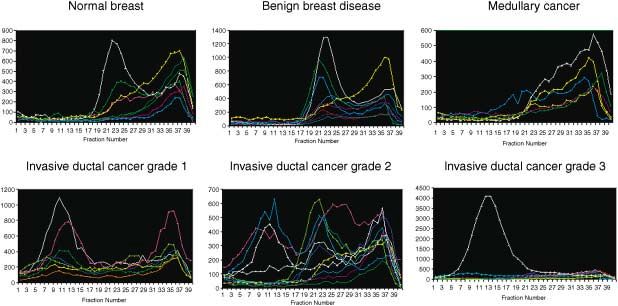
Palmieri et al.: ERβ in breast cancer

breast and in breast cancer and present a part of our large,       the proliferating cells are not the ones which express ERα.
as yet unpublished, study on ERβ in archived breast cancer         In immunohistochemical studies, markers of proliferation,
samples.                                                           such as Ki67 and cyclin A, are not found in cells which
                                                                   express ERα (Clarke et al. 1997a,b, Zeps et al. 1999). In the
                                                                   rodent mammary gland, ERβ-containing cells can proliferate
Estrogen and breast cancer
                                                                   but the majority of cells expressing proliferation markers do
Estrogen is a modulator of cellular growth and differentiation     not express either ER (Saji et al. 2000). This is in direct
in the mammary gland. It mediates most of its functions            contrast to estrogen action in breast cancer cell lines where
through two members of the nuclear receptor superfamily,           ERα-positive cells proliferate in response to estradiol
ERα and ERβ. These receptors are hormone-dependent tran-           (Allegra & Lippman 1978, Chalbos et al. 1982). Further-
scriptional regulators which, in the presence of appropriate       more, when ERα is introduced into ERα-negative cell lines,
ligands, bind to estrogen-response elements (EREs) on DNA          proliferation is often inhibited in response to estrogen and in
(Tsai & O’Malley 1994 for review ) or interact with proteins       many cases cell death results (Garcia et al. 1992, Jiang &
in other pathways (Batistuzzo de Medeiros 1997, Galien-            Jordan 1992, Jeng et al. 1994, Levenson & Jordan 1994).
and & Garcia 1997, Paech et al. 1997, Porter et al. 1999)          There appears to be a fundamental difference between nor-
and affect transcription of specific genes. Numerous animal         mal breast epithelium and breast cancer cell lines in their
studies show that estrogen can induce and promote breast           response to estrogen. What has been unclear is whether or
cancer, and removal of the ovaries or administration of anti-      not cells within a breast tumor respond to estradiol in a
estrogens can oppose this (Vignon et al. 1987, Hulka & Stark       fashion similar to breast cancer cell lines.
1995). However, the mechanisms through which estrogen ex-              If ERα-containing cells do not proliferate, the biological
erts its proliferative effects are not understood. One of the      significance of the well-documented induction of cyclin D by
prevailing concepts is that the proliferative action of estrogen   estrogen and the interaction of ERα with cyclin D (Neuman
on the epithelium is indirect, i.e. that estrogen stimulates se-   et al. 1997, Zwijsen et al. 1997), is difficult to understand.
cretion of growth factors from breast stroma and these factors     One possible, but untested, explanation for the absence of
stimulate epithelial cells to proliferate (Wiesen et al. 1999).    ERs in proliferating cells in the mammary gland, is that es-
     One of the main reasons for the uncertainty about the         trogen triggers proliferation in these cells but that ERs must
mechanism of estrogen-mediated proliferation in the mam-           be down-regulated for progress through the cell cycle to
mary gland, is that, although there is clearly epithelial growth   occur. Recent data suggest that ERα is rapidly down-
in mammary glands in response to estrogen, it appears that         regulated in response to estrogen in a ubiquitin-mediated

Figure 1 ERβ splice variants. The reported exon–intron junctions of human ERβ sequences are indicated. Models of each
variant protein and the location of primers are shown.

2                                                                               Downloaded from Bioscientifica.com at 06/29/2023 05:26:01AM
                                                                                                            www.endocrinology.org
                                                                                                                            via free access
Endocrine-Related Cancer (2002) 9 1–13
degradation pathway (Lonard et al. 2000). One interpretation of ERα in breast at the time of diagnosis of breast cancer is
of this phenomenon is that ERα must be removed from the cell taken as an indication of hormone responsiveness (Allegra &
in order for the cell to progress through the cell cycle. The ab- Lippman 1980) and on this basis treatment with anti-estrogen
sence of ERα in cells expressing proliferation markers such as therapy, such as tamoxifen, is commenced. It is well known
cyclin A and Ki67 would then have a different meaning. It that about two-thirds of ERα-positive patients respond to
would mean that proliferation is initiated in ERα-containing tamoxifen and that some patients, classified as ERα-negative,
cells but that ERα is down-regulated early in the cell cycle. do benefit from tamoxifen therapy (McGuire et al. 1982).
Such a mechanism could explain why, during the highest pro- There are several possible explanations for how tamoxifen
liferative phase of the breast, i.e. pregnancy, there is very little might produce its beneficial effects in ERα-positive cancers:
expression of ERα and high expression of ERβ, but most of (i) ERα-containing cells may produce growth factors which
the cells which express the proliferating cell antigen contain stimulate proliferation of surrounding cancer cells and
neither receptor (Saji et al. 2000), and why there is lower ex- tamoxifen may inhibit production of these growth factors and
pression of ERα in the breasts of women during the luteal hence proliferation of the tumor; (ii) tamoxifen may be lethal
phase of the menstrual cycle when proliferative activity in the to ERα-containing cells, which, although they may not be
breast is highest (Ricketts et al. 1991). If this interpretation of the proliferating cells, represent the bulk of some tumors;
the data is correct, the difference between normal and cancer (iii) ERα-containing cells may be the malignant cells but
cell lines would then be that progress through the cell cycle can when they enter the cell cycle ERα is down-regulated and
occur in the presence of ERs in cancer cells. tamoxifen targets these cells when they are in G0 or early in
Surprisingly, we have found (Jensen et al. 2001) that un- the cell cycle; and (iv) tamoxifen may prevent metastatic
like breast cancer cell lines, ERα-expressing cells in breast spread by reducing production of estrogen-regulated pro-
cancers do not express proliferation markers. This observ- teases required for invasion.
ation has raised questions about what exactly is the mech- In addition to questions about the role of ERα in estro-
anism of anti-estrogen therapy in breast cancer. The presence gen-induced growth of the breast, there is also the question
Figure 2 Whole mounts of WT and BERKO breasts. Whole mount preparations of the fourth abdominal mammary glands of
BERKO and WT female mice showing the development of the mammary ductal tree. The upper panels show mammary glands
of mature (4-month-old) intact mice. BERKO mice do not have regular estrous cycles and never show the branching seen in the
WT mammary glands. Lower panels show mammary glands of ovariectomized (Ovx) pubertal mice, treated with estrogen and
progesterone (E/P) (1:1000) for 15 days, starting at the age of 30 days. This hormone treatment produced extensive branching
in both WT and BERKO mouse mammary glands.
www.endocrinology.org 3
Downloaded from Bioscientifica.com at 06/29/2023 05:26:01AM
via free accessPalmieri et al.: ERβ in breast cancer
of whether ERβ plays a role in the breast. Some answers to case, some aspects of the mammary gland in ERβ-/- mice
this question have come from mice in which the ERβ gene may be due to unrestrained ERα activity.
is inactivated. In prepubertal female ERβ-/- mice, there is normal ductal
development. This confirms what has already been observed
i.e. that mammary ductal elongation, which is known to be
Breast phenotype in ER␣-/- and ER-/-
estrogen-driven, is ERα-mediated (Couse & Korach 1999).
mice Normal terminal end buds were also seen in the growing
In the rat mammary gland, ERβ is more abundant than ERα mammary ducts of both ERβ-/- and age-matched wild-type
(Saji et al. 2001) but the role of ERβ in this organ is still not (WT) glands. Sexually mature ERβ-/- female mammary
clear. Even though ERβ-/- mice are available (Krege et al. glands are less developed (fewer side branches and alveoli)
1998), there are two confounding factors which make it dif- than those of WT littermates (Fig. 2). In less than half of the
ficult to define the role of ERβ in the mammary gland. One ERβ-/- mice was there any evidence of estrous cycles and
is ovarian dysfunction in ERβ-/- mice (Cheng et al. 2002), this might explain the poor development of the ERβ-/- mam-
which manifests as absence of corpora lutea, lack of progest- mary gland. When ovariectomized mice were implanted s.c.
erone and lack of cyclical growth in the mammary gland. with pellets composed of estrogen and progesterone (1:1000)
The other confounding factor is the presence of substantial for 15 days, mammary glands in the ERβ-/- mice were indis-
quantities of the ERβ splice variant ERβ ins (Fig. 1) in the tinguishable from those of similarly treated WT mice (Fig.
rat mammary gland (Saji et al. 2001). In ERβ ins, there is an 2). This indicates that ERβ is not necessary for estrogen-
insertion in the ligand-binding domain of an 18 amino acid induced proliferation of the mammary gland. Interestingly,
sequence which reduces the affinity of the receptor for estra- in ERα-/- mice, when pituitary hormones are neutralized, es-
diol by 40-fold (Chu & Fuller 1997, Maruyama et al. 1998). trogen elicits normal growth of the mammary gland (Couse
This variant is co-expressed in cells with ERα and may func- et al. 2000, Korach 2000). Thus the results from the ER
tion as a repressor of ERα (Saji et al. 2001). If this is the knockout mice reveal that neither ERα-/- nor ERβ-/-,
Figure 3 Histological appearance of the mammary gland of ERβ-/- mouse at 2 years of age. Mammary glands were excised
and fixed in 4% paraformaldehyde. The section illustrates multiple cysts. These are found throughout the BERKO mammary
gland.
4 Downloaded from Bioscientifica.com at 06/29/2023 05:26:01AM
www.endocrinology.org
via free accesswww.endocrinology.org
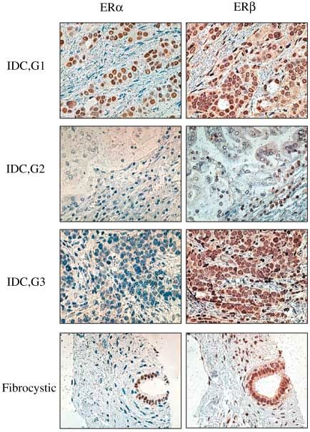
Figure 4 Estradiol binding in 8S (ERα) and 4S (ERβ) peaks in normal and breast tumors by sucrose density gradient assay. Tissue was frozen in liquid nitrogen and
pulverized in a dismembrator (Braun, Melsungen, Germany) for 45 s at 1800 r.p.m. Pulverized tissue was added to a buffer composed of 10 mM Tris–chloride, pH 7.5, 1.5
mM EDTA and 5 mM sodium molybdate, using 1 ml/100 µg tissue. Cytosol was obtained by centrifugation of the homogenate at 204 000 g for 1 h in a 70Ti rotor at 4 °C.
MCF-7 cell cytosol (for calibration of the gradient) was a generous gift from Abbott Laboratories. Breast tissue extracts were incubated for 3 h at 0 °C with 10 nM
[3H]estradiol-17β in either the presence or absence of a 50-fold excess of radio-inert 17β-estradiol, and the bound and unbound steroids were separated with dextran-coated
charcoal. Sucrose density gradients (10–30% (w/v) sucrose) were prepared in a buffer containing 10 mM Tris–HCl, 1.5 mM EDTA, 1 mM α-monothioglycerol, 10 mM KCl.
Samples of 200 µl were layered on 3.5 ml of gradients and centrifuged at 4 °C for 16 h at 300 000 g in a Beckman (Palo Alto, CA, USA) L-79K ultracentrifuge with an
SW-60Ti rotor. The figure shows high expression of ERβ in normal breasts, in benign breast disease and in medullary cancer. ERα expression in these samples is very low.
In invasive ductal cancer grade 1, ERα expression is high and ERβ low. In invasive ductal cancer type 2, both ERα and ERβ are highly expressed. In invasive ductal cancer
grade 3, the level of both ERs tends to be reduced, except for one sample which had extremely high levels of ERα.
Endocrine-Related Cancer (2002) 9 1–13
5
via free access
Downloaded from Bioscientifica.com at 06/29/2023 05:26:01AMPalmieri et al.: ERβ in breast cancer
individually, is necessary for mammary gland growth. It is the expression of another ERβ splice variant, ERβcx (Ogawa
still not clear whether there can be estrogen-induced growth et al. 1998a), a dominant negative repressor of ERα. In this
in the absence of both receptors. Such information will come variant, an alternative exon 8 is utilized (Fig. 1). This recep-
from studies on the double knockout mice. Even if ERs are tor has no measurable affinity for estradiol and, if expressed
not necessary for proliferation in the mammary gland, they together with ERα in cells, it silences ERα function (Ogawa
can still control both normal and malignant growth through et al. 1998b). It is not known whether the physiological func-
their influences on growth factor-mediated growth and tion of ERβcx is to repress ERα or whether there are, so far
apoptosis. As ERβ-/- mice age, they develop severe cystic unidentified, ligands which will confer novel functions on
breast disease, which is not found in their WT littermates. At this receptor. Whatever its functions, its expression in breast
2 years of age, all ERβ-/- mice mammary glands are cystic. cancer (see below) shows that it is one additional factor, in
A typical gland is shown in Fig. 3. the already complex estrogen story, which cannot be ignored.
Many functions have been suggested for ERβ in the
breast (Gustafsson & Warner 2000, Knowlden et al. 2000,
ER␣ and ER in the normal and
Speirs & Kerin 2000, Warner et al. 2000) but no clear picture
malignant human breast has emerged about its role in breast cancer. One group sug-
In the human breast the role of ERβ is even less well under- gested that it contributes to the initiation and progression of
stood than it is in rodents. One major confounding factor is chemical carcinogen-induced neoplastic transformation in
Figure 5 Detection of ERβ by Western blotting analysis. For Western blotting, sucrose gradient fractions were first precipitated
with TCA, and the precipitate resuspended in methanol. Samples were placed on dry ice for 30 min and the protein recovered
by centrifugation. Protein pellets were dissolved in SDS sample buffer and resolved by SDS-PAGE in 4–20% gradient gels with
a Tris–glycine buffer system. Transfer to ProBlot (Applied Biosystems, Foster City, CA, USA) membranes was done by
electroblotting. Signals were developed using SuperSignal West Femto Maximum Sensitivity Substrate from Pierce (Rockford,
IL, USA). The ERβ antibody was raised in rabbits against the ligand-binding domain of ERβ. The ERα antibody was H222 from
Abbott Laboratories. The panel illustrates the high expression of ERβ in fibrocystic disease and medullary cancer.
6 Downloaded from Bioscientifica.com at 06/29/2023 05:26:01AM
www.endocrinology.org
via free accessEndocrine-Related Cancer (2002) 9 1–13
breast because expression was induced in chemical carcino- or alongside ERα. Regarding the use of ERβ as a potential
gen-transformed human breast epithelial cells (Hu et al. novel target in the treatment of breast cancer, it is currently
1998). Other groups have shown the expression mRNAs of unclear whether agonists or antagonists will be useful. The
both ERs in normal and malignant human breast tissue by potential role of ERβ splice variants, to act as natural anti-
RT-PCR (Leygue et al. 1997, Vladusic et al. 1998, 2000, estrogens and repress ERα function, needs further investiga-
Spiers et al. 2000). Of three published immunohistochemical tion and evaluation, as this could affect the way we currently
studies, one (Leygue et al. 1997) demonstrated that those interpret ERα immunostaining to guide treatment with anti-
tumors that co-expressed ERα and ERβ were node-positive estrogens.
and tended to be of higher grade; another (Jarvinen et al.
2000), found that ERβ was often co-expressed with ERα and Unpublished results from our breast
progesterone receptor in breast cancer and that ERβ was sig-
cancer study
nificantly associated with negative axillary node status and
low tumor grade; while the third study (Mann et al. 2001) In an attempt to resolve some of the issues concerning the
found that expression of ERβ in more than 10% of cancer role of ERβ in breast cancer, we have initiated a large study
cells was associated with better survival. In one RT-PCR of ER profiles in primary and recurrent breast cancer. So far,
study there was increased expression of ERβ mRNA in ta- 63 frozen samples obtained from the histopathology archive
moxifen-resistant breast cancer patients (Speirs & Kerin at Charing Cross Hospital, London, UK have been processed.
2000) and in another study (Roger et al. 2001), there was They were composed of 33 invasive ductal carcinomas, five
decreased expression of ERβ protein in proliferative prein- medullary cancers, 14 samples from fibrocystic disease, six
vasive mammary tumors. samples of normal tissue adjacent to cancer and two in situ
Because of technical difficulties with antibodies and the ductal cancers. All samples were previously typed for ERα
need for fresh or frozen samples, there are very few studies by ligand-binding assays and/or immunohistochemistry. An-
where ERs are measured by Western blotting. However, one other twenty-four paraffin-embedded samples were from the
such study has been published and showed that full-length Helsinki University Central Hospital, Finland. They were
ERβ protein could be detected in three human breast tumors composed of five benign tumors, 25 invasive ductal cancers
of unspecified histopathology (Fuqua et al. 1999). From all and four lobular cancers. Information on patient’s age, meno-
of the data published so far, it is not yet clear how ERβ can pausal status, pathological diagnosis and differentiaton grade
be used as a routine prognostic indicator either independently was recorded. We are using multiple techniques to measure
Figure 6 Breast sample with non-estrogen-binding form of ERα. In the DCIS sample, there was estrogen binding in the 8S
peak and a corresponding ERα band on Western blots in the 8S fractions. In the fibrocystic sample there was no estrogen
binding in the 8S peak but there was a band of the correct size on Western blots.
www.endocrinology.org 7
Downloaded from Bioscientifica.com at 06/29/2023 05:26:01AM
via free accessPalmieri et al.: ERβ in breast cancer
Figure 7 Breast cancers expressing ERβcx. The two ductal cancers (LH panel, grade 1; RH panel, grade 3) shown in this
figure had no estrogen-binding 4S peak. ERβ, as detected by the LBD antibody, was found throughout the gradient. With an
ERβcx-specific antibody, ERβcx distribution in the gradient coincided with that found with the LBD antibody.
ERα and ERβ proteins in the frozen samples. Expression dient centrifugation. There is a clear difference in sedimen-
levels and binding capacity of the two ERs were measured tation profiles of the two receptors. As depicted in Fig. 4, in
by sucrose density gradient centrifugation of low-salt tissue the presence of 10 nM [3H]estradiol there are two distinct
extracts with estradiol as ligand. The proteins were analyzed peaks of estradiol binding detectable in low-salt extracts of
by Western blotting and localized in the tissues by immuno- human breast samples. As expected (Greene et al. 1977), the
histochemistry. The various ERβ isoforms were measured by 8S peak contained ERα. The unexpected finding was that the
RT-PCR. The fixed samples were examined by immuno- 4S estradiol-binding peak contained ERβ, exclusively (Fig.
histochemistry and this study has been published (Jensen et 5). Since there were clear differences between various breast
al. 2001). samples in the quantity of estradiol bound and the ratio of
One of the most unexpected and useful differences the two peaks, this method appears to be a simple and effi-
between ERα and ERβ was revealed by sucrose density gra- cient method to assess the presence of the two ERs. The
Figure 8 Immunohistochemistry. Paraffin sections (4 µm) were dewaxed in xylene and rehydrated through graded alcohol to
water. Endogenous peroxidase was blocked by incubation for 30 min with a solution of 1% hydrogen peroxide and antigen
retrieval was performed by microwaving sections in 0.01 M citrate buffer, pH 6.0, for 20 min at 800 W. ERα monoclonal
antibody (6F11) was from Novocastra (Newcastle, UK); ERβ rabbit polyclonal (06–629) was from Upstate Biotechnology (Lake
Placid, NY, USA). Biotinylated secondary antibodies, goat anti-mouse IgG, and goat anti-rabbit IgG and avidin-biotin (ABC) kits
were obtained from Vector Laboratories, Inc., Burlingame, CA, USA. Tissue sections were incubated for 1 h at 4 °C with normal
goat serum diluted 1:10 in PBS. Antibodies were diluted individually in PBS containing 3% BSA. Dilution for ERβ was 1:100 and
for ERα antibody 1:500. Sections were incubated with antibodies overnight at 4 °C. For negative controls, the primary antibody
was replaced with PBS or with primary antibodies after absorption with the corresponding antigens. Prior to addition of the
secondary antibody, sections were rinsed in PBS containing 0.05% Tween 20. The ABC method was used to visualize the
signal according to the manual provided by the manufacturer (Vector). Sections were incubated in biotinylated goat anti-rabbit
or goat anti-mouse immunoglobulin (1:200 dilution) for 2 h at room temperature, followed by washing with PBS and incubation
in avidin-biotin-horseradish peroxidase for 1 h. After thorough washing in PBS, sections were developed with 3,3′-diamino-
benzidine tetra-hydrochloride substrate (Dako, Glostrup, Denmark), slightly counterstained with Mayer’s hematoxylin, followed
by dehydration through an ethanol series, xylene and mounting. The percentage of positively stained cells was averaged after
counting the positively and negatively stained cells from four high-magnification fields with the software Image-Pro Plus (version
4.1), Media Cybernetics, Silver Spring, MD, USA. The pictures show that there are different patterns of ERα and ERβ
expression in invasive ductal cancer. In the fibrocystic tumor, ERα is expressed in the epithelial cells while ERβ is expressed in
both epithelial and stromal cells.
8 Downloaded from Bioscientifica.com at 06/29/2023 05:26:01AM
www.endocrinology.org
via free accessEndocrine-Related Cancer (2002) 9 1–13
Figure 8 See opposite page for legend.
www.endocrinology.org 9
Downloaded from Bioscientifica.com at 06/29/2023 05:26:01AM
via free accessPalmieri et al.: ERβ in breast cancer
higher sedimentation rate of ERα suggests that it is in a pressed in fractions which had positive signals with the anti-
larger multi-protein complex than is ERβ and careful exam- body raised against the ligand-binding domain of ERβ (Fig.
ination of the ERβ complex could provide useful information 7). Clearly, ERβcx is expressed as a protein in breast cancer.
about the proteins which associate with it in the cell. Furthermore, since there was no estradiol binding in the 4S
Of the breast cancer samples, 13% had an 8S peak only, peak of the sucrose gradient, it appears that no ligand-binding
42% had a 4S peak only and 31% had both peaks. In 11% form of ERβ1 is present. It is not clear whether there is no
of the samples no specific estradiol binding was observed. ERβ1 or if it is present but its binding to estradiol is inhibited
The 8S peak, ERα, was prominent (range 13–3700 fmol/mg by ERβcx. The presence of ERβcx was confirmed by RT-
protein) in invasive ductal cancer, but was not detected in PCR (Fig. 9). The ratio of ERβ1 to ERβcx mRNA varied
normal tissue or in benign breast disease. The 4S peak, ERβ, considerably between samples and in some cases ERβcx was
(range 20–475 fmol/mg protein) was present in both normal the only ERβ isoform detected.
and malignant breasts (80% of these samples were positive We conclude from these studies that ERβ is usually ex-
for ERβ). All grade 1 invasive ductal carcinomas were de- pressed in normal breasts and in benign and malignant breast
void of ERβ but had high levels of ERα. As discussed above, disease. The important remaining question is whether the
the presence of ERα in a cell might be an indication of a presence of ERβ in breast cancer could influence treatment
non-proliferative state, and this might explain why the grade of the disease. The answer to this question awaits identifica-
1 invasive ductal cancers are less aggressive. Grade 2 and 3 tion of the array of genes which are regulated by this recep-
invasive ductal cancers had various ratios of the two recep- tor. Identification of such genes is important if we are to
tors and in some samples no estrogen binding could be understand the actions of this nuclear receptor and the effect
detected. of its dysregulation in the human breast. Currently there is
From our estrogen binding data, 44% of the breast cancer not enough information available for decisions on whether
samples would be classified as ERα-positive. Interestingly, ERα and ERβ complement or oppose each other’s actions on
although ERα was present only in the 8S peak, never in the certain target genes within the breast. It is not known whether
4S peak, there were some samples in which no estradiol these receptor proteins form heterodimers in vivo or which
binding could be detected, but which had distinct and specific ERβ isoforms co-localize with ERα in cells. If ERβcx is
bands of the correct size on Western blots with H222 anti- expressed in cells with ERα, this may represent one mecha-
body (Abbott Laboratories, North Chicago, IL, USA) (Fig. nism by which ERα-positve cancers are resistant to the
6). There could be several reasons for lack of estrogen bind- actions of tamoxifen. On the other hand, if tamoxifen exerts
ing in samples which do express ERα protein such as point its beneficial effects in the treatment of breast cancer by op-
mutations, post-transcriptional modifications of the receptor posing the action of ERα, the question of whether ERβcx
or the presence of ERβ splice variants which prevent estro- functions as an ‘anti-estrogen’ in vivo must be examined. Of
gen binding by forming heterodimers with ERα (Palmieri et course, before this question can be answered, we need to
al. 2000). The reason for the lack of estradiol binding has know whether ERβ splice variants have alternative ligands
not been investigated but it does suggest that discrepancies or alternative routes of activation and whether they silence
between the ligand-binding assay and the immunodetection ERα action at sites of interaction other than on EREs.
assay can occur. This therefore requires further attention.
Western blotting confirmed what was found with the
Unanswered questions
binding data. ERα was highly expressed in invasive ductal
cancer but its expression was low in normal breasts and in There are important questions which need to be addressed
fibrocystic disease (Fig. 5). The 4S estradiol-binding peak with regard to ERβ and its splice variants in breast cancer.
always contained ERβ with a molecular mass of 60–63 kDa (i) Can their measurement be used to improve the prognostic
(Figs 5–7). value of ERα or could it even be used as an independent
Immunohistochemistry (Fig. 8) confirmed that ERα was prognostic indicator? (ii) What is the functional significance
highly expressed in grade 1 invasive ductal cancer. Its of ERβ splice variants such as ERβcx and can their presence
expression varied in grades 2 and 3 invasive ductal cancer be used to predict the likely response to hormonal therapy?
and was low in fibrocystic disease. Despite the lack of estro- (iii) Does ERβ represent a novel target in breast cancer and
gen binding in the 4S peak in grade 1 invasive ductal cancer, thus potentially allow the development of additional hor-
there was abundant, specific ERβ staining in sections of monal therapies that can add to effectiveness of existing
breasts from patients with this disease. Western blotting with agents? The answer to these questions will depend on studies
these samples confirmed that although there was no 4S estra- that look at the expression of ERβ and its variants by West-
diol-binding peak, there was abundant expression of ERβ ern blotting, PCR and immunohistochemistry. The response
protein. In order to understand this result, we developed an- to hormonal therapy and subsequent outcome will then need
other antibody, one specific for the cx exon. Western blotting to be analyzed in such patients. Such studies should, where
with this antibody showed that ERβcx was abundantly ex- possible, examine samples from patients being treated in the
10 Downloaded from Bioscientifica.com at 06/29/2023 05:26:01AM
www.endocrinology.org
via free accessEndocrine-Related Cancer (2002) 9 1–13
Figure 9 Identification of ERβ splice variants by RT-PCR in breast cancer. Total RNA was extracted from frozen breast tissue
sections using RNAwiz (Ambion, Austin, TX, USA) according to the manufacturer’s instructions, and quantified
spectrophotometrically. Five micrograms of total RNA were treated with 5 units RNase-free DNase (Promega, Madison, WI,
USA) for 60 min at 37 °C to remove genomic DNA from the RNA samples. After inactivation of DNase for 10 min at 65 °C,
samples were reverse-transcribed using SuperScript preamplification system (Life Technologies, Inc., Gaithersburg, MD, USA)
in a final volume of 20 µl according to the manufacturer’s instructions. Six primer pairs were designed to detect the following
ERβ isoforms: (1) the exon 5 deletion isoform (δ5); (2) ERβ ins; (3) ERβcx and β5. The primer sets for ERβ variants were as
follows: ERβ LBD U, 5′-GAGCTCAGCCTGTTCGACC; ERβ LBD L, 5′-GGCCTTGACACAGAGATATTC; ERβ δ5 U,
5′-ATGATGATGTCCCTGACCAAG; ERβ 1U, 5′-CGATGCTTTGGTTTGGGTGAT; E ERβ 1L, 5′-GCCCTCTTTGCTTTTACTGTC
and ERβ 2L, 5′-CTTTAGGCCACCGAGTTGATT. The location of each primer and the size of respective products are shown on
the left-hand side of the figure. β-Actin primers were 5’-CTGGCACCACACCTTCTAC for sense and
5′-GGGCACAGTGTGGGTGAC for antisense. The conditions for the PCR were as follows: 95 °C for 5 min, then 36 cycles (or
25 cycles for the β-actin primer set) at 95 °C for 30 s, 60 °C for 30 s and 72 °C for 60 s, finally 72 °C for 5 min. PCR products
were resolved on a 2% agarose gel containing ethidium bromide in 0.5 × TBE running buffer. In the negative controls the cDNA
template was replaced with DNase-treated RNA, and the identity of the positive band was confirmed by direct sequencing of the
PCR product or by cloning the PCR product into a TA cloning vector, selection of clones and sequencing of the insert from the
vector. None of the tested samples had LBD insertion isoform. The δ5 variant of ERβ was detected in samples 5, 16 and 21,
which had no estradiol binding on sucrose gradients, as well as in 2 which had no estradiol binding in the 8S peak. Triple
primer PCR, used for evaluating the ratio of the various isoforms (middle panel), revealed that the expression of C-terminally
truncated isoforms is common. Patient 9 (invasive ductal carcinoma, grade 3) had exclusively ERβcx and had very high binding
of estradiol in the 8S peak. Sample 14, which had estradiol binding in the 4S peak on sucrose gradients, had no WT C-terminal
sequence.
neoadjuvant, adjuvant and metastatic setting, as well as based on targeting ERβ, but before this can happen the above
patients who develop recurrent breast cancer during or after questions need to be answered.
completing adjuvant hormonal treatment. With results from
such detailed studies, it may be possible in the future that,
based on ERα, ERβ and ERβ splice variant status, patients
Acknowledgements
can be stratified for their likely response to hormonal therapy.
In addition, there is the potential for such information to as- The patient and very skilful work of Christina Thulin-
sist in identification of patients who may develop hormone- Andersson is gratefully acknowledged. This research was
independent breast cancer. The future also holds the supported by grants from the Swedish Cancer Fund, the UK
possibility of a new era in the therapy of hormonal treatment Cancer Research Campaign, and by KaroBio AB, Sweden.
www.endocrinology.org 11
Downloaded from Bioscientifica.com at 06/29/2023 05:26:01AM
via free accessPalmieri et al.: ERβ in breast cancer
References Greene GL, Closs LE, Fleming H, DeSombre ER & Jensen EV
1977 Antibodies to estrogen receptor: immunochemical
Ali S & Coombes RC 2000 Estrogen receptor alpha in human similarity of estrophilin from various mammalian species. PNAS
breast cancer: occurrence and significance. Journal of Mammary 74 3681–3685.
Gland Biology and Neoplasia 5 271–281. Gustafsson J-Å & Warner M 2000 Estrogen receptor beta in the
Allegra JC & Lippman ME 1978 Growth of a human breast cancer breast: role in estrogen responsiveness and development of
cell line in serum-free hormone-supplemented medium. Cancer breast cancer. Journal of Steroid Biochemistry and Molecular
Research 38 3823–3829. Biology 74 245–248.
Allegra JC & Lippman ME 1980 Estrogen receptor determination Hanstein B, Liu H, Yancisin M & Brown M 1999 Functional
predicts response to tamoxifen therapy. Recent Results in Cancer analysis of a novel estrogen receptor-β isoform. Molecular
Research 71 16–19. Endocrinology 13 129–137.
Batistuzzo de Medeiros SR, Krey G, Hihi AK & Wahli W 1997 Hu YF, Lau KM, Ho SM & Russo J 1998 Increased expression of
Functional interaction between the estrogen receptor and the estrogen receptor beta in chemically transformed human breast
transcription activator Sp1 regulate the estrogen-dependent epithelial cells. International Journal of Oncology 12 1225–
transcriptional activity of the vitellogenin A1 promoter. Journal 1228.
of Biological Chemistry 272 18250–18260. Hulka BS & Stark AT 1995 Breast cancer: cause and prevention.
Chalbos D, Vignon F, Keydar I & Rochefort H 1982 Estrogens Lancet 346 883–887.
stimulate cell proliferation and induce secretory proteins in a Iwao K, Miyoshi Y, Egawa C, Ikeda N, Tsukamoto F & Noguchi
human breast cancer cell line (T47D). Journal of Clinical S 2000 Quantitative analysis of estrogen receptor-alpha and -beta
Endocrinology and Metabolism 55 276–283. messenger RNA expression in breast carcinoma by real-time
Cheng GJ, Mäkinen S, Weihua Z, Mäkelä S, Saji S, Warner M, polymerase chain reaction. Cancer 89 1732–1738.
Gustafsson J-Å & Hovatta O 2002. A role for the androgen Jarvinen TA, Pelto-Huikko M, Holli K & Isola J 2000 Estrogen
receptor in follicular atresia of estrogen receptor beta knockout receptor beta is coexpressed with ERalpha and PR and
mouse ovary. Biology of Reproduction 66 77–84. associated with nodal status, grade, and proliferation rate in
Chu S & Fuller PJ 1997 Identification of a splice variant of the rat breast cancer. American Journal of Pathology 156 29–35.
estrogen receptor beta gene. Molecular and Cellular Jeng MH, Jiang SY & Jordan VC 1994 Paradoxical regulation of
Endocrinology 132 195–199. estrogen-dependent growth factor gene expression in estrogen
Clarke RB, Howell A, Potten CS & Anderson E 1997a receptor (ER)-negative human breast cancer cells stably
Dissociation between steroid receptor expression and cell expressing ER. Cancer Letters 82 123–128.
proliferation in the human breast. Cancer Research 57 4987– Jensen EV, Cheng G, Palmieri C, Saji S, Mäkelä S, Van Noorden
4991. S, Wahlstrom T, Warner M, Coombes RC & Gustafsson J-Å
Clarke RB, Howell A & Anderson E 1997b Estrogen sensitivity of 2001 Estrogen receptors and proliferation markers in primary
normal human breast tissue in vivo and implanted into athymic and recurrent breast cancer. PNAS 98 15197–15202.
nude mice: analysis of the relationship between estrogen-induced Jiang SY & Jordan VC 1992 Growth regulation of estrogen
proliferation and progesterone receptor expression. Breast receptor-negative breast cancer cells transfected with
Cancer Research and Treatment 45 121–133. complementary DNAs for estrogen receptor. Journal of the
Couse JF & Korach KS 1999 Estrogen receptor null mice: what National Cancer Institute 84 580–591.
have we learned and where will they lead us? Endocrine Knowlden JM, Gee JM, Robertson JF, Ellis IO & Nicholson RI
Reviews 20 358–417. 2000 A possible divergent role for the oestrogen receptor alpha
Couse JF, Curtis Hewitt S & Korach KS 2000 Receptor null mice and beta subtypes in clinical breast cancer. International Journal
reveal contrasting roles for estrogen receptor alpha and beta in of Cancer 89 209–212.
reproductive tissues. Journal of Steroid Biochemistry and Korach KS 2000 Estrogen receptor knock-out mice: molecular and
Molecular Biology 74 287–296. endocrine phenotypes. Journal of the Society for Gynecologic
Cullen R, MaGuire T, Diggin P, Hill A, McDermott E, O’Higgins Investigation 7 S16–S17.
N & Duffy MJ 2000 Detection of estrogen receptor-beta mRNA Krege JH, Hodin JB, Couse JF, Enmark E, Warner M, Mahler JF,
in breast cancer using RT-PCR. International Journal of Sar M, Korach KS, Gustafsson J-Å & Smithies O 1998
Biological Markers 15 114–115. Generation and reproductive phenotypes of mice lacking
Dotzlaw H, Leygue E, Watson PH & Murphy LC 1997 Expression estrogen receptor beta. PNAS 95 15677–15682.
of estrogen receptor-beta in human breast tumors. Journal of Kuiper GG, Enmark E, Pelto-Hhuikko M, Nilsson S & Gustafsson
Clinical Endocrinology and Metabolism 82 2371–2374. J-Å 1996 Cloning of a novel receptor expressed in rat prostate
Fuqua SA, Schiff R, Parra I, Friedrichs WE, Su JL, McKee DD, and ovary. PNAS 93 5925–5930.
Slentz-Kesler K, Moore LB, Willson TM & Moore JT 1999 Levenson AS & Jordan VC 1994 Transfection of human estrogen
Expression of wild-type estrogen receptor beta and variant receptor (ER) cDNA into ER-negative mammalian cell lines.
isoforms in human breast cancer. Cancer Research 59 5425– Journal of Steroid Biochemistry and Molecular Biology 51 229–
5428. 239.
Galienand R & Garcia T 1997 Estrogen receptor impairs Leygue E, Watson PH & Murphy LC 1997 Expression of estrogen
interleukin-6 expression by preventing protein binding on the receptor-beta in human breast tumors. Journal of Clinical
NF-kappaB site. Nucleic Acids Research 25 2424–2429. Endocrinology and Metabolism 82 2371–2374.
Garcia M, Derocq D, Freiss G & Rochefort H 1992 Activation of Lonard DM, Nawaz Z, Smith CL & O’Malley BW 2000 The 26S
estrogen receptor transfected into a receptor-negative breast proteasome is required for estrogen receptor-alpha and
cancer cell line decreases the metastatic and invasive potential of coactivator turnover and for efficient estrogen receptor-alpha
the cells. PNAS 89 11538–11542. transactivation. Molecular Cell 5 939–948.
12 Downloaded from Bioscientifica.com at 06/29/2023 05:26:01AM
www.endocrinology.org
via free accessEndocrine-Related Cancer (2002) 9 1–13
McGuire WL, Osborne CK, Clark GM & Knight WA III 1982 Roger P, Sahla ME, Makela S, Gustafsson J-Å, Baldet P &
Steroid hormone receptors and carcinoma of the breast. Rochefort H 2001 Decreased expression of estrogen receptor
American Journal of Physiology 243 E99–E102. beta protein in proliferative preinvasive mammary tumors.
Mann S, Laucirica R, Carlson N, Younes PS, Ali N, Younes A, Li Cancer Research 61 2537–2541.
Y & Younes M 2001 Estrogen receptor beta expression in Saji S, Jensen EV, Nilsson S, Rylander T, Warner M & Gustafsson
invasive breast cancer. Human Pathology 32 113–118. J-Å 2000 Estrogen receptors alpha and beta in the rodent
Maruyama K, Endoh H, Sasaki-Iwaoka H, Kanou H, Shimaya E, mammary gland. PNAS 91 337–342.
Hashimoto S, Kato S & Kawashima H 1998 A novel isoform of Saji S, Sakaguchi H, Andersson S, Warner M & Gustafsson J-Å
rat estrogen receptor beta with 18 amino acid insertion in the 2001 Quantitative analysis of estrogen receptor proteins in rat
ligand binding domain as a putative dominant negative regulator mammary gland. Endocrinology 142 3177–3186.
of estrogen action. Biochemical and Biophysical Research Speirs V & Kerin MJ 2000 Prognostic significance of oestrogen
Communications 246 142–147. receptor beta in breast cancer. British Journal of Surgery 87
Moore JT, McKee DD, Slentz-Kesler K, Moore LB, Jones SA, 405–409.
Horne EL, Su JL, Kliewer SA, Lehmann JM & Willson TM Speirs V, Malone C, Walton DS, Kerin MJ & Atkin SL 1999
1998 Cloning and characterization of human estrogen receptor Increased expression of estrogen receptor beta mRNA in
beta isoforms. Biochemical and Biophysical Research tamoxifen-resistant breast cancer patients. Cancer Research 59
Communications 247 75–78. 5421–5424.
Neuman E, Ladh MH, Lin N, Upton TM, Miller SJ, Direnzo J, Tsai M & O’Malley BW 1994 Molecular mechanisms of action of
Pestell RG, Hinds PW, Dowdy SF, Brown M & Ewen ME 1997 steroid/thyroid receptor superfamily members. Annual Review of
Cyclin D1 stimulation of estrogen receptor transcriptional Biochemistry 63 451–486.
activity independent of cdk4. Molecular and Cellular Biology 17 Vignon F, Bouton MM & Rochefort H 1987 Antiestrogens inhibit
5338–5347. the mitogenic effect of growth factors on breast cancer cells in
Ogawa S, Inoue S, Watanabe T, Orimo A, Hosoi T, Ouchi T & the total absence of estrogens. Biochemical and Biophysical
Muramatsu 1998a Molecular cloning and characterization of Research Communications 146 1502–1508.
human estrogen receptor βcx: a potential inhibitor of estrogen Vladusic EA, Hornby AE, Guerra-Vladusic FK & Lupu R 1998
action in human. Nucleic Acids Research 26 3505–3512. Expression of estrogen receptor beta messenger RNA variant in
Ogawa S, Inoue S, Watanabe S, Hiroi H, Orimo A, Hosoi T, breast cancer. Cancer Research 58 210–214.
Ouchi Y & Muramatsu M 1998b The complete primary structure Vladusic EA, Hornby AE, Guerra-Vladusic FK, Lakins J & Lupu
of human estrogen receptor β(hEβ) and its heterodimerization R 2000 Expression and regulation of estrogen receptor beta in
with ERα in vivo and in vitro. Biochemical and Biophysical human breast tumors and cell lines. Oncology Reports 7 157–
Research Communications 243 122–126. 167.
Ogawa S, Inoue S, Orimo A, Hosoi T, Ouchi Y & Muramatsu M Warner M, Saji S & Gustafsson J-Å 2000 The normal and
1998c Cross-inhibition of both estrogen receptor alpha and beta malignant mammary gland: a fresh look with ER beta on
pathways by each dominant negative mutant. FEBS Letters 423 board. Journal of Mammary Gland Biology and Neoplasia 5
129–132. 289–294.
Omoto Y, Inoue S, Ogawa S, Toyama T, Yamashita H, Muramatsu Weihua Z, Mäkelä S, Andersson LC, Salmi S, Saji S, Webster I,
M, Kobayashi S & Iwase H 2001 Clinical value of the wild-type Jensen EV, Nilsson S, Warner M & Gustafsson J-Å 2001 A role
estrogen receptor beta expression in breast cancer. Cancer for estrogen receptor β in the regulation of growth of the ventral
Letters 163 207–212. prostate. PNAS 98 6330–6335.
Paech K, Webb P, Kuiper GG, Nilsson S, Gustafsson J-Å, Kushner Wiesen JF, Young P, Werb Z & Cunha GR 1999 Signaling
PJ & Scalan TS 1997 Differential ligand activation of estrogen through the stromal epidermal growth factor receptor is
receptors ERalpha and ERbeta at AP1 sites. Science 277 1508– necessary for mammary ductal development. Development 126
1510. 335–344.
Palmieri C, Saji S, Gustafsson J-Å & Coombes RC 2000 False Zeps N, Bentel JM, Papadimitriou JM & Dawkins HJ 1999 Murine
negatives in oestrogen-receptor assay. Lancet 356 944. progesterone receptor expression in proliferating mammary
Porter W, Saville B, Hoivik D & Safe S 1999 Functional synergy epithelial cells during normal pubertal development and
between the transcription factor Sp1 and the estrogen receptor. adult estrous cycle. Association with ERalpha and ERbeta
Molecular Endocrinology 11 1569–1580. status. Journal of Histochemistry and Cytochemistry 47 1323–
Ricketts D, Turnbull L, Ryall G, Bakhshi R, Rawson NS, Gazet 1330.
JC, Nolan C & Coombes RC 1991 Estrogen and progesterone Zwijsen RM, Wientjens E, Klompmaker R, Van der Sman J,
receptors in the normal female breast. Cancer Research 51 Bernards R & Michalides RJ 1997 CDK-independent activation
1817–1822. of estrogen receptor by cyclin D1. Cell 88 405–415.
www.endocrinology.org 13
Downloaded from Bioscientifica.com at 06/29/2023 05:26:01AM
via free accessYou can also read