Therapeutic Effect of Rapamycin-Loaded Small Extracellular Vesicles Derived from Mesenchymal Stem Cells on Experimental Autoimmune Uveitis
←
→
Page content transcription
If your browser does not render page correctly, please read the page content below
ORIGINAL RESEARCH
                                                                                                                                               published: 29 March 2022
                                                                                                                                       doi: 10.3389/fimmu.2022.864956

                                           Therapeutic Effect of Rapamycin-
                                           Loaded Small Extracellular Vesicles
                                           Derived from Mesenchymal Stem
                                           Cells on Experimental
                                           Autoimmune Uveitis
                                           Huan Li †, Zhihui Zhang †, Yongtao Li †, Lin Su , Yanan Duan , Hui Zhang , Jinying An ,
                                           Tianwen Ni , Xiaorong Li and Xiaomin Zhang *

                                           Tianjin Key Laboratory of Retinal Functions and Diseases, Tianjin Branch of National Clinical Research Center for Ocular
                                           Disease, Eye Institute and School of Optometry, Tianjin Medical University Eye Hospital, Tianjin, China
                           Edited by:
                        Guangming Li,
 University of Maryland, United States
                                           Autoimmune uveitis is a major cause of vision loss and glucocorticoids are major
                       Reviewed by:
                                           traditional medications, which may induce serious complications. Rapamycin has been
                            Liyun Zou,
         Army Medical University, China    demonstrated to exhibit immunosuppressive effects and is promising to be used in
                                  He Li,   treating uveitis by intravitreal injection. However, repeated and frequent intravitreal
          Sun Yat-sen University, China
                                           injections increase the risk of severe ocular complications, while the efficacy of
                    *Correspondence:
                        Xiaomin Zhang
                                           subconjunctival injection of rapamycin is low since it is difficult for rapamycin to
                 xzhang08@tmu.edu.cn       penetrate eyeball. Recently, small extracellular vesicles (sEVs) have attracted
     †
      These authors have contributed       considerable research interest as natural drug delivery systems that can efficiently cross
                 equally to this work
                                           tissues and biological membranes. SEVs derived from mesenchymal stem cells (MSC-
                  Specialty section:
                                           sEVs) also can exert immunosuppressive effect and ameliorate experimental autoimmune
        This article was submitted to      uveitis (EAU). The aim of this study was to construct a Rapamycin-loaded MSC-sEVs
Vaccines and Molecular Therapeutics,       delivery system (Rapa-sEVs) and investigate its therapeutic effect on EAU by
              a section of the journal
             Frontiers in Immunology       subconjunctival injection. Rapa-sEVs were prepared by sonication and characterized by
           Received: 29 January 2022       nanoparticle tracking analysis, transmission electron microscopy, and western blotting.
            Accepted: 07 March 2022        Clinical and histological scores were obtained to assess the treatment efficacy.
           Published: 29 March 2022
                                           Additionally, T cell infiltration was evaluated by flow cytometry. The results indicated
                              Citation:
             Li H, Zhang Z, Li Y, Su L,
                                           that Rapa-sEVs could reach the retinal foci after subconjunctival injection. Compared to
    Duan Y, Zhang H, An J, Ni T, Li X      sEVs and rapamycin alone, Rapa-sEVs can produce a more marked therapeutic effect
      and Zhang X (2022) Therapeutic       and reduce ocular inflammatory cell infiltration. Overall, MSC-sEVs have significant
   Effect of Rapamycin-Loaded Small
         Extracellular Vesicles Derived    potential for the delivery of rapamycin to treat EAU. Subconjunctival injection of Rapa-
    from Mesenchymal Stem Cells on         sEVs may be contender for efficacious steroid-sparing immunomodulatory therapy.
   Experimental Autoimmune Uveitis.
          Front. Immunol. 13:864956.       Keywords: experimental autoimmune uveitis (EAU), uveitis, mesenchymal stem cells, small extracellular vesicles
   doi: 10.3389/fimmu.2022.864956          (sEVs), rapamycin

Frontiers in Immunology | www.frontiersin.org                                      1                                         March 2022 | Volume 13 | Article 864956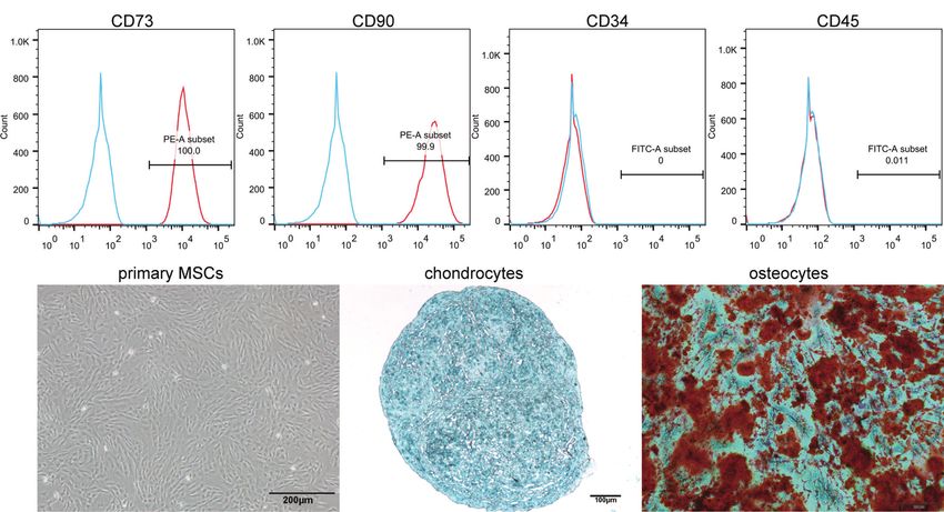
Li et al.                                                                                                 Rapamycin-Loaded sEVs Alleviated EAU

INTRODUCTION                                                              effects of MSC-sEVs in various animal models. In our previous
                                                                          studies, we showed that MSC-sEVs could ameliorate
Autoimmune uveitis is a common inflammatory ocular disease                 experimental autoimmune uveitis (EAU) in rats (33).
that often causes vision loss or blindness due to macular edema,          Therefore, they are ideal carriers for drugs targeting the retina
ocular hypertonia, and retinal ischemia (1). It is an intractable         and can be used to deliver drugs to treat autoimmune uveitis. In
problem in the treatment of uveitis in the chronic recurrent stage.       this study, we attempted to formulate Rapamycin-loaded sEVs
Glucocorticoids (GCs) are traditional medications. However,               (Rapa-sEVs) and investigate their effects in treating EAU.
prolonged use of high-dose GCs may induce systemic side
effects, including hypertension, hyperlipidemia, and diabetes (2,
3). Local application of GCs allows a smaller dose and reduces
systemic side effects, but patients may develop cataracts, ocular         MATERIALS AND METHODS
hypertension, or glaucoma (2–4). These adverse reactions limit the
clinical use of GCs in uveitis. Thus, there is a pressing need for        Animals
efficacious steroid-sparing immunomodulatory therapies.                    Female C57BL/6J mice (6–8 weeks old) with no ocular or
    Rapamycin (also known as sirolimus) is a bacteria-derived             systemic diseases were purchased from Vital River (Beijing,
immunosuppressing agent. It is an inhibitor of mammalian                  China). All protocols involving mice were approved by the
targets of rapamycin (mTOR), which can suppress T-cell                    Animal Care and Use committee of Tianjin Medical University
proliferation by binding to FK-506-binding protein (FKBP)-12              Eye Hospital and conformed to the ARVO Statement for the Use
(5). Therefore, it is thought to be useful for the treatment              of Animals in Ophthalmic and Vision Research.
autoimmune uveitis (6, 7). Unfortunately, systemic administration
of rapamycin is associated with adverse effects, including                Culture and Identification of MSCs
cytotoxicity, particularly in hematologic diseases (8–13). In             Human umbilical cord MSCs were provided by Beijing Beilai
recent years, some clinical studies have assessed the efficacy             Biological Co., Ltd. (China), and MSC isolation and culture were
and safety of intravitreal rapamycin for the management of                performed as previously described (33). Umbilical cords
uveitis (14–17). Many patients with uveitis have a prolonged              obtained from normal pregnancies without complications after
course and are vulnerable to recurrence. They may necessitate             cesarean section delivery were placed immediately in saline
repeated and frequent intravitreal injections, which can result           containing penicillin (100U/mL) and streptomycin (100mg/mL)
in several complications such as endophthalmitis, retinal tears or        (Gibco, USA), then transported to the laboratory within 6 h.
detachment, and vitreous hemorrhage (18). Subconjunctival                 After removing residual blood and the blood vessels, the
injection can reduce the aforementioned risks, but this route             obtained Wharton’s jelly was cut into 1-3 mm3 pieces and
of drug delivery may not be effective. Douglas et al. found that          digested with 0.1% type-2 collagenase (Gibco) and 0.125%
rapamycin could only be detected in the vitreous humor of                 trypsin (Gibco) at 37°C for 1 h. The suspension was then
horses until 21 days after subconjunctival injection (19).                filtered through a 100-mesh screen to remove the undigested
Therefore, the ability of rapamycin to penetrate the intraocular          tissue. The supernatant from the filtration was centrifuged and
tissues should be enhanced when subconjunctival injection                 washed three times with PBS. The cell precipitate was
is applied.                                                               resuspended in Dulbecco’s modified Eagle’s medium/nutrient
    In recent years, some studies have attempted to employ drug           mixture F12 (DMEM/F12, Invitrogen, USA) complete medium.
carriers to improve rapamycin’s ability to penetrate the blood-           The medium contained 10% fetal bovine serum (FBS;
retinal barrier, thereby enhancing its therapeutic efficacy for            Invitrogen), 100 U/ml penicillin, and 100 mg/ml streptomycin.
ocular posterior segment diseases. These carriers include                 The cells were seeded in a T175 flask and cultured at 37°C in a
micelles, liposomes, niosomes, and different polymeric vesicles           5% CO2 incubator. The medium was changed every 3 days.
(20–22). Nonetheless, these carriers are derived from xenobiotic          When cell fusion reached 80%, the passage was carried out at a
materials and are vulnerable to clearance by the mononuclear              subculture ratio of 1:2, and cells from P3 to P5 were used
phagocyte system. Small extracellular vesicles (sEVs) are                 for experiments.
biological nanoparticles with a bilipid membrane secreted by                  According to the criteria proposed by ISCT (34), flow
different cell types with a diameter under 200 nm, the main               cytometry was used to identify the surface markers of MSCs,
components of which are exosomes. They are ideal carriers for             including CD73 (eBioscience, USA), CD90 (eBioscience), CD45
drug delivery because of their distinctive advantages, including          (eBioscience), and CD34 (eBioscience). Meanwhile, the
small particle size, high biocompatibility, biological barrier            osteogenic and chondrogenic differentiation abilities of MSCs
penetration, and low immunogenicity (23–25). SEVs have                    were demonstrated by staining in vitro.
already been successfully tested for loading several drugs, such
as curcumin (26, 27), paclitaxel (28, 29), and doxorubicin (30,           Isolation of MSC-sEVs
31). Mesenchymal stem cells (MSCs) are the most prolific                   To generate exosome-free FBS, FBS was centrifuged overnight
producers of sEVs (32). SEVs derived from MSCs (MSC-sEVs)                 (approximately 14 h) at 110,000 × g at 4°C. When cell fusion
have similar functions as MSCs, including promoting tissue                reached 60%, the cells were cultured in complete medium with
regeneration, inhibiting autoimmune responses, and protecting             10% exosome-free FBS for 24 h. Then, supernatants were
neuron function. There have been some encouraging therapeutic             collected and sEVs were isolated by ultracentrifugation at 4°C.

Frontiers in Immunology | www.frontiersin.org                         2                                 March 2022 | Volume 13 | Article 864956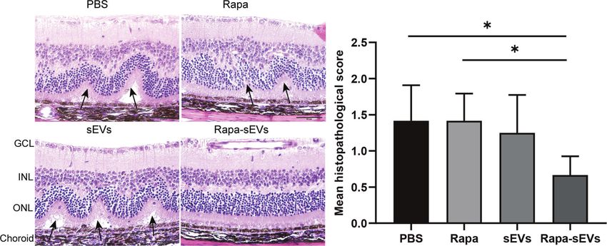
Li et al.                                                                                               Rapamycin-Loaded sEVs Alleviated EAU

Specific steps include 300 × g for 10 min, 2000 × g for 20 min,          water (Vacetonitrile/Vwater= 65/35) were used at a flow rate
10,000 × g for 30 min, and 110,000 × g for 2h, followed by              of 1ml/min. The detection wavelength was 278 nm, and the
filtration through a 0.22-mm filter. In all experiments, sEVs were        column temperature was 62°C. The drug loading capacity was
used immediately after ultracentrifugation. The BCA protein             calculated according to the formula: Loading Capacity (%) =
assay kit (Solarbio, China) was used to measure the total protein       (Drug entrapped)/(Drug entrapped + The total mass of sEVs) ×
content of the sEVs.                                                    100%. The encapsulation efficiency was calculated as follows:
                                                                        Encapsulation Efficiency (%) = (Drug entrapped)/(Total amount
Drug Entrapment Into sEVs                                               of drug) × 100%.
Rapa-sEVs were prepared by sonication. Rapamycin (Sigma-
Aldrich, USA) was mixed with sEVs at a 9:1 concentration ratio.         Induction of EAU and Treatment Methods
The mixture was incubated for 10 min and sonicated using an             Mice were immunized with a subcutaneous injection of 200 ml
ultrasonic cell crusher (25% power, 6 cycles of a 30 s pulse/30 s       emulsion containing 250 mg human interphotoreceptor retinoid
pause). The mixture was incubated at 37°C for 1 h to allow the          binding protein peptide [LAQGAYRTAVDLESLASQLT
recovery of the sEV membrane. Ultrafiltration centrifugation was         (hIRBP651-670), Shanghai Hanhong Chemical Co., Ltd.,
used to remove the free rapamycin.                                      Shanghai, China] and 3.5 mg Mycobacterium tuberculosis (BD
                                                                        Biosciences) emulsified with complete Freund’s adjuvant (CFA,
Characterization of sEVs and Rapa-sEVs                                  Sigma Aldrich). Pertussis toxin (PTX, List Biological
SEVs and Rapa-sEVs were characterized according to the criteria         Laboratories Inc.) was administered intraperitoneally at 0.5 mg/
proposed by members of four societies (SOCRATES, ISEV, ISCT             dose, and repeated 24 h later. The mice were injected
and ISBT) (35). Nanoparticle-tracking analysis (NTA) was used           subconjunctivally with sEVs and Rapa-sEVs at day 11 and day
to analyze the size distribution of sEVs and Rapa-sEVs. The             16 following immunization. Approximately 10 µL of suspension
particle size was analyzed using NTA software (version 3.3,             (the protein concentration of sEVs was 1mg/ml) was delivered
Nanosight). To further observe the size and morphology of               with a 33 gauge needle per injection. Mice in the control group
these particles, transmission electron microscopy (TEM) was             were injected with the same volume of PBS and rapamycin the
performed. First, samples were fixed with paraformaldehyde for           drug content was same as Rapa-sEV treated group.
5 min and loaded onto Formvar-coated grids. Phosphotungstic
acid was used for negative staining. Finally, the samples were          Trace of sEVs and Rapa-sEVs
dried and observed by TEM at 80.0Kv. Markers on the surface of          SEVs were labelled with PKH26 fluorescent dye (PKH26
sEVs and Rapa-sEVs, such as CD9, CD63, and TSG-101, were                fluorescent cell linker kit, Sigma-Aldrich). The immunized
detected by western blot. Proteins were extracted from normal           mice were injected subconjunctivally with sEVs and Rapa-sEVs
sEVs (N-sEVs), sonicated sEVs (S-sEVs), and Rapa-sEVs using a           on day 18 following immunization. The mice were sacrificed
cell lysis buffer. The BCA Protein Assay Kit was used to                after 24 h (n=3) and 48 h (n=3), and the eyes were processed for
determine the total protein concentrations of the three                 frozen sections. After staining with DAPI (Sigma-Aldrich), the
samples, according to the manufacturer’s recommendations.               sections were observed under a confocal microscope.
The samples were then boiled at 95°C for 5 min. 20 mg of
protein was electrophoresed and transferred to a polyvinylidene         Clinical and Histological Assessment
difluoride (PVDF) membrane. Membranes were blocked with                  On day 9 post-immunization, the fundi of the mice (each group,
5% non-fat dried milk and incubated with primary antibodies             n=6) were observed by mydriatic binocular indirect ophthalmoscopy
overnight at 4°C. The primary antibodies included antibodies            on alternate days. On day 21 post-immunization, the mice (each
against CD9 (Abcam, UK), CD63 (Abcam), TSG-101 (Abcam),                 group, n=6) were sacrificed by cranio-cervical dislocation. Their
and b-actin (Abcam). The membranes were then incubated with             eyes were collected and embedded in paraffin, sectioned
secondary antibodies for 2 h. Protein bands were visualized using       (4mm), and stained with hematoxylin and eosin (HE). Clinical
Western Lightening Chemiluminescence reagents.                          and histological grades were scored according to the criteria
   The concentration of rapamycin in Rapa-sEVs was detected             reported by Caspi et al (36).
by high performance liquid chromatography (HPLC) (Thermo
Scientific UltiMate 3000 series). A series of rapamycin at               Optical Coherence Tomography
concentrations ranging from 0 to 50 µg/mL were prepared to              On day 18 post-immunization, the mice (each group, n=6) were
generate the standard curve. An aliquot of 20 ml of standard            systemically anesthetized. The pupils were anesthetized and
solution and sample solution was injected into the HPLC system          dilated with 0.1% tropicamide. The mice were weared using a
for analysis. Taking the average peak area as transverse                corneal contact lens. Spectralis optical coherence tomography
coordinate and the concentration of rapamycin as longitudinal           (OCT) (Heidelberg, Germany) was used to scan the retina.
coordinate, the standard curve was drawn and the regression             Scores were recorded according to the criteria described by
equation was calculated. Acetonitrile was added to Rapa-sEVs to         Gadjanski’s group (37).
precipitate the proteins of sEVs and extract rapamycin. This
solution was centrifuged, and the supernatant was used for              Flow Cytometry Analysis
HPLC. The chromatographic column was a C18 reverse phase                On day 18 post-immunization, eyes and cervical draining lymph
column. For the mobile phase, HPLC-grade acetonitrile and               nodes of EAU mice (each group, n=3) were collected, and cell

Frontiers in Immunology | www.frontiersin.org                       3                                 March 2022 | Volume 13 | Article 864956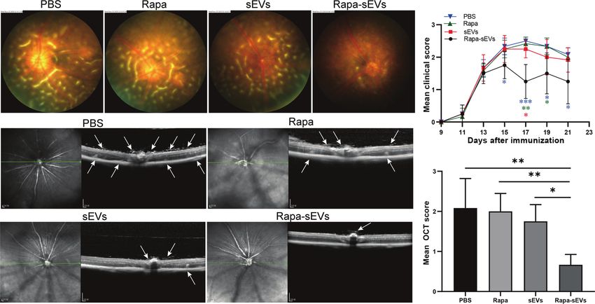
Li et al. Rapamycin-Loaded sEVs Alleviated EAU
suspensions were prepared. The cells were surface stained with in different differentiation media (Figure 1B). The sizes of the
anti-CD4 antibody (BioLegend) for 30 min at 4°C. After fixation sEVs were analyzed by NTA. The size distribution of N-sEVs, S-
and permeabilization, the cells were stained with an anti-Foxp3 sEVs and Rapa-sEVs ranged from 50 to 200nm (Figure 2A).
antibody (BioLegend) to detect Foxp3+ cells. For intracellular TEM showed that sEVs were cup-shaped in all three groups
staining of interferon (IFN) -g and interleukin (IL)-17, cells were (Figure 2B).
were pretreated for 4 to 6 h with 50 ng/ml photoblog 12-myristate Western blot analysis confirmed that the vesicles expressed
13w-acetate, 1mg/ml ionomycin, and 1mg/ml brefeldin A (Sigma- sEVs markers including CD63, CD9, and TSG101 (Figure 2C),
Aldrich), and then incubated with antibodies against IFN-g and indicating that most of the vesicles were sEVs.
IL-17 (BioLegend) after fixation and overnight permeabilization. The drug loading capacity and encapsulation efficiency were
Data collection was performed on a FACS Calibur flow cytometer analyzed by HPLC. The results showed that a good shape peak
(BD Biosciences, USA), and analyzed using flow cytometry could be detected at 16.773 min (Figure 3B). The linear
software (FlowJo, USA). regression equation of rapamycin standard curve was
Y=0.0008X+0.0006, R2 = 0.9985(Figure 3A). The drug loading
Statistical Analysis capacity of rapamycin in Rapa-sEVs was 45.7 ± 1.3%, and the
For all experiments, data are presented as mean ± standard encapsulation efficiency was 82.1 ± 4.3%.
deviation (SD). Tests for significance of differences between the
groups were performed using the Kruskal-Wallis test or one-way SEVs Penetrated Ocular Wall and Reached
analysis of variance (one-way ANOVA) in GraphPad Prism 8.0 the Retina Rapidly
(GraphPad Software, USA). A minimum p value of 0.05 was SEVs are the natural drug carriers. The ability of sEVs to deliver
chosen as the significance level. drugs to retina is essential for therapeutic efficiency. 24 h after
subconjunctival injection of sEVs (Figure 4A) and Rapa-sEVs
(Figure 4B), red fluorescence (PKH-26) was detected in the
RESULTS retina foci by confocal microscopy. It was still visualized 48 h
post-injection (Figures 4C, D) but decayed compared to 24 h
Identification of MSCs and sEVs after injection.
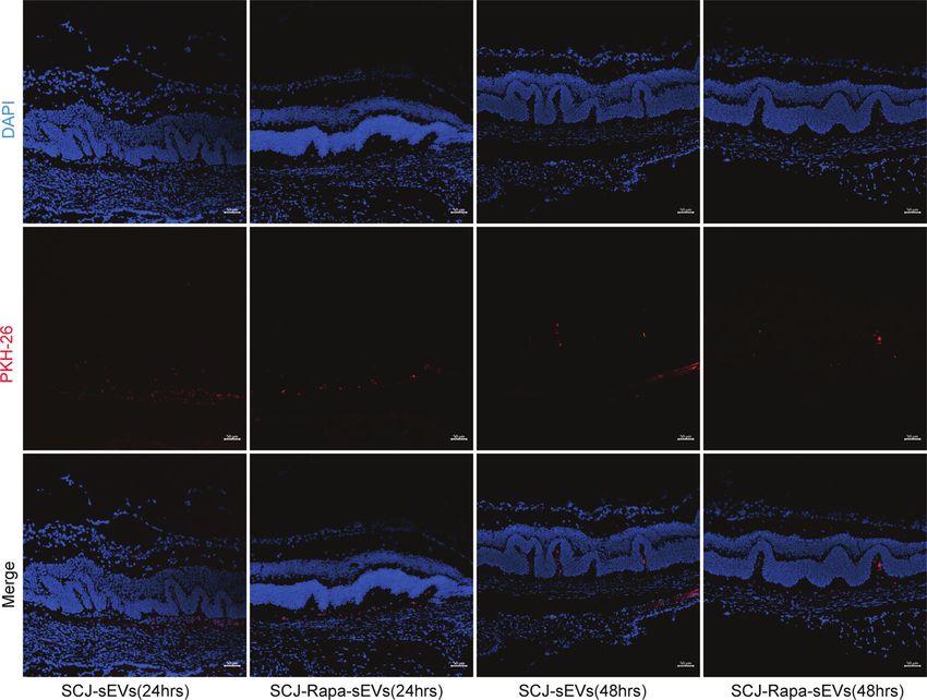
The surface antigens of MSCs were identified by flow cytometry.
The expression of CD73 and CD90, and the absence of CD34 and Subconjunctival Injection of Rapa-sEVs
CD45 were confirmed (Figure 1A). Under light microscopy, the Ameliorated Uveitis in Mice
primary MSCs were plastic-adherent and spindle-shaped EAU mice were treated twice at day 11 and 16 post
(Figure 1B). In addition, MSCs were functionally characterized immunization. The clinical scores of mice treated with Rapa-
by differentiation into osteogenic and chondrogenic phenotypes sEVs decreased significantly from day 15 compared to those in
A
B
FIGURE 1 | Identification of MSCs. (A) Immunophenotypic characterization of hMSCs was performed by flow cytometry. The vast majority of cells (>99%) were
positive for CD73 and CD90, but a few cells (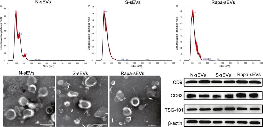
Li et al.                                                                                                                Rapamycin-Loaded sEVs Alleviated EAU

            A

            B                                                                                     C

  FIGURE 2 | Identification of sEVs. (A) NTA displayed that the diameters of most sEVs in each group were around 100nm. (B) TEM showed that sEVs in each group
  were 50–200 nm disc-like vesicles with bilayer membrane. (C) Western blot analysis showed that sEVs of all groups express CD9, CD63, and TSG101.

all other groups (Kruskal-Wallis test) (Figure 5B). Representive                   were in accordance with the OCT findings (Kruskal-Wallis
pictures of the fundus at the peak stage were shown in Figure 5A,                  test) (Figure 6). Few inflammatory cells infiltrated in the retina
which demonstrated that multiple linear and large confluent                         and no retinal folders and detachments were found in the Rapa-
lesions were observed in the fundus of mice in the PBS and Rapa                    sEV treated group. These results indicated that subconjunctival
control groups, and some confined lesions were found in the                         injection of Rapa-sEVs attenuated ocular inflammation and
MSC-sEV treated group, while only very few small focal lesions                     protected retinal structure.
near the optic disk were detected in the fundus of mice treated
with Rapa-sEVs. On OCT (Figure 5C) performed at the same                           Rapa-sEVs Treatment Reduced
point as fundus photography, remarkable inflammatory cell                           Inflammatory Cell Infiltration in the Rye
infiltration in the vitreous and retinal disorders, including                       IFN-g and IL-17 double‐positive T cells are involved in a variety
retinal folders and detachment were found in Rapa and PBS                          of autoimmune diseases, such as rheumatoid arthritis (38),
groups, while very few inflammatory cells were found in the                         multiple sclerosis (39), and inflammatory bowel disease (40).
vitreous and the retinal structure was almost normal in the mice                   Compared with the classical Th17 cells, CD4+IFN-g+IL-17+ cells
treated with Rapa-sEVs. The scores of OCT changes in Rapa-sEV                      are more pathogenic and are considered as pathogenic T cells in
group were significantly lower than those of the other three                        autoimmune response (41).To further explore the therapeutic
groups (Kruskal-Wallis test) (Figure 5D). In addition, the                         effects of Rapa-sEVs on retinal inflammation, we assessed the
histopathological evaluations on day 21 after immunization                         frequency of CD4+IFN-g+IL-17+, CD4+Foxp3+cells in the eyes

            A                                                          B

  FIGURE 3 | (A) The linear regression equation of rapamycin standard curve. (B) The chromatographic peak of rapamycin in Rapa-sEVs detected by HPLC.

Frontiers in Immunology | www.frontiersin.org                                  5                                      March 2022 | Volume 13 | Article 864956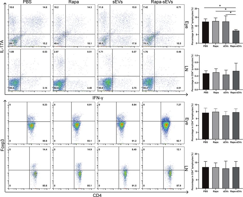
Li et al.                                                                                                                      Rapamycin-Loaded sEVs Alleviated EAU

                                        A                            B                            C                             D

  FIGURE 4 | Trace of sEVs. Distribution of sEVs labeled with PKH-26 in the retina was observed at 24 h and 48 h after subconjunctival injection. 24 h after the
  injection of sEVs (A) and Rapa-sEVs (B), fluorescence was detected in the retina. A mild reduction in red fluorescence was observed 48 h after the injection of sEVs
  (C) and Rapa-sEVs (D).

and lymph nodes on day 18 post-immunization by flow                                     inhibits protein translation initiation, and results in
cytometry. As shown in Figure 7, Rapa-sEV treatment                                    antiproliferative effects (5).
downregulated the proportions of CD4+IFN-g+IL-17+ cells in                                 It has been demonstrated that rapamycin exhibits therapeutic
the retina (one-way ANOVA) (Figure 7B), but did not affect T                           potential for uveitis in animal experiments and clinical trials.
cells in lymph nodes (one-way ANOVA) (Figure 7C). No                                   Several preclinical studies have demonstrated the ability of
differences of the percentage of CD4+ Foxp3+ Treg cells among                          rapamycin to significantly inhibit experimental uveitis (42, 43).
all groups were found (one-way ANOVA) (Figure 7D, E).                                  In addition, rapamycin showed a synergetic effect with other
                                                                                       agents, such as tacrolimus, cyclosporine, and corticosteroids (44–
                                                                                       46). A retrospective clinical study evaluated the therapeutic role
DISCUSSION                                                                             of oral low-dose rapamycin in active uveitis (47). The results
                                                                                       showed that rapamycin might have a limited role in the
In this study, rapamycin was loaded into sEVs by sonication.                           treatment of severe uveitis (47). Although low-dose rapamycin
This formulation was administrated to EAU mice via                                     appeared to be well tolerated, the incidence of adverse effects was
subconjunctival injection at the onset and peak of disease. The                        high. Topical applications can reduce the systemic side effects.
results of the present study reveal that Rapa-sEVs can alleviate                       An experimental study using New Zealand white rabbits revealed
retinal inflammation and protect the retinal structure of mice                          that intravitreal injection of rapamycin had good safety,
with EAU. In conclusion, sEVs play an important role in drug                           tolerance, and stability (48). In another study, rapamycin was
delivery, especially for hydrophobic drugs.                                            detected quickly in the aqueous humor and vitreous of horses
    Rapamycin was considered an antifungal agent in the early                          with equine recurrent uveitis after intravitreal injection (19).
1970s. However, the pharmacological potential of rapamycin was                         However, until 21 days after subconjunctival injection,
not discovered until years later, including immunosuppressive,                         rapamycin in the aqueous humor could not be detected (19).
antiangiogenic, and antiproliferative effects. In contrast to other                    This study demonstrated that intravitreal injection may be an
immunosuppressive agents, including tacrolimus and                                     efficient route of administration. Intravitreal injection of
cyclosporine, rapamycin inhibits mTOR by binding to FKBP-                              rapamycin has entered phase III clinical trials. A clinical study,
12. This prevents the cell cycle transition from G1 to S phase,                        known as SAKURA, was performed to evaluate the therapeutic

Frontiers in Immunology | www.frontiersin.org                                      6                                        March 2022 | Volume 13 | Article 864956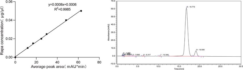
Li et al.                                                                                                                          Rapamycin-Loaded sEVs Alleviated EAU

               A                                                                                                   B

               C

                                                                                                                   D

  FIGURE 5 | Subconjunctival injection of Rapa-sEVs ameliorated uveitis in mice. Fundus images and OCT at day 18 post-immunization showed ocular inflammation
  in PBS, Rapa, and sEV treated groups (A, C). In contrast, Rapa-sEV treatment led to decreased ocular inflammation (A, C). (B) Clinical observation of EAU mice
  treated with PBS, Rapa, sEVs, and Rapa-sEVs. ***P < 0.001, **P < 0.01, *P < 0.5, Kruskal-Wallis test (D) OCT was performed at day 18 post-immunization, and
  results are expressed quantitatively as OCT scores. The inflammatory cells in the vitreous, retinal folders, and retinal detachment near the optic disk were indicated
  by white arrows. Values are expressed as the mean ± SD of six mice (6 eyes) per group. **P < 0.01, *P < 0.5, Kruskal-Wallis test.

effects of intravitreal rapamycin (three different doses: 44 mg, 440                     administrated by subconjunctival injection. As drug carriers, sEVs
mg, and 880 mg) in noninfectious uveitis (17). The results                               have many advantages. The lipid bilayers of sEVs can effectively
indicated that intravitreal rapamycin 440 mg ameliorated                                 protect loaded drugs from degradation and allow them to remain
ocular inflammation (17). However, further studies are                                    stable for a long period of time. Moreover, it can provide the
required to identify its safety and efficacy.                                             advantages of nanotechnology for efficient drug transport capable
    Repeated intravitreal injections may increase the risk of                            of overcoming various biological barriers (49), such as the blood-
infection and the financial and psychological burden of patients.                         ocular barrier (Figure 4). SEVs are naturally occurring membrane
In the present study, rapamycin was loaded into the sEVs and                             vesicles that are secreted by nearly all cell types in all body fluids

                   A                                                                          B

  FIGURE 6 | Histological assessment of the retina in EAU. (A) Representative H&E staining images of PBS, Rapa, sEV, and Rapa-sEV treated groups. The black
  arrows represented the retinal folds and detachments near the optic disk. GCL, ganglion cell layer; INL, inner nuclear layer; ONL, outer nuclear layer. (B) Results are
  expressed quantitatively as histopathological scores. Values are expressed as the mean ± SD of six mice (6 eyes) per group. *P < 0.05, Kruskal-Wallis test.

Frontiers in Immunology | www.frontiersin.org                                        7                                          March 2022 | Volume 13 | Article 864956
Li et al. Rapamycin-Loaded sEVs Alleviated EAU
A B
C
D
E
FIGURE 7 | Analysis of T cell subsets from eye and lymph node in EAU. (A) Representive figures of intracellular staining of IFN-g+IL-17+, Foxp3+ in eyes and lymph
nodes derived CD4+T cells from PBS, Rapa, sEV, and Rapa-sEV treated mice with EAU. (B) The percentage of CD4+IFN-g+IL-17+ cells in eyes. (C) The percentage
of CD4+IFN-g+IL-17A+ cells in cervical draining lymph nodes. (D) The percentage of CD4+Foxp3+ cells in eyes. (E) The percentage of CD4+Foxp3+ cells in cervical
draining lymph nodes. Values are expressed as the mean ± SD of three mice per group. *P < 0.05, one-way ANOVA.
demonstrating excellent biocompatibility. SEVs have been shown conclusion, sEVs as delivery vehicles are attractive and
to reduce inflammation in EAU. Therefore, Rapa-sEVs may play a promising for autoimmune uveitis. Moreover, to promote its
dual role in therapeutic actions. In addition, sEVs may be clinical transformation, further investigations regarding the
repeatedly freeze thawed while retaining their original potency and toxicology of Rapa-sEVs are necessary.
morphology and other characteristics. Finally, sEVs have a low
risk of immunogenicity and tumorigenicity compared to cells.
Therapeutic agents can be incorporated into sEVs using
different approaches, including incubation, sonication, DATA AVAILABILITY STATEMENT
extrusion, and electroporation. A study investigated two
methods: incubation at 37°C and sonication for PTX loading The original contributions presented in the study are included in
(50). The results revealed that sonication resulted in the greatest the article/supplementary material. Further inquiries can be
loading efficiency (50). Kim et al. compared three methods of directed to the corresponding author.
loading sEVs with PTX and arrived at the same conclusion (51).
In addition, this method does not significantly affect the
membrane-bound proteins of sEVs, which is the same as that ETHICS STATEMENT
reported in our study.
However, there are several deficiencies in the present study. The animal study was reviewed and approved by the Animal
Further research should be conducted to investigate in detail the Care and Use committee of Tianjin Medical University
drug release kinetics and drug toxicity of Rapa-sEVs. In Eye Hospital.
Frontiers in Immunology | www.frontiersin.org 8 March 2022 | Volume 13 | Article 864956Li et al. Rapamycin-Loaded sEVs Alleviated EAU
AUTHOR CONTRIBUTIONS FUNDING
XZ and HL designed the research and interpreted data. HL, ZZ and This work was supported by National Natural Science
YL performed experiments and analyzed data. LS, YD, HZ, JA and Foundation of China (81870651, 82171042). Tianjin Science
TN provided methodological guidance. HL wrote the manuscript. and Technology Support Plan (20YFZCSY00990),
XZ reviewed and approved the final manuscript. XZ and XL Natural Science Foundation of Tianjin (20JCZDJC00100)
provided financial and administrative support. All authors and Tianjin Key Medical Discipline (Specialty)
contributed to the article and approved the submitted version. Construction Project.
16. Nguyen QD, Merrill PT, Clark WL, Banker AS, Fardeau C, Franco P, et al.
REFERENCES Intravitreal Sirolimus for Noninfectious Uveitis: A Phase III Sirolimus Study
1. Dick AD, Tundia N, Sorg R, Zhao C, Chao J, Joshi A, et al. Risk of Ocular Assessing Double-Masked Uveitis Treatment (SAKURA). Ophthalmology
Complications in Patients With Noninfectious Intermediate Uveitis, Posterior (2016) 123(11):2413–23. doi: 10.1016/j.ophtha.2016.07.029
Uveitis, or Panuveitis. Ophthalmology (2016) 123(3):655–62. doi: 10.1016/ 17. Merrill PT, Clark WL, Banker AS, Fardeau C, Franco P, LeHoang P, et al.
j.ophtha.2015.10.028 Efficacy and Safety of Intravitreal Sirolimus for Noninfectious Uveitis of the
2. Jabs DA, Rosenbaum JT, Foster CS, Holland GN, Jaffe GJ, Louie JS, et al. Posterior Segment: Results From the Sirolimus Study Assessing Double-
Guidelines for the Use of Immunosuppressive Drugs in Patients With Ocular Masked Uveitis Treatment (SAKURA) Program. Ophthalmology (2020) 120
Inflammatory Disorders: Recommendations of an Expert Panel. Am J (10):1405–15. doi: 10.1016/j.ophtha.2020.03.033
Ophthalmol (2000) 130(4):492–513. doi: 10.1016/s0002-9394(00)00659-0 18. Shikari H, Silva PS, Sun JK. Complications of Intravitreal Injections in
3. Heo J, Sepah Y, Yohannan J, Renner M, Akhtar A, Gregory A, et al. The Role Patients With Diabetes. Semin Ophthalmol (2014) 29(5-6):276–89.
of Biologic Agents in the Management of Non-Infectious Uveitis. Expert Opin doi: 10.3109/08820538.2014.962167
Biol Ther (2012) 12(8):995–1008. doi: 10.1517/14712598.2012.688021 19. Douglas LC, Yi NY, Davis JL, Salmon JH, Gilger BC. Ocular Toxicity and
4. Sen H, Vitale S, Gangaputra S, Nussenblatt R, Liesegang T, Levy-Clarke G, Distribution of Subconjunctival and Intravitreal Rapamycin in Horses. J Vet
et al. Periocular Corticosteroid Injections in Uveitis: Effects and Pharmacol Ther (2008) 31(6):511–6. doi: 10.1111/j.1365-2885.2008.00986.x
Complications. Ophthalmology (2014) 121(11):2275–86. doi: 10.1016/ 20. Wu W, He Z, Zhang Z, Yu X, Song Z, Li X. Intravitreal Injection of
j.ophtha.2014.05.021 Rapamycin-Loaded Polymeric Micelles for Inhibition of Ocular
5. Napoli K, Taylor P. From Beach to Bedside: History of the Development of Inflammation in Rat Model. Int J Pharm (2016) 513(1-2):238–46.
Sirolimus. Ther Drug Monit (2001) 23(5):559–86. doi: 10.1097/00007691- doi: 10.1016/j.ijpharm.2016.09.013
200110000-00012 21. Suri R, Neupane YR, Mehra N, Jain GK, Kohli K. Sirolimus Loaded Polyol
6. Powell J, Pollizzi K, Heikamp E, Horton M. Regulation of Immune Responses Modified Liposomes for the Treatment of Posterior Segment Eye Diseases.
by Mtor. Annu Rev Immunol (2012) 30:39–68. doi: 10.1146/annurev- Med Hypotheses (2020) 136:109518. doi: 10.1016/j.mehy.2019.109518
immunol-020711-075024 22. Haeri A, Osouli M, Bayat F, Alavi S, Dadashzadeh S. Nanomedicine
7. Blair J, Barry R, Moore DJ, Denniston AK. A Comprehensive Review of Mtor- Approaches for Sirolimus Delivery: A Review of Pharmaceutical Properties
Inhibiting Pharmacotherapy for the Treatment of Non-Infectious Uveitis. and Preclinical Studies. Artif Cells Nanomed Biotechnol (2018) 46(sup1):1–14.
Curr Pharm Des (2017) 23(20):3005–14. doi: 10.2174/138161282366 doi: 10.1080/21691401.2017.1408123
6170111125550 23. Srivastava A, Babu A, Filant J, Moxley K, Ruskin R, Dhanasekaran D, et al.
8. Roberts RJ, Wells AC, Unitt E, Griffiths M, Tasker AD, Allison ME, et al. Exploitation of Exosomes as Nanocarriers for Gene-, Chemo-, and Immune-
Sirolimus-Induced Pneumonitis Following Liver Transplantation. Liver Therapy of Cancer. J BioMed Nanotechnol (2016) 12(6):1159–73.
Transpl (2007) 13(6):853–6. doi: 10.1002/lt.21141 doi: 10.1166/jbn.2016.2205
9. Pilotte AP, Hohos MB, Polson KM, Huftalen TM, Treister N. Managing 24. Barile L, Vassalli G. Exosomes: Therapy Delivery Tools and Biomarkers of Diseases.
Stomatitis in Patients Treated With Mammalian Target of Rapamycin Pharmacol Ther (2017) 174:63–78. doi: 10.1016/j.pharmthera.2017.02.020
Inhibitors. Clin J Oncol Nurs (2011) 15(5):E83–9. doi: 10.1188/11.Cjon.E83-e89 25. Armstrong J, Stevens M. Strategic Design of Extracellular Vesicle Drug
10. Ravaud A. Treatment-Associated Adverse Event Management in the Delivery Systems. Adv Drug Deliv Rev (2018) 130:12–6. doi: 10.1016/
Advanced Renal Cell Carcinoma Patient Treated With Targeted Therapies. j.addr.2018.06.017
Oncologist (2011) 16 Suppl 2(Suppl 2):32–44. doi: 10.1634/theoncologist. 26. Sun D, Zhuang X, Xiang X, Liu Y, Zhang S, Liu C, et al. A Novel Nanoparticle
2011-S2-32 Drug Delivery System: The Anti-Inflammatory Activity of Curcumin Is
11. Soefje SA, Karnad A, Brenner AJ. Common Toxicities of Mammalian Target Enhanced When Encapsulated in Exosomes. Mol Ther (2010) 18(9):1606–
of Rapamycin Inhibitors. Target Oncol (2011) 6(2):125–9. doi: 10.1007/ 14. doi: 10.1038/mt.2010.105
s11523-011-0174-9 27. Zhuang X, Xiang X, Grizzle W, Sun D, Zhang S, Axtell R, et al. Treatment of
12. Sofroniadou S, Goldsmith D. Mammalian Target of Rapamycin (Mtor) Brain Inflammatory Diseases by Delivering Exosome Encapsulated Anti-
Inhibitors: Potential Uses and a Review of Haematological Adverse Effects. Inflammatory Drugs From the Nasal Region to the Brain. Mol Ther: J Am
Drug Saf (2011) 34(2):97–115. doi: 10.2165/11585040-000000000-00000 Soc Gene Ther (2011) 19(10):1769–79. doi: 10.1038/mt.2011.164
13. Balagula Y, Rosen A, Tan BH, Busam KJ, Pulitzer MP, Motzer RJ, et al. 28. Yang T, Martin P, Fogarty B, Brown A, Schurman K, Phipps R, et al. Exosome
Clinical and Histopathologic Characteristics of Rash in Cancer Patients Delivered Anticancer Drugs Across the Blood-Brain Barrier for Brain Cancer
Treated With Mammalian Target of Rapamycin Inhibitors. Cancer (2012) Therapy in Danio Rerio. Pharm Res (2015) 32(6):2003–14. doi: 10.1007/
118(20):5078–83. doi: 10.1002/cncr.27505 s11095-014-1593-y
14. Nguyen QD, Ibrahim MA, Watters A, Bittencourt M, Yohannan J, Sepah YJ, 29. Pascucci L, Cocce V, Bonomi A, Ami D, Ceccarelli P, Ciusani E, et al. Paclitaxel
et al. Ocular Tolerability and Efficacy of Intravitreal and Subconjunctival Is Incorporated by Mesenchymal Stromal Cells and Released in Exosomes That
Injections of Sirolimus in Patients With Non-Infectious Uveitis: Primary 6- Inhibit In Vitro Tumor Growth: A New Approach for Drug Delivery. J Control
Month Results of the SAVE Study. J Ophthalmic Inflamm Infect (2013) 3 Release (2014) 192:262–70. doi: 10.1016/j.jconrel.2014.07.042
(1):32. doi: 10.1186/1869-5760-3-32 30. Tian Y, Li S, Song J, Ji T, Zhu M, Anderson GJ, et al. A Doxorubicin Delivery
15. Ibrahim MA, Sepah YJ, Watters A, Bittencourt M, Vigil EM, Do DV, et al. Platform Using Engineered Natural Membrane Vesicle Exosomes for
One-Year Outcomes of the SAVE Study: Sirolimus as a Therapeutic Approach Targeted Tumor Therapy. Biomaterials (2014) 35(7):2383–90. doi: 10.1016/
for Uveitis. Transl Vis Sci Technol (2015) 4(2):4. doi: 10.1167/tvst.4.2.4 j.biomaterials.2013.11.083
Frontiers in Immunology | www.frontiersin.org 9 March 2022 | Volume 13 | Article 864956Li et al. Rapamycin-Loaded sEVs Alleviated EAU
31. Jang S, Kim O, Yoon C, Choi D, Roh T, Park J, et al. Bioinspired Exosome- 44. Ikeda E, Hikita N, Eto K, Mochizuki M. Tacrolimus-Rapamycin Combination
Mimetic Nanovesicles for Targeted Delivery of Chemotherapeutics to Therapy for Experimental Autoimmune Uveoretinitis. Jpn J Ophthalmol
Malignant Tumors. ACS Nano (2013) 7(9):7698–710. doi: 10.1021/nn402232g (1997) 41(6):396–402. doi: 10.1016/s0021-5155(97)00083-x
32. Yeo RW, Lai RC, Zhang B, Tan SS, Yin Y, Teh BJ, et al. Mesenchymal Stem 45. Martin DF, DeBarge LR, Nussenblatt RB, Chan CC, Roberge FG. Synergistic
Cell: An Efficient Mass Producer of Exosomes for Drug Delivery. Adv Drug Effect of Rapamycin and Cyclosporin a in the Treatment of Experimental
Deliv Rev (2013) 65(3):336–41. doi: 10.1016/j.addr.2012.07.001 Autoimmune Uveoretinitis. J Immunol (1995) 154(2):922–7.
33. Bai L, Shao H, Wang H, Zhang Z, Su C, Dong L, et al. Effects of Mesenchymal 46. Roberge FG, Martin DF, Xu D, Chen H, Chan CC. Synergism Between
Stem Cell-Derived Exosomes on Experimental Autoimmune Uveitis. Sci Rep Corticosteroids and Rapamycin for the Treatment of Intraocular
(2017) 7(1):4323. doi: 10.1038/s41598-017-04559-y Inflammation. Ocul Immunol Inflamm (1995) 3(3):195–202. doi: 10.3109/
34. Dominici M, Le Blanc K, Mueller I, Slaper-Cortenbach I, Marini F, 09273949509069112
Krause D, et al. Minimal Criteria for Defining Multipotent Mesenchymal 47. Phillips BN, Wroblewski KJ. A Retrospective Review of Oral Low-Dose
Stromal Cells. The International Society for Cellular Therapy Position Sirolimus (Rapamycin) for the Treatment of Active Uveitis. J Ophthalmic
Statement. Cytotherapy (2006) 8(4):315–7. doi: 10.1080/14653240600855905 Inflamm Infect (2010) 1(1):29–34. doi: 10.1007/s12348-010-0015-5
35. Witwer KW, Van Balkom BWM, Bruno S, Choo A, Dominici M, Gimona M, 48. Mudumba S, Bezwada P, Takanaga H, Hosoi K, Tsuboi T, Ueda K, et al.
et al. Defining Mesenchymal Stromal Cell (MSC)-Derived Small Extracellular Tolerability and Pharmacokinetics of Intravitreal Sirolimus. J Ocul Pharmacol
Vesicles for Therapeutic Applications. J Extracell Vesicles (2019) 8(1):1609206. Ther (2012) 28(5):507–14. doi: 10.1089/jop.2011.0226
doi: 10.1080/20013078.2019.1609206 49. Hajrasouliha A, Jiang G, Lu Q, Lu H, Kaplan H, Zhang H, et al. Exosomes
36. Caspi R. Experimental Autoimmune Uveoretinitis in the Rat and Mouse. From Retinal Astrocytes Contain Antiangiogenic Components That Inhibit
Curr Protoc Immunol (2003) Chapter 15:Unit 15.6. doi: 10.1002/ Laser-Induced Choroidal Neovascularization. J Biol Chem (2013) 288
0471142735.im1506s53 (39):28058–67. doi: 10.1074/jbc.M113.470765
37. Gadjanski I, Williams S, Hein K, Sättler M, Bähr M, Diem R. Correlation of 50. Salarpour S, Forootanfar H, Pournamdari M, Ahmadi-Zeidabadi M, Esmaeeli
Optical Coherence Tomography With Clinical and Histopathological M, Pardakhty A. Paclitaxel Incorporated Exosomes Derived From
Findings in Experimental Autoimmune Uveoretinitis. Exp Eye Res (2011) Glioblastoma Cells: Comparative Study of Two Loading Techniques. Daru
93(1):82–90. doi: 10.1016/j.exer.2011.04.012 (2019) 27(2):533–9. doi: 10.1007/s40199-019-00280-5
38. van Hamburg JP, Tas SW. Molecular Mechanisms Underpinning T Helper 17 51. Kim MS, Haney MJ, Zhao Y, Mahajan V, Deygen I, Klyachko NL, et al.
Cell Heterogeneity and Functions in Rheumatoid Arthritis. J Autoimmun Development of Exosome-Encapsulated Paclitaxel to Overcome MDR in Cancer
(2018) 87:69–81. doi: 10.1016/j.jaut.2017.12.006 Cells. Nanomedicine (2016) 12(3):655–64. doi: 10.1016/j.nano.2015.10.012
39. van Langelaar J, van der Vuurst de Vries RM, Janssen M, Wierenga-Wolf AF,
Spilt IM, Siepman TA, et al. T Helper 17.1 Cells Associate With Multiple Conflict of Interest: The authors declare that the research was conducted in the
Sclerosis Disease Activity: Perspectives for Early Intervention. Brain (2018) absence of any commercial or financial relationships that could be construed as a
141(5):1334–49. doi: 10.1093/brain/awy069 potential conflict of interest.
40. Harbour S, Maynard C, Zindl C, Schoeb T, Weaver C. Th17 Cells Give Rise to
Th1 Cells That Are Required for the Pathogenesis of Colitis. Proc Natl Acad Publisher’s Note: All claims expressed in this article are solely those of the authors
Sci USA (2015) 112(22):7061–6. doi: 10.1073/pnas.1415675112 and do not necessarily represent those of their affiliated organizations, or those of
41. Basdeo SA, Cluxton D, Sulaimani J, Moran B, Canavan M, Orr C, et al. Ex- the publisher, the editors and the reviewers. Any product that may be evaluated in
Th17 (Nonclassical Th1) Cells Are Functionally Distinct From Classical Th1 this article, or claim that may be made by its manufacturer, is not guaranteed or
and Th17 Cells and Are Not Constrained by Regulatory T Cells. J Immunol endorsed by the publisher.
(2017) 198(6):2249–59. doi: 10.4049/jimmunol.1600737
42. Ohia E, Mancino M, Kulkarni P. Effects of Steroids and Immunosuppressive Copyright © 2022 Li, Zhang, Li, Su, Duan, Zhang, An, Ni, Li and Zhang. This is an
Drugs on Endotoxin-Uveitis in Rabbits. J Ocul Pharmacol (1992) 8(4):295– open-access article distributed under the terms of the Creative Commons Attribution
307. doi: 10.1089/jop.1992.8.295 License (CC BY). The use, distribution or reproduction in other forums is permitted,
43. Roberge F, Xu D, Chan C, de Smet M, Nussenblatt R, Chen H. Treatment of provided the original author(s) and the copyright owner(s) are credited and that the
Autoimmune Uveoretinitis in the Rat With Rapamycin, an Inhibitor of original publication in this journal is cited, in accordance with accepted academic
Lymphocyte Growth Factor Signal Transduction. Curr Eye Res (1993) 12 practice. No use, distribution or reproduction is permitted which does not comply with
(2):197–203. doi: 10.3109/02713689308999487 these terms.
Frontiers in Immunology | www.frontiersin.org 10 March 2022 | Volume 13 | Article 864956You can also read