Scheffler, K., Giannini, F., Lemonnier, T. A. A., & Mogessie, B. (2022). The prophase oocyte nucleus is a homeostatic G-actin buffer. Journal of ...
←
→
Page content transcription
If your browser does not render page correctly, please read the page content below
Scheffler, K., Giannini, F., Lemonnier, T. A. A., & Mogessie, B. (2022).
The prophase oocyte nucleus is a homeostatic G-actin buffer. Journal
of Cell Science, 135(6), [ jcs259807].
https://doi.org/10.1242/jcs.259807

Publisher's PDF, also known as Version of record
License (if available):
CC BY
Link to published version (if available):
10.1242/jcs.259807

Link to publication record in Explore Bristol Research
PDF-document

This is the final published version of the article (version of record). It first appeared online via The Company of
Biologists at https://doi.org/10.1242/jcs.259807 . Please refer to any applicable terms of use of the publisher.

University of Bristol - Explore Bristol Research
General rights

This document is made available in accordance with publisher policies. Please cite only the
published version using the reference above. Full terms of use are available:
http://www.bristol.ac.uk/red/research-policy/pure/user-guides/ebr-terms/
© 2022. Published by The Company of Biologists Ltd | Journal of Cell Science (2022) 135, jcs259807. doi:10.1242/jcs.259807
RESEARCH ARTICLE
The prophase oocyte nucleus is a homeostatic G-actin buffer
Kathleen Scheffler1,*, Federica Giannini1, *, Tom Lemonnier1, * and Binyam Mogessie1,2,‡
ABSTRACT filaments that oversee long-range vesicle transport (Schuh, 2011)
Formation of healthy mammalian eggs from oocytes requires and asymmetric division in mammalian oocytes (Holubcova et al.,
specialised F-actin structures. F-actin disruption produces 2013; Schuh and Ellenberg, 2008; Azoury et al., 2008). In addition,
aneuploid eggs, which are a leading cause of human embryo oocyte meiotic spindles in several mammalian species contain actin
deaths, genetic disorders and infertility. We found that oocytes filaments that aid microtubule fibres in chromosome separation
contain prominent nuclear F-actin structures that are correlated with (Mogessie and Schuh, 2017). After completion of meiosis, the actin
meiotic developmental capacity. We demonstrate that nuclear F-actin cytoskeleton also plays a crucial role in pronuclear-stage zygotes,
is a conserved feature of healthy mammalian oocytes and declines where it directly participates in the unification of parental genomes
significantly with female reproductive ageing. Actin monomers used (Scheffler et al., 2021).
for nuclear F-actin assembly are sourced from an excess pool in the In this study, we show that actin filaments and bundles frequently
oocyte cytoplasm. Increasing monomeric G-actin transfer from the occupy the nucleus in prophase-arrested mammalian oocytes.
cytoplasm to the nucleus or directly enriching the nucleus with Furthermore, we demonstrate that, unlike in other experimental
monomers led to assembly of stable nuclear F-actin bundles that systems, endogenous nuclear actin filaments are assembled in
significantly restrict chromatin mobility. By contrast, reducing G-actin healthy oocytes without any manipulation. By combining
monomer transfer by blocking nuclear import triggered assembly of a cytoskeletal disruption tools with nuclear import inhibition and
dense cytoplasmic F-actin network that is incompatible with healthy high-resolution microscopy assays, we provided evidence for a
oocyte development. Overall, our data suggest that the large oocyte homeostatic balance between cytoplasmic and nuclear F-actin
nucleus helps to maintain cytoplasmic F-actin organisation and that structures in mammalian oocytes. Importantly, three-dimensional
defects in this function are linked with reproductive age-related female (3D) volumetric analyses of F-actin structures revealed that the
infertility. amount and complexity of nuclear actin filaments declines
significantly with increasing reproductive age in females.
This article has an associated First Person interview with Federica
Giannini, joint first author of the paper. RESULTS
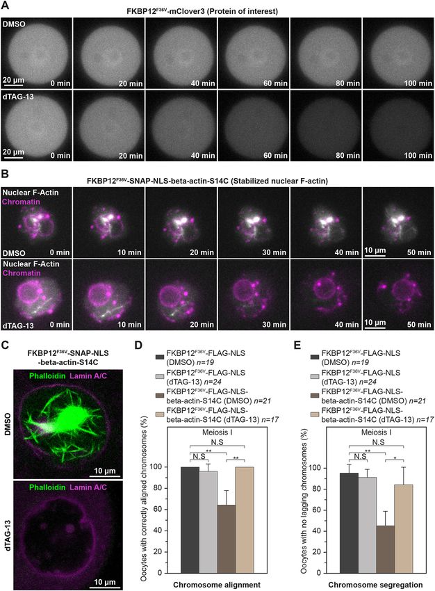
Mammalian oocyte nuclei contain prominent F-actin
KEY WORDS: Oocyte, Nuclear F-actin, Meiosis, Aneuploidy, structures
Chromosome segregation, Infertility We have found that the nucleus of prophase-arrested, non-
manipulated mouse oocytes contains prominent actin filaments
INTRODUCTION (Fig. 1A). Using fluorescent phalloidin, we detected actin filaments
Mammalian eggs are formed when oocyte chromosomes are and bundles in the nucleoplasm of 80% of fixed oocytes (Fig. 1B and
segregated during meiosis (Mogessie et al., 2018), successful C). Notably, this observation was strain- and species-independent, as
completion of which is a prerequisite for healthy embryogenesis and we could detect nuclear F-actin structures in oocytes isolated from
development. Meiotic chromosome segregation errors in oocytes outbred and inbred mouse (Fig. 1D and E) and sheep ovaries
are a leading cause of aneuploidies that underlie human infertility (Fig. 1F). To visualise these structures in live cells, we microinjected
and genetic disorders, such as Down’s syndrome (Herbert et al., prophase-arrested mouse oocytes with low to high concentrations of
2015). The incidence of oocyte and egg aneuploidy increases in vitro transcribed mRNAs encoding a fluorescence-labelled actin
almost exponentially with advancing female reproductive age nanobody (nuclear actin chromobody) (Baarlink et al., 2017) and
(Hassold and Hunt, 2001; Nagaoka et al., 2012). performed fast, super-resolution live imaging of nuclear F-actin. This
Distinct F-actin polymers assembled from soluble G-actin approach revealed that these nuclear F-actin structures are highly
monomers ensure the production of healthy eggs from oocytes. mobile – filaments continuously move in non-directed fashion within
These actin-rich drivers include a network of cytoplasmic actin the nucleoplasm (Fig. 2A and B; Movie 1 and Movie 2). To validate
that low expression of the nuclear actin chromobody in these
experiments did not grossly stabilise nuclear F-actin structures, we
Journal of Cell Science
1
School of Biochemistry, University of Bristol, Bristol BS8 1TD, UK. 2Department of
Molecular, Cellular and Developmental Biology, Yale University, New Haven, CT fixed microinjected oocytes after super-resolution live microscopy,
06511, USA. labelled actin with fluorescent phalloidin and visualised the F-actin
*These authors contributed equally to this work content of oocytes. This confirmed that nuclear F-actin structures in
‡
Author for correspondence (binyam.mogessie@yale.edu) our live imaging experiments were not morphologically distinct
from endogenous nuclear actin filaments (Fig. 1A and D, Fig. 2C).
B.M., 0000-0002-0702-6356 Importantly, the presence of nuclear F-actin was remarkably
This is an Open Access article distributed under the terms of the Creative Commons Attribution associated with distinct organisation of chromatin surrounding the
License (https://creativecommons.org/licenses/by/4.0), which permits unrestricted use, nucleolus (Fig. S1A and B), a marker of high oocyte meiotic
distribution and reproduction in any medium provided that the original work is properly attributed.
competence and developmental capacity (Zuccotti et al., 1998).
Handling Editor: Michael Way These results collectively demonstrated that nuclear F-actin
Received 24 January 2022; Accepted 24 January 2022 structures are a common feature of healthy, non-manipulated
1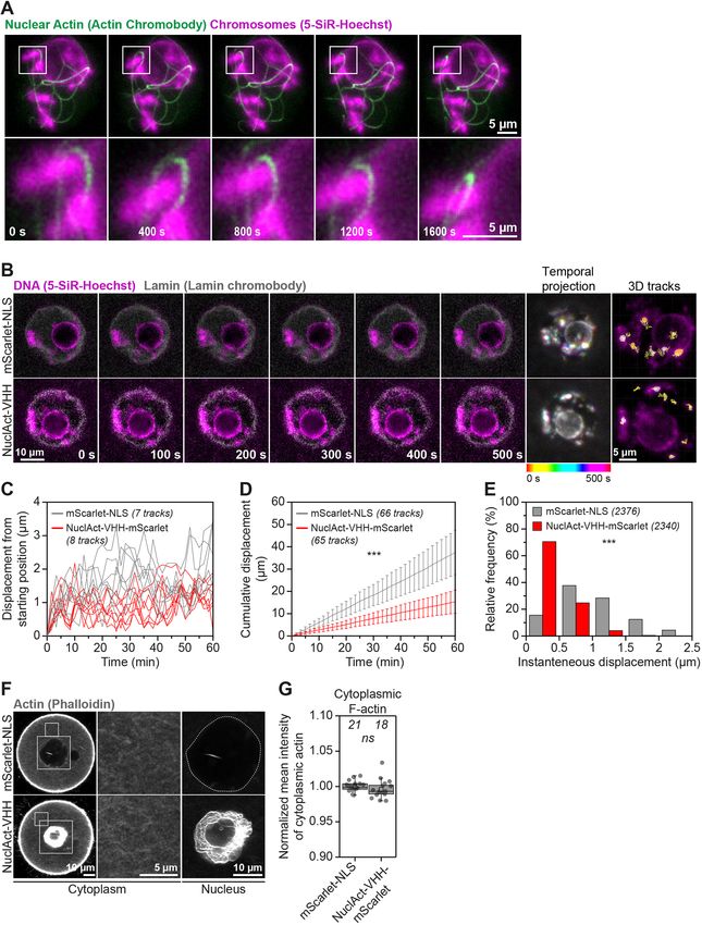
RESEARCH ARTICLE                                                                          Journal of Cell Science (2022) 135, jcs259807. doi:10.1242/jcs.259807

Fig. 1. Mammalian prophase oocyte nuclei contain prominent actin filaments. (A) Phalloidin-labelled nuclear actin filaments (grey) and chromosomes
(Hoechst, magenta) in three prophase-arrested mouse oocytes. Single confocal sections spaced 2 μm apart and corresponding maximum intensity projections
are shown. (B) Pipeline for 3D isosurface reconstruction nuclear membrane (green) and nuclear F-actin (grey). DNA is shown in magenta. (C) Quantification of
nuclear F-actin presence in prophase-arrested mouse oocytes. Data are from three independent experiments. (D) Phalloidin-labelled nuclear actin filaments
(grey) and chromosomes (Hoechst, magenta) in prophase-arrested oocyte isolated from 129S6/SvEvTac mouse (inbred) strain. Single confocal sections spaced
2 μm apart and corresponding maximum intensity projections are shown. (E) Quantification of nuclear F-actin presence in prophase-arrested 129S6/SvEvTac
strain mouse oocytes. Data are from three independent experiments. (F) Maximum intensity projection (nine confocal sections) images of phalloidin labelled
nuclear F-actin (grey), DNA (magenta) and nuclear membrane (green) in two sheep oocytes. Single confocal sections spaced 2 μm apart are shown. Isosurface
reconstruction of actin (white) demonstrates prominent nuclear actin filaments. Sheep oocytes from two independent experiments are shown.

mammalian oocytes. Furthermore, we were able to detect nuclear                 the oocyte cytoplasmic actin network by Cytochalasin D (Schuh,
F-actin structures by targeting F-tractin (Melak et al., 2017) and the         2011) treatment, instead, triggered excessive nuclear F-actin
                                                                                                                                                                  Journal of Cell Science

calponin homology domain of utrophin (UtrCH) (Mogessie and                     assembly (Fig. 3A). Nuclei were also more likely to contain
Schuh, 2017) to the nucleus (Fig. S1C and D). These probes                     F-actin structures when oocytes were treated with Cytochalasin D
generally stabilised and reduced the mobility of nuclear actin                 (Fig. 3B). To confirm this, we imaged fluorescent phalloidin-
filaments (Movies 3 and 4), indicating that they can be used to                labelled nuclear actin filaments by high-resolution microscopy,
experimentally increase the F-actin content of mammalian oocyte                selectively reconstructed them in 3D and quantified their volume
nuclei.                                                                        inside nuclei – marked with antibodies against nuclear membrane
                                                                               proteins – of oocytes treated with DMSO (Control) or Cytochalasin
Cytoplasmic F-actin disruption induces excessive nuclear                       D (Fig. 3A). This showed a near fortyfold increase in nuclear F-actin
F-actin assembly                                                               volume after disruption of the cytoplasmic actin network (Fig. 3C).
We next sought to disrupt nuclear F-actin structures by using                  Actin polymerises into filaments when monomer concentration
cytoskeletal drugs. Unexpectedly, we observed that disruption of               exceeds critical concentration (Doolittle et al., 2013). We therefore

                                                                                                                                                             2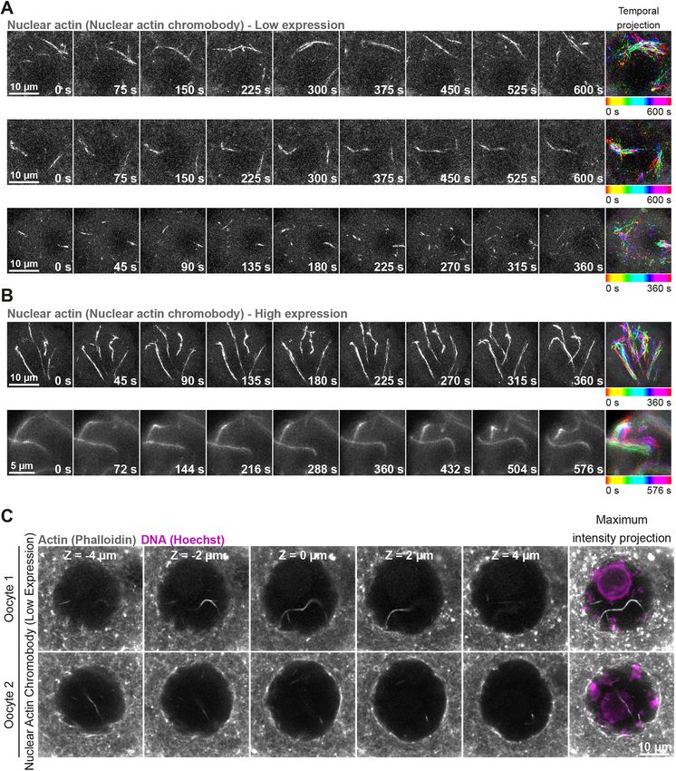
RESEARCH ARTICLE                                                                              Journal of Cell Science (2022) 135, jcs259807. doi:10.1242/jcs.259807

Fig. 2. Oocyte nuclear F-actin structures are dynamic. (A) Super-resolution live imaging of nuclear actin filaments in three prophase-arrested mouse oocytes.
Nuclear F-actin is labelled by low level expression of a fluorescent nanobody (nuclear actin chromobody). Colour-coded temporal projection images (built from 20
                                                                                                                                                                      Journal of Cell Science

frames at 30 s intervals for top and middle panels, 25 frames at 15 s intervals for bottom panel) indicate mobility of filaments during the observation time.
(B) Super-resolution live imaging of nuclear actin filaments in two prophase-arrested mouse oocytes. Nuclear F-actin is labelled by high level expression of a
fluorescent nanobody (nuclear actin chromobody). Colour-coded temporal projection images (built from nine frames at 45 s intervals for top panel, 33 frames at
18 s intervals for bottom panel) indicate mobility of filaments during the observation time. (C) Phalloidin-labelled nuclear actin filaments (grey) and chromosomes
(Hoechst, magenta) in two prophase-arrested mouse oocytes fixed after low-level expression and live imaging of nuclear actin chromobody. Single confocal
sections spaced 2 μm apart and the corresponding maximum intensity projections are shown.

reasoned that bulk transfer of Cytochalasin D-generated G-actin                    1986). We tested this possibility by using Latrunculin, which disrupts
monomers from a large oocyte cytoplasm to a smaller nuclear                        the cytoplasmic actin network by sequestering monomers and
volume could drive excess F-actin polymerisation. Such filaments                   preventing their addition to actin filaments, to complement our
are likely to be drug resistant because Cytochalasin D is generally                Cytochalasin D studies in a mechanistically distinct manner (Fig. 3D)
less effective at high actin monomer concentration (Carlier et al.,                (Spector et al., 1983; Coue et al., 1987; Morton et al., 2000). Under

                                                                                                                                                                 3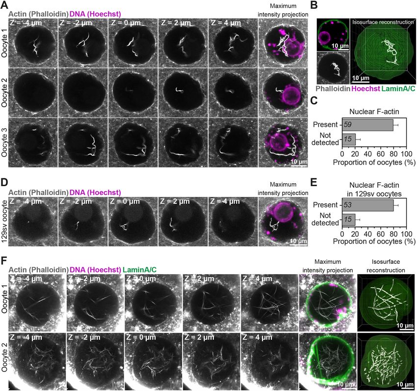
RESEARCH ARTICLE                    Journal of Cell Science (2022) 135, jcs259807. doi:10.1242/jcs.259807

                                                                                                            Journal of Cell Science

Fig. 3. See next page for legend.

                                                                                                       4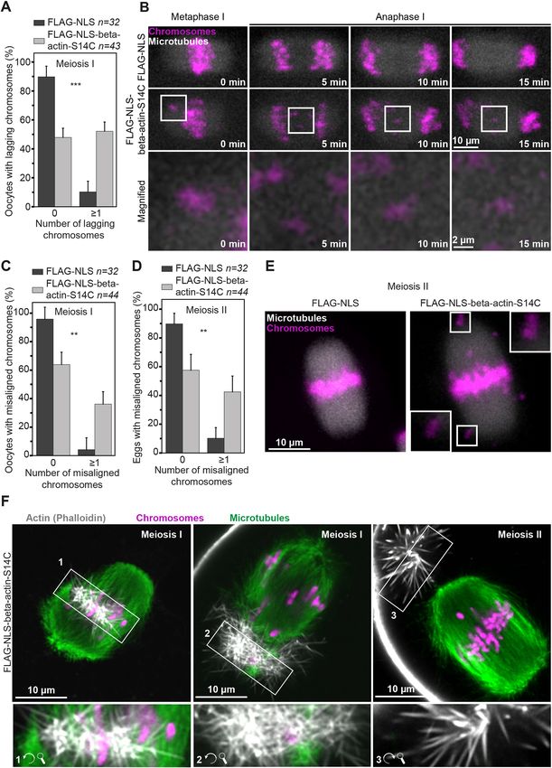
RESEARCH ARTICLE Journal of Cell Science (2022) 135, jcs259807. doi:10.1242/jcs.259807
Fig. 3. Excess G-actin causes uncontrolled cytoplasmic and nuclear fluorescence signal increased notably before the nuclear assembly
F-actin assembly. (A) Single-section Airyscan images of phalloidin-labelled of F-actin bundles (Fig. S2D, Movie 5). Excess amount of
cytoplasmic F-actin and maximum intensity projections (nine confocal
cytoplasmic actin monomers can therefore be transferred to the
sections) of nuclear actin filaments (grey), DNA (magenta) and nuclear
membrane (green) in DMSO- or Cytochalasin D-treated mouse oocytes. oocyte nucleus, where they participate in nuclear F-actin
Boxed areas indicate regions within the oocyte cytoplasm that are shown polymerisation. Consistent with our data, increasing cellular
magnified to the right of the respective Airyscan images. (B) Quantification of G-actin by using cytoskeletal drugs in frog egg extracts (Parisis
nuclear F-actin presence in DMSO- or Cytochalasin D-treated mouse oocytes. et al., 2017) or in cultured mammalian cells (Pendleton et al., 2003)
Data are from three independent experiments. (C) Quantification of nuclear can cause nuclear F-actin polymerisation. Excess transfer of
F-actin volumes from isosurface reconstructions shown in A, in DMSO- or
cytosolic actin to the nucleus might thus be an actin homeostasis
Cytochalasin D-treated mouse oocytes. Data are from three independent
experiments. (D) Single-section Airyscan images of Phalloidin-labelled
feature that is conserved in other cell types.
cytoplasmic F-actin and maximum intensity projections (nine confocal We next directly tested whether high monomeric G-actin
sections) of nuclear actin filaments (grey), DNA (magenta) and nuclear concentration is sufficient to induce F-actin polymerisation in
membrane (green) in DMSO- or Latrunculin B-treated mouse oocytes. Boxed oocytes. We exceeded the nuclear monomeric G-actin concentration
areas indicate regions within the oocyte cytoplasm that are shown magnified to in mouse oocytes by targeting FLAG-tagged beta-actin (FLAG-
the right of the respective Airyscan images. (E) Quantification of nuclear beta-actin) to the nucleus by using a nuclear localisation signal
F-actin presence in DMSO- or Latrunculin B-treated mouse oocytes. Data are
from three independent experiments. (F) Quantification of nuclear F-actin
(NLS). This led to a tenfold increase in the volume of nuclear
volumes from isosurface reconstructions in D in DMSO- or Latrunculin F-actin in FLAG-beta-actin-NLS expressing oocytes, which were
B-treated mouse oocytes. Data are from three independent experiments. also more likely to contain F-actin than FLAG-NLS-expressing
(G) Maximum intensity projection (nine confocal sections) images of Control oocytes (Fig. 3G–I). Therefore, the concentration of
phalloidin-labelled nuclear F-actin (grey), DNA (magenta) and nuclear monomeric G-actin in the nucleus indeed dictates the extent of
membrane (green) in control and wild-type or S14C mutant actin expressing nuclear F-actin polymerisation.
prophase-arrested oocytes. Excess nuclear F-actin in wild-type and S14C-
overexpressing oocytes is demonstrated by presenting overexposed (when
nuclear F-actin is highly visible in controls) or moderately overexposed (when Maintenance of oocyte cytoplasmic F-actin network
nuclear F-actin is poorly visible in controls) images. (H) Quantification of organisation requires nuclear import activity
nuclear F-actin presence in control and wild-type or S14C actin mutant- To further examine the transfer of G-actin monomers to the nucleus,
expressing mouse oocytes. Data are from three independent experiments. we blocked nuclear import by using a combination of Importazole
(I) Quantification of nuclear F-actin volumes from isosurface reconstructions in and Ivermectin (Soderholm et al., 2011; Wagstaff et al., 2012)
G in control and wild-type or S14C actin mutant-expressing mouse oocytes.
(Fig. S3A), before disrupting the cytoplasmic actin network.
Data are from three independent experiments. (J) Single-section Airyscan
images of phalloidin-labelled cytoplasmic F-actin and maximum intensity
Treatment of oocytes with Cytochalasin D did not cause
projections (nine confocal sections) of nuclear actin filaments (grey), DNA excessive nuclear F-actin assembly when nuclear import was
(magenta) and nuclear membrane (green) in DMSO- or Importazole/ blocked (Fig. 3C,J,K; Fig. S3B). In addition, nuclear import
Ivermectin-treated mouse oocytes that were then treated with Cytochalasin blockage caused distinct accumulation of actin on the surface of
D. Boxed areas indicate regions within the oocyte cytoplasm that are shown oocyte nuclei (Fig. S3D). Surprisingly, Cytochalasin D treatment of
magnified to the right of the respective Airyscan images. (K) Quantification of oocytes defective in nuclear import resulted in a significantly denser
nuclear F-actin volumes from isosurface reconstructions in J of DMSO- or
Importazole/Ivermectin-treated mouse oocytes that were then treated with
cytoplasmic actin network, and one that was composed of drug-
Cytochalasin D. Data are from three independent experiments. resistant filaments (Fig. 3J,L; Fig. S3C). This is consistent with a
(L) Quantification of cytoplasmic F-actin network intensity in DMSO- or significant rise in cytosolic monomeric G-actin concentration
Importazole/Ivermectin-treated mouse oocytes that were then treated with caused by blocked nuclear import, which reduces Cytochalasin D
Cytochalasin D. Data are from three independent experiments. Statistical activity (Carlier et al., 1986) in the cytoplasm and causes assembly
significance was tested using Fisher’s exact test (B, E and H) and two-tailed of drug-resistant filaments. Prompt transfer of excess G-actin
Student’s t-test (C, F, I, K and L).
monomers to the nucleus could be necessary to maintain the
organisation of cytoplasmic F-actin network in oocytes. This is
this circumstance, more cytosolic monomers would still be supported by the observation that blocking nuclear import in
transferred to the nucleus but would be unable to participate in oocytes not previously treated with cytoskeletal drugs significantly
actin polymerisation. In stark contrast to treatment with Cytochalasin increases cytoplasmic actin network density (Fig. S3D and E).
D, nuclei in Latrunculin B-treated oocytes were not more likely to Indeed, monomeric actin is known to continuously shuttle between
contain actin filaments (Fig. 3E) and only showed a twofold increase the cytoplasm and the nucleus in other experimental systems (Dopie
in the volume of F-actin (Fig. 3F; Fig. S2A). Thus, the concentration et al., 2012; Stuven et al., 2003). Thus, notwithstanding the role of
of polymerisation-ready monomers imported from the cytoplasm is other nuclear import cargoes in these observations, our data support
likely to determine the degree of F-actin assembly in the oocyte the notion that excess actin monomer import helps to maintain
nucleus. Consistent with these perturbations, treatment of oocytes cytoplasmic F-actin network organisation in mouse oocytes. We
Journal of Cell Science
with the Arp2/3 inhibitor compound CK-666 also significantly propose that shuttling of cytosolic monomers into a large (∼30 µm
increased polymerisation of nuclear F-actin (Fig. S2B and C). diameter) nucleus is a physiological G-actin-buffering process that
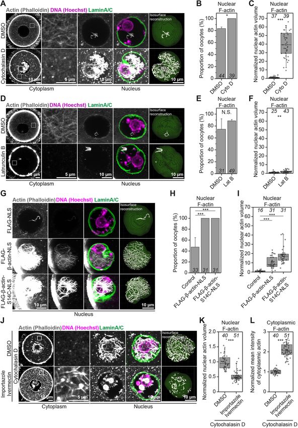
We next took an approach to directly observe the transfer of oocytes use to continuously modulate the cytoplasmic actin network.
excess actin monomers from the cytoplasm to the nucleus. For this,
we first overexpressed SNAP-tagged beta-actin (SNAP-beta-actin) Stable nuclear actin filaments restrict chromatin mobility in
in prophase-arrested mouse oocytes. We then performed high- oocytes
resolution time-lapse microscopy to assess the fluorescence of To investigate the consequences of stable assembly of nuclear actin
cytoplasmic and nuclear beta-actin immediately after addition of filaments, we induced excess cytosolic monomers by treating
Cytochalasin D to the oocyte culture medium. This live imaging oocytes with Cytochalasin D (Fig. 3A) or directly enriched oocyte
experiment revealed that cytoplasmic fluorescence of beta-actin nuclei with monomers by expressing a nuclear actin mutant (FLAG-
rapidly decreased after Cytochalasin D addition, while the nuclear NLS-beta-actin-S14C) that is more able to polymerise (Posern et al.,
5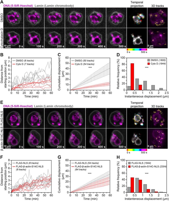
RESEARCH ARTICLE                                                                            Journal of Cell Science (2022) 135, jcs259807. doi:10.1242/jcs.259807

Fig. 4. Excess nuclear actin filaments severely restrict oocyte chromatin mobility. (A) Still images from representative time-lapse movies of chromatin
movement in DMSO- or Cytochalasin D-treated mouse oocytes. Chromatin (SiR-5-Hoechst) is shown in magenta and nuclear membrane (lamin chromobody) is
shown in grey. Colour-coded temporal projection images indicate the degree of chromatin mobility during the 500 s observation time. 3D tracks represent the
                                                                                                                                                                    Journal of Cell Science

spatial coverage of prominent chromatin spots over a 60-min observation period. (B) Distance from starting position of prominent chromatin spots in 3D over a 60-
min observation period in DMSO- or Cytochalasin D-treated mouse oocytes. Data are from three independent experiments. (C) Cumulative instantaneous
displacement of prominent chromatin spots in 3D over a 60-min observation period in DMSO- or Cytochalasin D-treated mouse oocytes. Data are from three
independent experiments. Two-way analysis of variance was used to test for significance. (D) Relative frequencies of chromatin spot instantaneous displacement
in DMSO- or Cytochalasin D-treated mouse oocytes. Data are from three independent experiments. Two-tailed Student’s t-test was used to evaluate statistical
significance. (E) Still images from representative time-lapse movies of chromatin movement in control or S14C actin mutant expressing mouse oocytes.
Chromatin (SiR-5-Hoechst) is shown in magenta and nuclear membrane (Lamin chromobody) is shown in grey. Colour-coded temporal projection images indicate
the degree of chromatin mobility in the 500 s observation time. 3D tracks represent the spatial coverage of prominent chromatin spots over a 60-min observation
period. (F) Distance from starting position of prominent chromatin spots in 3D over a 60-min observation period in control or S14C actin mutant expressing mouse
oocytes. Data are from three independent experiments. (G) Cumulative instantaneous displacement of prominent chromatin spots in 3D over a 60-min
observation period in control or S14C actin mutant expressing mouse oocytes. Data are from three independent experiments. Two-way analysis of variance was
used to evaluate statistical significance. (H) Relative frequencies of chromatin spot instantaneous displacement in control or S14C actin mutant expressing mouse
oocytes. Data are from three independent experiments. Two-tailed Student’s t-test was used to evaluate statistical significance.

                                                                                                                                                               6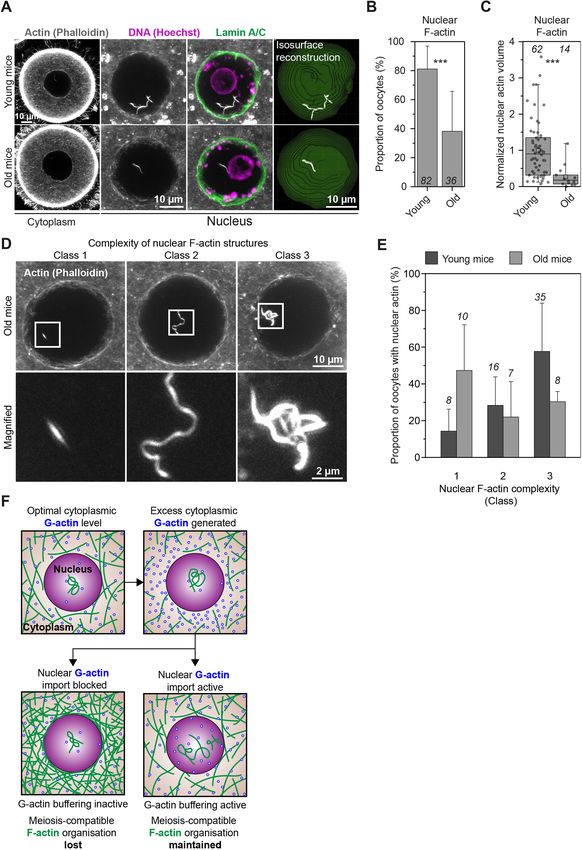
RESEARCH ARTICLE                                                             Journal of Cell Science (2022) 135, jcs259807. doi:10.1242/jcs.259807

                                                                                                                                                     Journal of Cell Science

Fig. 5. See next page for legend.

2004; Wei et al., 2020) (Fig. 3G–I). We then visualised chromatin    excess nuclear actin filaments led to notably reduced chromatin
(by staining with 5-SiR-Hoechst) and the nuclear envelope            mobility (Fig. 4A and E; Movies 6–9). We investigated this further
(by using fluorescent nuclear membrane nanobodies) at high-          by automated 3D tracking of prominent chromatin spots throughout
temporal resolution. Initial analysis of these data indicated that   the nucleoplasm (Fig. 4A and E). Indeed, nuclei containing excess

                                                                                                                                                7
RESEARCH ARTICLE Journal of Cell Science (2022) 135, jcs259807. doi:10.1242/jcs.259807
Fig. 5. Targeted nuclear F-actin stabilisation diminishes chromatin and support transcription through interactions between actin and
mobility. (A) Still images from a time-lapse movie of chromatin (5-SiR- RNA polymerase II (Dopie et al., 2012; Serebryannyy et al., 2016a).
Hoechst, magenta) and stabilised nuclear F-actin (overexpressed nuclear actin
chromobody, green). Boxed areas are shown magnified in images below.
(B) Still images from representative time-lapse movies of chromatin movement Actin mutants severely impact oocyte chromosomal
in control (mScarlet-NLS) or nuclear actin chromobody overexpressing alignment and segregation
(NuclAct-VHH) mouse oocytes. Chromatin (SiR-5-Hoechst) is shown in Importantly, some disease-causing actin mutants also lead to
magenta and nuclear membrane (Lamin chromobody) is shown in grey. assembly of persistent nuclear actin filaments in interphase nuclei
Colour-coded temporal projection images indicate the degree of chromatin that typically do not contain F-actin (Serebryannyy et al., 2016b).
mobility in the 500 s observation time. 3D tracks represent the spatial coverage
In this context, we probed the lasting effect of nuclear beta-actin-
of prominent chromatin spots over a 60-min observation period. (C) Distance
from starting position of prominent chromatin spots in 3D over a 60-min
S14C mutants on oocyte meiosis by live imaging of metaphase
observation period in control (mScarlet-NLS) or nuclear actin nanobody- chromosome dynamics in oocytes that contained excess nuclear
(NuclAct-VHH-mScarlet) overexpressing mouse oocytes. Data are from three F-actin bundles during meiotic prophase arrest. These experiments
independent experiments. (D) Cumulative instantaneous displacement of revealed striking defects in chromosome alignment and segregation
prominent chromatin spots in 3D over a 60-min observation period in control in oocytes expressing mutant beta-actin-S14C. Compared to ∼90%
(mScarlet-NLS) or nuclear actin nanobody (NuclAct-VHH-mScarlet) of control oocytes that progressed normally through meiosis I,
overexpressing mouse oocytes. Data are from three independent experiments.
Two-way analysis of variance was used to evaluate statistical significance.
∼50% of oocytes expressing actin mutants showed at least one
(E) Relative frequencies of chromatin spot instantaneous displacement in lagging chromosome during anaphase I (Fig. 6A and B; Movies 13
control (mScarlet-NLS) or nuclear actin nanobody- (NuclAct-VHH-mScarlet) and 14). In addition, expression of the 14C actin mutant caused
overexpressing mouse oocytes. Data are from three independent experiments. significant metaphase chromosome misalignment in oocytes and
Two-tailed Student’s t-test was used to evaluate statistical significance. metaphase II-arrested eggs (Fig. 6C–E; Movies 13 and 14). Super-
(F) Single-section Airyscan images of phalloidin-labelled cytoplasmic F-actin resolution immunofluorescence microscopy of F-actin,
and maximum intensity projections (nine confocal sections) of nuclear actin
microtubules and chromosomes in oocytes expressing actin
filaments (grey) in Control (mScarlet-NLS) or nuclear actin nanobody-
(NuclAct-VHH-mScarlet) overexpressing mouse oocytes. Boxes in the oocyte mutants further suggested that, after nuclear envelope
cytoplasm and surrounding the nucleus mark regions that are magnified in disassembly, stable F-actin structures become embedded in
insets. (G) Quantification of cytoplasmic F-actin network intensity in Control meiotic spindles where they are likely to obstruct chromosomal
(mScarlet-NLS) or nuclear actin nanobody- (NuclAct-VHH-mScarlet) organisation (Fig. 6F).
overexpressing mouse oocytes. Data are from three independent experiments.
Statistical significance was evaluated using two-tailed Student’s t-test.
Targeted actin mutant degradation restores meiotic fidelity
in oocytes
levels of F-actin showed significantly less movement of To directly confirm whether chromosome alignment and separation
chromatin spots over time (Fig. 4B–D,F–H). These data are defects arise from actin mutants that are retained in the cytoplasm
consistent with recent observations that Cytochalasin D treatment after nuclear envelope disassembly, we sought to rapidly degrade
dampens chromatin mobility in mouse oocytes (Almonacid et al., these mutants after releasing oocytes from prophase arrest. We thus
2019). adapted the degradation tag (dTAG) system, a recently innovated
Our results strongly indicated that reduced chromatin movement method for targeted and rapid degradation of FKBP12F36V-tagged
in cytoskeletal perturbation assays are more attributed to increased proteins (Nabet et al., 2018, 2020) (Fig. S4A), for use in mouse
nuclear F-actin levels than with disruption of the cytoplasmic oocytes. Our proof-of-concept experiments using the fluorescent
actin networks. This prediction was supported by live imaging protein mClover3 showed that the small molecule dTAG-13 (see
experiments, revealing that stable nuclear actin filaments (induced Materials and Methods) can degrade overexpressed FKBP12F36V-
by high expression of nuclear actin chromobody) can physically mClover3 within 100 min of addition (Fig. 7A; Movies 15 and 16).
entrap chromatin (marked with histone H2B) (Fig. 5A; Movie 10). Live imaging experiments further demonstrated that dTAG-13 is
We therefore used this nuclear actin chromobody to stabilise F-actin highly efficient in rapidly degrading SNAP-NLS-FKBP12F36V-
inside the oocyte nucleus and track the mobility of chromatin in 3D. beta-actin-S14C mutants, thereby effectively eliminating stable
Consistent with our Cytochalasin D addition and nuclear-specific nuclear F-actin cables as early as 40 min after the addition of small
actin monomer enrichment experiments, stabilisation of nuclear molecules (Fig. 7B; Movies 17 and 18). Phalloidin staining of
F-actin by using this approach diminished chromatin movement DMSO- and dTAG-13-treated oocytes confirmed the complete
(Fig. 5B–E; Movies 11 and 12). Importantly, this manipulation did degradation of stable nuclear F-actin bundles when this approach
not alter cytoplasmic F-actin networks (Fig. 5F and G). These had been used (Fig. 7C). We therefore generated and expressed
results demonstrate that nuclear F-actin stabilisation is sufficient to FKBP12F36V-FLAG-NLS (Control) and FKBP12F36V-FLAG-
restrict chromatin movement (Fig. 3I–K; Movies 11 and 12) and NLS-beta-actin-S14C mRNA in prophase-arrested oocytes.
provide new explanation for why drugs that disrupt cytoplasmic Consistent with our previous experiments, this led to chromosome
Journal of Cell Science
F-actin also impact chromatin mobility. Dynamic movement of alignment and segregation errors in oocytes that contained S14C
chromatin within the nucleus has recently been linked to basal level mutants (Fig. 7D and E; Movies 19 and 20). dTAG-mediated
of transcription in mouse oocytes (Almonacid et al., 2019). We thus degradation of actin mutants before meiotic spindle assembly
expect stable nuclear F-actin bundles that arise from excess successfully rescued defects of chromosome dynamics in oocytes
monomer import and drastically reduce chromatin mobility to that expressed FKBP12F36V-FLAG-NLS-beta-actin-S14C (Fig. 7D
ultimately impact transcriptional dynamics in oocytes. Induction of and E; Movies 21 and 22). This confirmed that actin mutants that
stable nuclear actin filaments in cultured cells that do not normally disrupt nuclear dynamics can also compromise later stages of
contain nuclear F-actin has also been shown to inhibit transcription mammalian oocyte meiosis. Future studies that explore
by RNA polymerase II (Serebryannyy et al., 2016a). However, the physiologically relevant actin mutants in the context of oocyte
underlying mechanisms in this model are thought to involve meiosis could therefore advance the current understanding of causes
sequestration of nuclear actin monomers that are actively imported of female infertility.
8RESEARCH ARTICLE Journal of Cell Science (2022) 135, jcs259807. doi:10.1242/jcs.259807
Journal of Cell Science
Fig. 6. See next page for legend.
9RESEARCH ARTICLE Journal of Cell Science (2022) 135, jcs259807. doi:10.1242/jcs.259807
Fig. 6. Actin mutants that disrupt nuclear dynamics compromise oocyte mechanotransduction of actin-based forces to the nucleus is known
meiosis. (A) Frequency of lagging chromosomes in control- (optimal nuclear to modulate nuclear mechanics and function in health and disease
F-actin in prophase) and S14C actin mutant-expressing (excess nuclear
(Isermann and Lammerding, 2013; Martino et al., 2018; Alisafaei
F-actin in prophase) mouse oocytes. Data are from four independent
experiments. (B) Still images from representative time-lapse movies of et al., 2019). However, a direct role of the nucleus itself in this
chromosome segregation during meiosis I in control or S14C actin mutant- process by regulating cytosolic G-actin concentration, and thus
expressing oocytes. Microtubules (EGFP-MAP4-MTBD) are shown in grey and F-actin assembly and force generation, should now be considered.
chromosomes (H2B-mRFP) are shown in magenta. Boxed areas are shown Finally, our data indicate that commonly used actin drugs
magnified in bottom panels. (C) Frequency of misaligned chromosomes during unintendedly stabilise nuclear F-actin and significantly affect
meiosis I in control (optimal nuclear F-actin in prophase) and S14C actin
nuclear mechanics. This will have important implications for past
mutant-expressing (excess nuclear F-actin in prophase) mouse oocytes. Data
are from four independent experiments. (D) Frequency of misaligned
and future studies of sub-cellular actin-based structures in single-
chromosomes during meiosis II in control (optimal nuclear F-actin in prophase) and multi-nucleated cells.
and S14C actin mutant-expressing (excess nuclear F-actin in prophase)
mouse eggs. Data are from four independent experiments. (E) Representative MATERIALS AND METHODS
images of fully aligned chromosomes in control (FLAG-NLS) and severely
Preparation and microinjection of mammalian oocytes
misaligned chromosomes (white arrows) in S14C actin mutant-expressing
All mice were maintained in a specific pathogen-free environment according
mouse eggs. Microtubules (EGFP-MAP4-MTBD) are shown in grey,
chromosomes (H2B-mRFP) are shown in magenta. Boxed areas are shown in
to UK Home Office regulations and the guidelines of the University of
insets magnified (5×). (F) Single-section Airyscan images of actin (grey), Bristol Animal Services Unit. Oocytes were isolated from ovaries of 129S6/
microtubules (green) and chromosomes (magenta) in S14C actin mutant- SvEvTac mice aged between 8 and 12 weeks (young CD-1 mice), 10 and
expressing (excess nuclear F-actin in prophase) oocytes during meiosis I and 12 months (old CD-1 mice) or 8 and 12 weeks, cultured and microinjected
in one egg during meiosis II. Numbered and boxed areas indicate stable actin with 6–8 pl of in vitro transcribed mRNA, as described in detail recently
filament bundles that are shown rotated and magnified (2×) in the insets below. (Mogessie, 2020).
Statistical significance was evaluated in A, C and D by using Fisher’s exact Sheep ovaries were obtained from a University of Bristol Veterinary
test. School slaughterhouse and transported to the laboratory, while stored at
37°C in M2 medium (Mogessie, 2020). Oocytes covered with several layers
of cumulus cells were collected from ovaries by aspiration with an 18-gauge
DISCUSSION needle and cultured in M2 medium supplemented with 750 μM N6,2′-O-
Transferring excess monomeric G-actin to the nucleus could be dibutyryladenosine 3′,5′-cyclic monophosphate sodium salt (dbcAMP)
physiologically important when prophase-arrested oocytes resume before fixation and processing.
meiosis, en route to becoming eggs. This process is accompanied by
a natural reduction in cytoplasmic F-actin network density that is Generation of expression constructs and mRNA synthesis
thought to support asymmetric cell division (Holubcova et al., To mark microtubules, an EGFP variant of the microtubule binding domain
2013). It is conceivable that such F-actin reduction, similarly to of mouse MAP4 (MAP4-MTBD, 659–1125 aa) (Mogessie et al., 2015) was
drug-mediated F-actin disruption, also releases actin monomers into generated as described by Mogessie and Schuh (2017). To label
the cytoplasm. Prompt monomer transfer to the large nucleus might chromosomes, the coding sequence of histone H2B was obtained from
therefore enable oocytes to maintain a cytoplasmic F-actin mouse cDNA and transferred by using Gibson assembly into the HindIII site
organisation that is compatible with healthy meiosis. Importantly, of pmRFP-N3 using primers 5′-GGACTCAGATCTCGAGCTCAATG-
we found that the amount and complexity of nuclear F-actin CCTGAGCCTGCGAAG-3′ and 5′-CCGTCGACTGCAGAATTCGACT-
TGGAGCTGGTGTACTTGG-3′. The fragment corresponding to H2B-
structures in non-manipulated mouse oocytes significantly declines
mRFP was then transferred into the XhoI-NotI site of pGEMHE (Schuh and
with increasing reproductive age in females – with oocytes from Ellenberg, 2007) to generate the final construct pGEM-H2B-mRFP. To
12-month-old mothers having only 27% of the nuclear F-actin label nuclear actin, fluorescent nuclear actin nanobody (nuclear actin
levels observed in younger (8–12 weeks old) mothers (Fig. 8A–E). chromobody) plasmid was purchased from ChromoTek and transferred into
This raises the intriguing possibility that loss of nuclear actin the HindIII-EcoRI site of pGEMHE. To label the nuclear envelope,
polymerisation capacity in oocytes could be linked with fluorescent lamin nanobody (lamin chromobody) plasmid was purchased
reproductive age-related female infertility. from ChromoTek and transferred into the NcoI-XbaI site of pGEMHE.
Homeostatic G-actin buffering (Fig. 8F) could be a widely Wild-type and S14C actin mutant expression constructs were generated
conserved feature of large mammalian oocyte nuclei. For instance, using a synthetic construct encoding the SV40 nuclear localisation signal
prophase nuclei in non-manipulated sheep oocytes can also contain (NLS) (5′-CCGCCTAAGAAAAAGCGGAAGGTG-3′) fused to mouse
beta-actin (NM_007393.5). pGEM-FLAG-NLS beta-actin was generated
prominent nuclear actin filaments (Fig. 1F). In frog oocytes, where
by PCR linearisation of pGEMHE with primers 5′-AATTCTGCAGTC-
the nucleus measures over 400 μm in diameter, F-actin maintains GACGGC-3′ and 5′-CGAAGCTTGAGCTCGAGATC-3′ and joined by
organisation of the nucleus by stabilising its contents against using Gibson assembly with NLS-beta-actin that was flanked by primers
gravitational forces (Feric and Brangwynne, 2013). Our data 5′-GATCTCGAGCTCAAGCTTCGATGGACTACAAGGACGACGAC-
therefore extend to mammalian experimental models the GACAAGGGGCCGCCTAAG-3′ and 5′-GGGCCGTCGACTGCAGA-
Journal of Cell Science
observation that F-actin is linked to the dynamics of large nuclei. ATTTTAGAAGCACTTGCGGTG-3′, with the coding sequence of the
Interestingly, nuclear F-actin is also known to assemble in a variety FLAG peptide DYKDDDDK shown in bold. pGEM-FLAG-NLS-beta-
of cellular contexts in non-gamete cells and embryos (Baarlink et al., actin-S14C mutant was generated by site-directed mutagenesis using
2017, 2013; Caridi et al., 2019; Plessner et al., 2015; Wang et al., primers 5′-GTCGTCGACAACGGCTGCGGCATGTGCAAAGCC-3′
2019; Kelpsch and Tootle, 2018; Wesolowska and Lenart, 2015; and 5′-GGCTTTGCACATGCCGCAGCCGTTGTCGACGAC-3′. pGEM-
FKBP12F36V-mClover3 was generated by linearising pGEM-mClover3
Okuno et al., 2020) with postulated functions ranging from DNA
using primers 5′-AATTCTGCAGTCGACGGC-3′ and 5′-CGAAGC-
repair to chromatin organisation. It will be important to explore TTGAGCTCGAGATC-3′, and transferring into it by Gibson assembly
whether the oocyte G-actin buffering process we described here is a (lower case nucleotides indicate vector homology sequence) 5′-ga-
universal feature of mammalian nuclei and non-mammalian models tctcgagctcaagcttcgATGGGAGTGCAGGTGGAAAC-3′ and 5′-gggcc-
in which F-actin structures are intimately associated with the gtcgactgcagaattGCCGCCTTCCAGTTTTAG-3′ flanked FKBP12F36V
nucleus (Mori et al., 2014; Wesolowska et al., 2020). In addition, synthetic coding sequence (Nabet et al., 2018). pGEM-FKBP12F36V-
10RESEARCH ARTICLE Journal of Cell Science (2022) 135, jcs259807. doi:10.1242/jcs.259807
Journal of Cell Science
Fig. 7. See next page for legend.
11RESEARCH ARTICLE Journal of Cell Science (2022) 135, jcs259807. doi:10.1242/jcs.259807
Fig. 7. Targeted degradation of stable nuclear actin filaments restores and 0.5% Triton X-100 (v/v) at 37°C for 25–30 min (mouse) or for 60 min
chromosome segregation fidelity in oocytes. (A) Panels from time-lapse after 10 s pre-permeabilisation in 0.4% Triton X-100 (v/v) in water (sheep).
movies of Control (DMSO) and dTAG-13-treated prophase-arrested mouse Oocytes were extracted in PBS supplemented with 0.3% Triton X-100 (v/v)
oocytes expressing FKBP12F36V-mClover3. (B) Rapid degradation of oocyte at 4°C overnight. Antibody, F-actin and chromosome staining were
nuclear F-actin bundles using the dTAG system. Panels from time-lapse performed for 2–2.5 h in PBS, 3% BSA (w/v), and 0.1% Triton X-100
movies of Control (DMSO) and dTAG-13-treated prophase-arrested mouse (v/v) at room temperature. The nuclear envelope was stained by using
oocytes expressing FKBP12F36V-SNAP-NLS-beta-actin-S14C are shown. primary rabbit anti-Lamin A/C antibody (ab133256, Abcam; 1:500) and
(C) Phalloidin labelling of nuclear F-actin in FKBP12F36V-SNAP-NLS-beta-
Alexa-Fluor-647-labelled secondary anti-rat (Molecular Probes; 1:400)
actin-S14C expressing prophase-arrested mouse oocytes fixed after treatment
antibodies. F-actin was stained with Rhodamine or Alexa-Fluor-488
with DMSO (Control) (DMSO) or dTAG-13. (D) Quantification of metaphase I
phalloidin (Molecular Probes; 1:20). DNA was stained with 5 µg/ml
oocytes expressing Control (FKBP12F36V-FLAG-NLS) or FKBP12F36V-FLAG-
NLS-beta-actin-S14C with correctly aligned chromosomes after maturation in
Hoechst dye 33342 (Molecular Probes).
DMSO or dTAG-13 (to degrade actin mutants). Data are from three Confocal and Airyscan super-resolution images were acquired with a
independent experiments. (E) Quantification of metaphase I oocytes Zeiss LSM800 confocal microscope equipped with a 40× C-Apochromat
expressing Control (FKBP12F36V-FLAG-NLS) or FKBP12F36V-FLAG-NLS- 1.2 NA water-immersion objective. Images under control and perturbation
beta-actin-S14C with correctly segregated chromosomes after maturation in conditions were acquired by using identical imaging conditions.
DMSO or dTAG-13 (to degrade actin mutants). Data are from three
independent experiments. Statistical significance was assessed using Fisher’s Drug addition and fluorescence labelling experiments
exact test in D and E. To disrupt actin, oocytes were treated for 1 h with Cytochalasin D (C8273-
1MG, Merck) at a final concentration of 5 μg/ml, Latrunculin B (428020-
1MG, Merck) at a final concentration of 5 μM or CK-666 (182515-25MG,
SNAP-NLS-beta-actin-S14C was generated by linearising pGEM with
Merck) at a final concentration of 200 μM in M2 medium supplemented
5′-ATGGACAAAGACTGCGAAATG-3′ and 5′-TGAGCTCGAGATCT-
with dbcAMP. To block nuclear import, oocytes were treated with a
GAGAC-3′, and transferring into it by Gibson assembly 5′-ggtctcagatctc-
combination of 100 μM Importazole (SML0341-5MG, Merck) and 30 μM
gagctcaATGGGAGTGCAGGTGGAAAC-3′ and 5′-atttcgcagtctttgtc-
Ivermectin (I8000010, Merck). We have found that a combination of these
catGCCGCCTTCCAGTTTTAG-3′ flanked FKBP12F36V. pGEM-
two drugs is a much tighter block of nuclear import in mouse oocytes. This
FKBP12F36V-SNAP-NLS was generated by linearising pGEM with
combination was thus used to achieve the maximal effect of inhibition in our
5′-AATTCTGCAGTCGACGGC-3′ and 5′-CGAAGCTTGAGCTCGA-
analyses. These final concentrations were maintained in experiments where
GATC-3′, and transferring into it by Gibson assembly 5′-gatctcgagct-
oocytes were simultaneously treated with Cytochalasin D, Importazole and
caagcttcgATGGGAGTGCAGGTGGAAAC-3′ and 5′-gggccgtcgactgca-
Ivermectin. For rapid protein degradation experiments, prophase-arrested
gaattTTACACCTTCCGCTTTTTCTTAGG-3′ flanked FKBP12F36V from
oocytes were treated with and matured in 100 nM dTAG-13 (SML2601,
pGEM-FKBP12F36V-SNAP-NLS-beta-actin-S14C. pGEM-FKBP12F36V-
Merck). All drugs were dissolved in DMSO (D2650-5X5ML, Merck).
FLAG-NLS was generated by linearising pGEM-FLAG-NLS with
Where DMSO was used as control, it was diluted in M2 medium
5′-GGGCCGCCTAAGAAAAAG-3′ and 5′-CTTGTCGTCGTCGTCCTT-
supplemented with dbcAMP, identically to corresponding experimental
G-3′, and transfer into it by Gibson assembly 5′-acaaggacgacgacgacaa-
conditions. SNAP-tagged proteins expressed in oocytes from microinjected
gATGGGAGTGCAGGTGGAAAC-3′ and 5′-cgctttttcttaggcggcccTTC-
mRNA were fluorescence labelled by incubating cells with 3 μM SNAP-
CAGTTTTAGAAGCTCCACATC-3′ flanked FKBP12F36V. pGEM-
Cell 647-SiR (S9102S, NEB) in M2 medium for 30 min and for a further
FKBP12F36V-FLAG-NLS-beta-actin-S14C was generated by linearising
30 min in M2 medium without substrate.
pGEM-FLAG-NLS-beta-actin-S14C with 5′-GGGCCGCCTAAGAAAA-
To fluorescence label nuclear actin structures, nuclear actin chromobody
AG-3′ and 5′-CTTGTCGTCGTCGTCCTTG-3′, and transferring into it by
was expressed in oocytes at variable concentrations. For low-level
Gibson assembly 5′-acaaggacgacgacgacaagATGGGAGTGCAGGTG-
expression, oocytes were microinjected with 6–8 pl of 0.02 μg/μl nuclear
GAAAC-3′ and 5′-cgctttttcttaggcggcccTTCCAGTTTTAGAAGCTCCA-
actin chromobody mRNA followed by expression for 1–3 h. For high-level
CATC-3′ flanked FKBP12F36V. pGEM-F-Tractin-NLS was generated by
expression, oocytes were microinjected with 6–8 pl of 0.2 μg/μl nuclear
linearising pGEM-mClover3 with 5′-AATTCTGCAGTCGACGGC-3′ and
actin chromobody mRNA followed by expression for 2 h.
5′-CGAAGCTTGAGCTCGAGATC-3′, and transferring into it by Gibson
assembly 5′-gatctcgagctcaagcttcgATGGCGCGGCCACGGGGC-3′ and
5′-gggccgtcgactgcagaattTGCCCTAGATCGCAAACCACTCACCTT- Chromosome alignment and segregation analysis
CCG-3′ flanked synthetic coding sequence of mouse F-Tractin (70–199 bp For chromosome alignment and segregation analyses, images were acquired
in NM_146125.2). pGEM-mEGFP-UtrCH-NLS was generated by inserting a at a temporal resolution of 5 min and with a Z-stack thickness of ∼40 µm at
BamHI-NotI flanked synthetic coding sequence of UtrCH-NLS into the 1.5 µm confocal sections. Chromosomes that were distinctly separate from
BamHI-NotI site of pGEM-mEGFP-C1 (Mogessie and Schuh, 2017). the metaphase plate chromosome mass at the time of anaphase onset (shown
Capped mRNA was synthesised using T7 polymerase (mMessage in Fig. 4D) were scored as misaligned chromosomes. Chromosomes that fell
mMachine kit, following the manufacturer’s instructions, Ambion). behind the main mass of segregating chromosomes for at least 10 min after
mRNA concentrations were determined by measurement on a Nanodrop anaphase onset were scored as lagging chromosomes. For both
spectrophotometer (Thermo Scientific). quantifications, maximum intensity projections of only those metaphase
spindles that were oriented parallel to the imaging plane at and during
Confocal and super-resolution live imaging anaphase were analysed.
Journal of Cell Science
Images were acquired with a Zeiss LSM800 microscope at 37°C. Oocytes
were imaged in M2 medium under mineral oil using a 40× C-Apochromat Isosurface reconstruction and 3D volume quantification of
1.2 NA water-immersion objective as described in more detail by Mogessie nuclear actin filaments
(2020). Super-resolution time-lapse images were acquired using the For 3D volume quantification of nuclear actin filaments, confocal images
Airyscan module on a Zeiss LSM800 microscope and processed post- of nuclei in fixed oocytes were typically acquired at a spatial resolution of
acquisition using ZEN2 software (Zeiss). 1 μm confocal sections covering 45 μm. Isosurfaces, i.e. 3D surface
representations of points with equal values in a 3D data distribution,
Immunofluorescence microscopy corresponding to nuclear membranes were reconstructed in 3D by using the
We have recently published detailed protocols for optimised staining of actin Cell module of Imaris software (Bitplane) and the immunofluorescence
filaments, which were used in this study to visualise nuclear F-actin signal of nuclear envelope antibodies. The nuclear isosurface was used to
structures (Mogessie, 2020). Mouse and sheep oocytes were fixed with mask F-actin signal and to remove cytoplasmic F-actin structures
100 mM HEPES, 50 mM EGTA, 10 mM MgSO4, 2% formaldehyde (v/v), surrounding the nuclei. In the masked region, 3D isosurfaces of
12RESEARCH ARTICLE Journal of Cell Science (2022) 135, jcs259807. doi:10.1242/jcs.259807
Journal of Cell Science
Fig. 8. See next page for legend.
13RESEARCH ARTICLE Journal of Cell Science (2022) 135, jcs259807. doi:10.1242/jcs.259807 Fig. 8. Oocyte nuclear F-actin abundance and complexity declines with (line), mean (small square), 5th, 95th (whiskers) and 25th and 75th female reproductive age. (A) Single-section Airyscan images of phalloidin- percentile (box enclosing 50% of the data) and are overlaid with individual labelled cytoplasmic F-actin and maximum intensity projections (nine confocal data points. Average (mean), standard deviation and statistical significance sections) of nuclear actin filaments (grey), DNA (magenta) and nuclear based on two-tailed Student’s t-test or Fisher’s exact test were calculated in membrane (green) in oocytes isolated from young and old mice. OriginPro software (OriginLab). All error bars represent standard (B) Quantification of nuclear F-actin presence in oocytes isolated from young deviations. Two-way analysis of variance was performed in Prism and old mice. Data are from three independent experiments. (C) Quantification software (GraphPad). Significance values are *P
RESEARCH ARTICLE                                                                                     Journal of Cell Science (2022) 135, jcs259807. doi:10.1242/jcs.259807

Herbert, M., Kalleas, D., Cooney, D., Lamb, M. and Lister, L. (2015). Meiosis and         Plessner, M., Melak, M., Chinchilla, P., Baarlink, C. and Grosse, R. (2015).
  maternal aging: insights from aneuploid oocytes and trisomy births. Cold Spring           Nuclear F-actin formation and reorganization upon cell spreading. J. Biol. Chem.,
  Harb Perspect Biol 7, a017970. doi:10.1101/cshperspect.a017970                            290, 11209-11216. doi:10.1074/jbc.M114.627166
Holubcova, Z., Howard, G. and Schuh, M. (2013). Vesicles modulate an actin                Posern, G., Miralles, F., Guettler, S. and Treisman, R. (2004). Mutant actins that
  network for asymmetric spindle positioning. Nat. Cell Biol. 15, 937-947.                  stabilise F-actin use distinct mechanisms to activate the SRF coactivator MAL.
  doi:10.1038/ncb2802                                                                       EMBO J. 23, 3973-3983. doi:10.1038/sj.emboj.7600404
Isermann, P. and Lammerding, J. (2013). Nuclear mechanics and                             Scheffler, K., Uraji, J., Jentoft, I., Cavazza, T., Monnich, E., Mogessie, B. and
  mechanotransduction in health and disease. Curr. Biol. 23, R1113-R1121.                   Schuh, M. (2021). Two mechanisms drive pronuclear migration in mouse
  doi:10.1016/j.cub.2013.11.009
                                                                                            zygotes. Nat. Commun. 12, 841. doi:10.1038/s41467-021-21020-x
Kelpsch, D. J. and Tootle, T. L. (2018). Nuclear actin: from discovery to function.
                                                                                          Schuh, M. (2011). An actin-dependent mechanism for long-range vesicle transport.
  Anat. Rec. 301, 1999-2013. doi:10.1002/ar.23959
                                                                                            Nat. Cell Biol. 13, 1431-1436. doi:10.1038/ncb2353
Martino, F., Perestrelo, A. R., Vinarsky, V., Pagliari, S. and Forte, G. (2018).
                                                                                          Schuh, M. and Ellenberg, J. (2007). Self-organization of MTOCs replaces
  Cellular mechanotransduction: from tension to function. Front Physiol 9, 824.
  doi:10.3389/fphys.2018.00824                                                              centrosome function during acentrosomal spindle assembly in live mouse
Melak, M., Plessner, M. and Grosse, R. (2017). Actin visualization at a glance.             oocytes. Cell 130, 484-498. doi:10.1016/j.cell.2007.06.025
  J. Cell Sci. 130, 525-530. doi:10.1242/jcs.204487                                       Schuh, M. and Ellenberg, J. (2008). A new model for asymmetric spindle
Mogessie, B. (2020). Visualization and functional analysis of spindle actin and             positioning in mouse oocytes. Curr. Biol. 18, 1986-1992. doi:10.1016/j.cub.2008.
  chromosome segregation in mammalian oocytes. Methods Mol. Biol. 2101,                     11.022
  267-295. doi:10.1007/978-1-0716-0219-5_17                                               Serebryannyy, L. A., Parilla, M., Annibale, P., Cruz, C. M., Laster, K., Gratton, E.,
Mogessie, B. and Schuh, M. (2017). Actin protects mammalian eggs against                    Kudryashov, D., Kosak, S. T., Gottardi, C. J. and De Lanerolle, P. (2016a).
  chromosome segregation errors. Science 357, eaal1647. doi:10.1126/science.                Persistent nuclear actin filaments inhibit transcription by RNA polymerase II.
  aal1647                                                                                   J. Cell Sci. 129, 3412-3425.
Mogessie, B., Roth, D., Rahil, Z. and Straube, A. (2015). A novel isoform of MAP4         Serebryannyy, L. A., Yuen, M., Parilla, M., Cooper, S. T. and De Lanerolle, P.
  organises the paraxial microtubule array required for muscle cell differentiation.        (2016b). The effects of disease models of nuclear actin polymerization on the
  eLife 4, e05697. doi:10.7554/eLife.05697                                                  nucleus. Front Physiol 7, 454. doi:10.3389/fphys.2016.00454
Mogessie, B., Scheffler, K. and Schuh, M. (2018). Assembly and positioning of the         Soderholm, J. F., Bird, S. L., Kalab, P., Sampathkumar, Y., Hasegawa, K.,
  oocyte meiotic spindle. Annu. Rev. Cell Dev. Biol. 34, 381-403. doi:10.1146/              Uehara-Bingen, M., Weis, K. and Heald, R. (2011). Importazole, a small
  annurev-cellbio-100616-060553
                                                                                            molecule inhibitor of the transport receptor importin-beta. ACS Chem. Biol. 6,
Mori, M., Somogyi, K., Kondo, H., Monnier, N., Falk, H. J., Machado, P.,
                                                                                            700-708. doi:10.1021/cb2000296
  Bathe, M., Nedelec, F. and Lenart, P. (2014). An Arp2/3 nucleated F-actin shell
                                                                                          Spector, I., Shochet, N. R., Kashman, Y. and Groweiss, A. (1983). Latrunculins:
  fragments nuclear membranes at nuclear envelope breakdown in starfish
  oocytes. Curr. Biol. 24, 1421-1428. doi:10.1016/j.cub.2014.05.019                         novel marine toxins that disrupt microfilament organization in cultured cells.
Morton, W. M., Ayscough, K. R. and Mclaughlin, P. J. (2000). Latrunculin alters             Science 219, 493-495. doi:10.1126/science.6681676
  the actin-monomer subunit interface to prevent polymerization. Nat. Cell Biol. 2,       Stuven, T., Hartmann, E. and Gorlich, D. (2003). Exportin 6: a novel nuclear export
  376-378. doi:10.1038/35014075                                                             receptor that is specific for profilin.actin complexes. EMBO J. 22, 5928-5940.
Nabet, B., Roberts, J. M., Buckley, D. L., Paulk, J., Dastjerdi, S., Yang, A.,              doi:10.1093/emboj/cdg565
  Leggett, A. L., Erb, M. A., Lawlor, M. A., Souza, A. et al. (2018). The dTAG            Wagstaff, K. M., Sivakumaran, H., Heaton, S. M., Harrich, D. and Jans, D. A.
  system for immediate and target-specific protein degradation. Nat. Chem. Biol. 14,        (2012). Ivermectin is a specific inhibitor of importin alpha/beta-mediated nuclear
  431-441. doi:10.1038/s41589-018-0021-8                                                    import able to inhibit replication of HIV-1 and dengue virus. Biochem. J. 443,
Nabet, B., Ferguson, F. M., Seong, B. K. A., Kuljanin, M., Leggett, A. L.,                  851-856. doi:10.1042/BJ20120150
  Mohardt, M. L., Robichaud, A., Conway, A. S., Buckley, D. L., Mancias, J. D.            Wang, Y., Sherrard, A., Zhao, B., Melak, M., Trautwein, J., Kleinschnitz, E. M.,
  et al. (2020). Rapid and direct control of target protein levels with VHL-recruiting      Tsopoulidis, N., Fackler, O. T., Schwan, C. and Grosse, R. (2019). GPCR-
  dTAG molecules. Nat. Commun. 11, 4687. doi:10.1038/s41467-020-18377-w                     induced calcium transients trigger nuclear actin assembly for chromatin dynamics.
Nagaoka, S. I., Hassold, T. J. and Hunt, P. A. (2012). Human aneuploidy:                    Nat. Commun. 10, 5271. doi:10.1038/s41467-019-13322-y
  mechanisms and new insights into an age-old problem. Nat. Rev. Genet. 13,               Wei, M., Fan, X., Ding, M., Li, R., Shao, S., Hou, Y., Meng, S., Tang, F., Li, C. and
  493-504. doi:10.1038/nrg3245
                                                                                            Sun, Y. (2020). Nuclear actin regulates inducible transcription by enhancing RNA
Okuno, T., Li, W. Y., Hatano, Y., Takasu, A., Sakamoto, Y., Yamamoto, M.,
                                                                                            polymerase II clustering. Sci. Adv. 6, eaay6515. doi:10.1126/sciadv.aay6515
  Ikeda, Z., Shindo, T., Plessner, M., Morita, K. et al. (2020). Zygotic nuclear F-
                                                                                          Wesolowska, N. and Lenart, P. (2015). Nuclear roles for actin. Chromosoma 124,
  Actin safeguards embryonic development. Cell Rep 31, 107824. doi:10.1016/j.
  celrep.2020.107824                                                                        481-489. doi:10.1007/s00412-015-0519-8
Parisis, N., Krasinska, L., Harker, B., Urbach, S., Rossignol, M., Camasses, A.,          Wesolowska, N., Avilov, I., Machado, P., Geiss, C., Kondo, H., Mori, M. and
  Dewar, J., Morin, N. and Fisher, D. (2017). Initiation of DNA replication requires        Lenart, P. (2020). Actin assembly ruptures the nuclear envelope by prying the
  actin dynamics and formin activity. EMBO J. 36, 3212-3231. doi:10.15252/embj.             lamina away from nuclear pores and nuclear membranes in starfish oocytes. eLife
  201796585                                                                                 9, e49774. doi:10.7554/eLife.49774
Pendleton, A., Pope, B., Weeds, A. and Koffer, A. (2003). Latrunculin B or ATP            Zuccotti, M., Giorgi Rossi, P., Martinez, A., Garagna, S., Forabosco, A. and
  depletion induces cofilin-dependent translocation of actin into nuclei of mast cells.     Redi, C. A. (1998). Meiotic and developmental competence of mouse antral
  J. Biol. Chem. 278, 14394-14400. doi:10.1074/jbc.M206393200                               oocytes. Biol. Reprod. 58, 700-704. doi:10.1095/biolreprod58.3.700

                                                                                                                                                                                  Journal of Cell Science

                                                                                                                                                                           15
You can also read