Complimentary and personal copy for - Christian Arning
←
→
Page content transcription
If your browser does not render page correctly, please read the page content below
Complimentary and personal copy for
Christian Arning

Brought to you by Thieme            www.thieme.com

                                    Ultrasound Criteria for
                                    Diagnosing Spontaneous
                                    Cervical Artery Dissections

                                    Ultraschall in der Medizin -
                                    European Journal of Ultra-
                                    sound
                                    2023
                                    2–26
                                    10.1055/a-2004-4986

                                    This electronic reprint is provided for non-
                                    commercial and personal use only: this
                                    reprint may be forwarded to individual
                                    colleagues or may be used on the author’s
                                    homepage. This reprint is not provided for
                                    distribution in repositories, including social
                                    and scientific networks and platforms.
Copyright & Ownership
© 2023. Thieme. All rights
reserved.
The journal Ultraschall in der
Medizin - European Journal of Ul-
trasound is owned by Thieme.
Georg Thieme Verlag KG,
Rüdigerstraße 14,
70469 Stuttgart, Germany
ISSN 0172-4614
Continuing Education

                                                              b
                                                            Ultrasound Criteria for Diagnosing Spontaneous
                                                            Cervical Artery Dissections

                                                            Authors                                                            nosis of dissection can contribute to stroke prevention –
                                                            Christian Arning                                                   through immediate therapy with anticoagulants or antiplate-
                                                                                                                               let drugs. This article describes the diagnostic criteria and typ-
                                                            Affiliations
                                                                                                                               ical findings of spontaneous dissection, in which no dissecting
                                                            Neurology, Praxis Neuro-Ultraschall, Hamburg, Germany
                                                                                                                               membrane is to be expected as in aortic dissection. Traumatic
                                                            Bibliography                                                       dissections following blunt or penetrating injuries also present
                                                            Ultraschall in Med                                                 with different findings. Examiners should be aware of possible
                                                            Published online: February 6, 2023                                 differential diagnoses whose ultrasound image may mimic a
                                                            DOI 10.1055/a-2004-4986                                            dissection. A frequently occurring but avoidable cause of mis-
                                                            ISSN 0172-4614                                                     diagnosis is idiopathic carotidynia. Ultrasound also enables
                                                            © 2023. Thieme. All rights reserved.                               differentiation between dissection and vasculitis or carotid
                                                                                                                               web and detection of normal variants such as fenestration of
Elektronischer Sonderdruck zur persönlichen Verwendung

                                                            Georg Thieme Verlag KG, Rüdigerstraße 14,
                                                            70469 Stuttgart, Germany                                           the vertebral artery. Further possibilities for misdiagnosis may
                                                                                                                               arise in the presence of a variant of the ascending pharyngeal
                                                            ABSTR AC T                                                         artery or in extracranial vasospasm. The different imaging
                                                            Spontaneous dissection of brain-supplying cervical arteries,       techniques for the detection of a dissection are complemen-
                                                            which also includes findings after minor injuries, is one of the   tary, as false-negative findings occur with all techniques; no
                                                            main causes of ischaemic strokes in young adults. Strokes due      method serves as the gold standard. In any case, ultrasound
                                                            to dissection are usually due to arterio-arterial embolism.        can make an important contribution to the detection of a dis-
                                                            They are rarely the first symptom of dissection because an in-     section, and it is worth knowing the diagnostic criteria.
                                                            traluminal thrombus must first develop. Therefore, early diag-

                                                                    Dissection of the cervical arteries supplying the brain is a          ical Symptoms“ information box. The clinical symptoms
                                                                    leading cause of ischemic stroke in young adults. Since               of dissections are described in detail in a recently pub-
                                                                    stroke rarely occurs as the initial symptom of dissection,            lished review [3]. Imaging techniques are necessary in
                                                                    early diagnosis and treatment can prevent strokes. Ima-               the second step for a definitive diagnosis.
                                                                    ging with ultrasound is beneficial, but there are some pit-
                                                                    falls to using this procedure. Examiners should have pre-
                                                                    cise knowledge of the diagnostic criteria and be informed                 I N F O R M AT I O N B O X : D I S S E C T I O N C L I N I C A L
                                                                    regarding differential diagnoses.                                         SYM P TOM S ( SEE ▶ FIG. 1A)
                                                                                                                                              1. Vascular wall symptoms
                                                                                                                                                  – Unilateral headache and/or facial pain asso-
                                                                    Importance of early Diagnosis                                                     ciated with dissection of the internal carotid
                                                                    Strokes resulting from dissection are most commonly                               artery
                                                                    due to arterioarterial embolism into a cerebral artery.                       – Unilateral neck occipital pain associated with
                                                                    However, the embolism does not manifest itself immedi-                            vertebral artery dissection
                                                                    ately with the development of the dissection, since an                    2. Vascular dilation to the outside
                                                                    intraluminal thrombus must first develop. For internal                        – Horner's syndrome or caudal cranial nerve le-
                                                                    carotid artery dissections, the mean time to onset of                             sion in internal carotid artery dissection
                                                                    cerebral symptoms is 8.8 days (median 96 hours) [1].                          – Cervical nerve root damage in vertebral artery
                                                                    Early diagnosis of dissection allows stroke prevention                            dissection (rare)
                                                                    through the use of anticoagulants or antiplatelet drugs                   3. Vascular stenosis due to compression of the
                                                                    [2].                                                                          vascular lumen
                                                                                                                                                  – Cerebral ischemia due to arterioarterial em-
                                                                    The diagnosis is made in two steps. The first step is the                         bolism originating from intraluminal thrombi
                                                                    suspected clinical diagnosis, refer to the „Dissection Clin-                      at the stenosis (delayed)

                                                                                                                               Arning C. Ultrasound Criteria for… Ultraschall in Med | © 2023. Thieme. All rights reserved.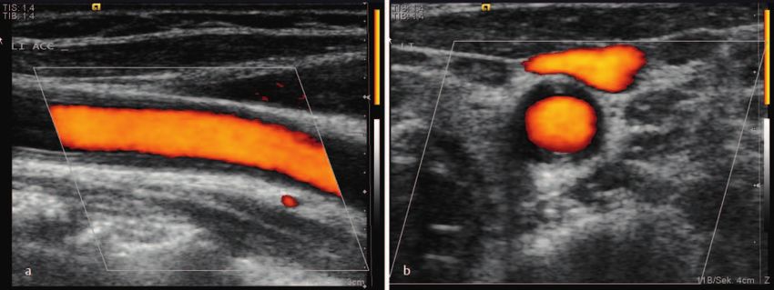
Continuing Education
b
Ultraschallkriterien zur Diagnose spontaner
Halsarterien-Dissektionen
Autorinnen/Autoren minaler Thrombus bilden muss. Daher kann die frühzeitige Di-
Christian Arning agnose der Dissektion zur Schlaganfallprävention beitragen –
durch sofortige gerinnungswirksame Therapie. Dieser Beitrag
Institute
beschreibt die diagnostischen Kriterien und typischen Befun-
Neurology, Praxis Neuro-Ultraschall, Hamburg, Germany
de der spontanen Dissektion, bei der ja keine Dissektions-
Bibliografie membran wie bei einer Aortendissektion zu erwarten ist.
Ultraschall in Med Auch traumatische Dissektionen nach stumpfen oder pene-
Published online: 2023 trierenden Verletzungen weisen oft andere Befunde auf. Un-
DOI 10.1055/a-2004-4986 tersucher sollten mögliche Differenzialdiagnosen kennen, de-
ISSN 0172-4614 ren Ultraschallbild eine Dissektion imitieren kann. Ultraschall
© 2023. Thieme. All rights reserved. erlaubt auch die Differenzierung zwischen Dissektion und
Vaskulitis oder Carotid Web und die Erkennung von Normva-
Elektronischer Sonderdruck zur persönlichen Verwendung
Georg Thieme Verlag KG, Rüdigerstraße 14,
70469 Stuttgart, Germany rianten wie Fensterung der A. vertebralis. Weitere Fehlermög-
lichkeiten können sich bei einer Abgangsvariante der Arteria
Z US A M M E N FA SS U N G pharyngea ascendens oder bei extrakraniellem Vasospasmus
Die spontane Dissektion hirnversorgender Halsarterien, die ergeben. Die verschiedenen bildgebenden Verfahren zum
auch Befunde nach Bagatellverletzungen einschließt, ist eine Nachweis einer Dissektion ergänzen sich, da bei allen Techni-
der Hauptursachen ischämischer Schlaganfälle bei jungen Er- ken falsch-negative Befunde vorkommen – keine Methode ist
wachsenen. Schlaganfälle bei Dissektion sind meist auf eine Goldstandard. Ultraschall kann jedenfalls einen wichtigen Bei-
arterioarterielle Embolie zurückzuführen und sind selten das trag zum Nachweis einer Dissektion leisten, und es lohnt sich,
erste Symptom einer Dissektion, da sich zunächst ein intralu- die diagnostischen Kriterien zu kennen.
Die Dissektion hirnversorgender Halsarterien ist eine der Dissektion“. In einer kürzlich publizierten Übersicht sind
Hauptursachen ischämischer Schlaganfälle bei jungen Er- die klinischen Symptome von Dissektionen detailliert be-
wachsenen. Da der Schlaganfall selten als erstes Symp- schrieben [3]. Für die definitive Diagnose sind im zweiten
tom der Dissektion auftritt, kann die frühzeitige Diagnose Schritt bildgebende Verfahren notwendig.
und Therapie Schlaganfällen vorbeugen. Bildgebung mit
Ultraschall ist vorteilhaft, es gibt aber einige Fallstricke.
Untersucher sollten die diagnostischen Kriterien genau INFOBOX: KLINI K DE R D ISS EK T ION
kennen und über Differenzialdiagnosen informiert sein. (S . ▶ ABB. 1A)
1. Symptome der Gefäßwand
– Einseitige Kopf- und/oder Gesichtsschmerzen
Relevanz frühzeitiger Diagnostik bei Dissektion der Arteria carotis interna
Schlaganfälle infolge einer Dissektion sind in den meisten – Einseitige Nacken-Hinterkopfschmerzen bei
Fällen auf eine arterioarterielle Embolie in einer Hirnarterie Dissektion der Arteria vertebralis
zurückzuführen. Die Embolie manifestiert sich aber nicht 2. Gefäßerweiterung nach außen
sofort mit Entstehung der Dissektion, denn zunächst – Horner-Syndrom oder kaudale Hirnnervenläsi-
muss sich ein intraluminaler Thrombus entwickeln. Bei Dis- on bei Dissektion der Arteria carotis interna
sektionen der Arteria carotis interna beträgt die mittlere – Zervikale Nervenwurzelschädigung bei Dis-
Zeit bis zum Auftreten zerebraler Symptome 8,8 Tage sektion der Arteria vertebralis (selten)
(Median 96 Stunden) [1]. Die frühe Diagnose der Dissekti- 3. Gefäßstenose durch Kompression des Gefäßlu-
on ermöglicht die Schlaganfallprävention durch Einsatz ei- mens
ner gerinnungshemmenden Medikation [2]. – Zerebrale Ischämie durch arterioarterielle Em-
bolie, ausgehend von intraluminalen Throm-
Die Diagnose wird in 2 Schritten gestellt. Erster Schritt ist ben an der Stenose (mit Verzögerung auftre-
die klinische Verdachtsdiagnose, s. Infobox „Klinik der tend)
Arning C. Ultrasound Criteria for… Ultraschall in Med | © 2023. Thieme. All rights reserved.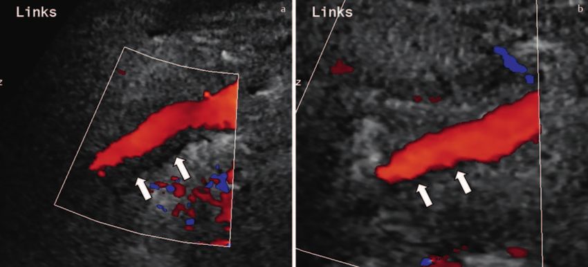
Continuing Education
b
imaging was carried out for a different issue [4]. Even
with targeted diagnostics with the issue of a dissection,
false-negative results occur with all imaging methods,
even with digital subtraction angiography, which allows
the best representation of the vascular lumen, but does
not depict the vascular wall and its thickening. According
to the joint statement of the American Heart Association
and American Stroke Association, no imaging modality is
the gold standard and superior to all others [2]. The Ger-
man guideline recommends combining two different ima-
ging modalities when dissection is suspected: MRI/MRA
plus ultrasound or CT/CTA plus ultrasound [5].
Finally, errors can also result from imaging – especially ul-
trasound – leading to a false-positive diagnosis of dissec-
tion. Some other vascular pathologies and normal variants
present findings that can be confused with a dissection.
Elektronischer Sonderdruck zur persönlichen Verwendung
Pathological Anatomy
Spontaneous dissection of the internal carotid artery or
▶ Fig. 1 Illustration of the pathological anatomy of dis- vertebral artery also includes dissections after minor inju-
section A Dissection of the internal carotid artery/verte- ries and is characterized by a mural hematoma that arises
bral artery with hematoma in the vessel wall due to rup- primarily without tearing the intima [6], probably due to
ture of vasa vasorum. An intramural hematoma can lead rupture of the vasa vasorum. The hematoma results in a
to a secondary intimal tear if the hematoma breaks
mass that causes outward vessel dilatation and inward lu-
through towards the lumen. B Dissection of the aorta with
rupture of the intima and formation of two lumina. Modi- men narrowing (▶ Fig. 1A). The intramural hematoma
fied according to [9]. can break through towards the lumen and lead to a sec-
ondary tear of the intima.
▶ Abb. 1 Pathologische Anatomie der Dissektion, sche-
matisch. a Dissektion der Arteria carotis interna/Arteria
vertebralis mit Hämatom in der Gefäßwand durch Ruptur Two other pathologies with dissection of the carotid ar-
von Vasa vasorum. Das intramurale Hämatom kann sekun- teries must be distinguished from spontaneous dissec-
där zu einem Einriss der Intima führen, wenn das Hämatom tion, in which both the pathological anatomy and the ul-
lumenwärts durchbricht. b Dissektion der Aorta mit Ruptur
trasound images differ fundamentally from the findings
der Intima und Bildung von 2 Lumina. Aus [9], modifiziert.
in spontaneous dissection; they are not considered in
the following discussion. On the one hand, there are trau-
matic dissections due to blunt or penetrating vascular in-
– Rarely, hemodynamically-caused ischemia
juries, also due to incorrect puncture in the neck with
with high-grade stenosis / vascular occlusion
highly variable pathological anatomy. The injury can
and insufficient collateral supply (then often
cause intimal ruptures and mural hematomas, vasocon-
early onset of ischemia immediately with
striction and pseudoaneurysms as well as AV fistulas; the
manifestation of dissection)
common carotid artery is particularly frequently affected.
– Unilateral pulse-synchronous tinnitus in dis-
Second, an aortic dissection can spread into the su-
section of the internal carotid artery
praaortic arteries and then be detectable in the common
carotid and/or subclavian arteries, often bilaterally. An
elongated double lumen is typical here (▶ Fig. 1B), with
the false lumen ending blindly or with a reentry [7].
Problems in the Diagnosis of Dissections
Dissections are often overlooked. If the suspected clinical In addition, late and residual findings of carotid artery
diagnosis is missing, the required imaging cannot be used dissection are not considered in this article. A pseudo-
in a targeted manner. If MRI is used indiscriminately, se- aneurysm can develop from the mural hematoma with
quences that would be critical for detecting the dissection vasodilatation to the outside. When it manifests in the
are missing, and with routine ultrasound diagnostics, vas- internal carotid artery, it is located very far cranially and
cular sections with a preferred localization of a dissection cannot be visualized directly with ultrasound. Suitable
might not be examined. It also happens that a dissection methods for detecting pseudoaneurysms of the internal
is visible in the image but is not recognized because the carotid artery are MRI/MRA or CT/CTA.
Arning C. Ultrasound Criteria for… Ultraschall in Med | © 2023. Thieme. All rights reserved.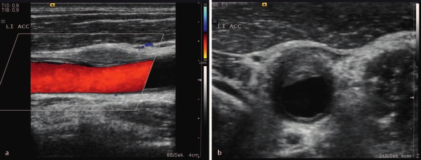
Continuing Education
b
Von der spontanen Dissektion abzugrenzen sind 2 weite-
– Seltener hämodynamisch bedingte Ischämien
re Krankheitsbilder mit Dissektion der Halsarterien, bei
bei hochgradiger Stenose/Gefäßverschluss
denen sich sowohl die pathologische Anatomie als auch
und unzureichender Kollateralversorgung
die Ultraschallbilder grundlegend von den Befunden bei
(dann oft frühes Auftreten der Ischämie, sofort
spontaner Dissektion unterscheiden – sie werden im Fol-
mit Manifestation der Dissektion)
genden nicht berücksichtigt. Zum einen sind dies trau-
– Einseitiges pulssynchrones Ohrgeräusch bei
matische Dissektionen durch stumpfe oder penetrieren-
Dissektion der Arteria carotis interna
de Gefäßverletzung, auch durch Fehlpunktion am Hals,
mit sehr variabler pathologischer Anatomie. Die Verlet-
zung kann Intimarupturen und Wandhämatome, Gefäß-
verengungen und Pseudoaneurysmen sowie AV-Fisteln
Probleme bei der Diagnostik verursachen; besonders häufig ist die Arteria carotis com-
von Dissektionen munis betroffen. Zweitens kann sich eine Aortendissekti-
Dissektionen werden nicht selten übersehen. Wenn die on in die supraaortalen Arterien ausbreiten und ist dann
klinische Verdachtsdiagnose fehlt, kann die notwendige in der Arteria carotis communis und/oder Arteria subcla-
Bildgebung nicht gezielt eingesetzt werden. Bei ungeziel- via nachweisbar, oft bilateral. Typisch ist hier ein langstre-
tem Einsatz der MRT fehlen Sequenzen, die zum Nach- ckiges Doppellumen (▶ Abb. 1B), wobei das falsche
weis der Dissektion wichtig wären, und bei routinemäßig Lumen blind oder mit einem Reentry endet [7].
durchgeführter Ultraschalldiagnostik werden Gefäßab-
Elektronischer Sonderdruck zur persönlichen Verwendung
schnitte mit Vorzugslokalisation einer Dissektion mögli- In diesem Beitrag bleiben außerdem späte und residuelle
cherweise nicht untersucht. Es kommt auch vor, dass Befunde einer Halsarterien-Dissektion unberücksichtigt.
eine Dissektion im Bild zwar zu sehen ist, aber nicht er- Aus dem Wandhämatom mit Gefäßerweiterung nach au-
kannt wird, weil die Bildgebung mit einer anderen Frage- ßen kann sich ein Pseudoaneurysma entwickeln, das bei
stellung erfolgte [4]. Auch bei gezielter Diagnostik mit Manifestation an der Arteria carotis interna sehr weit kra-
der Frage nach einer Dissektion kommen bei allen bildge- nial lokalisiert und mit Ultraschall nicht direkt darstellbar
benden Verfahren falsch-negative Ergebnisse vor, sogar ist. Geeignete Methoden zum Nachweis von Pseudoaneu-
bei der digitalen Subtraktionsangiografie, die zwar die rysmen der Arteria carotis interna sind MRT/MRA oder
beste Darstellung des Gefäßlumens erlaubt, aber die CT/CTA.
Gefäßwand und ihre Verdickung nicht abbildet. Gemäß
gemeinsamer Stellungnahme der American Heart Asso-
ciation und der American Stroke Association ist keine Lokalisation von Dissektionen
bildgebende Methode Goldstandard und somit allen an- Typische Lokalisationen spontaner Dissektionen sind da-
deren überlegen [2]. Die deutsche Leitlinie empfiehlt, durch gekennzeichnet, dass die Gefäße von einem beweg-
bei Verdacht auf eine Dissektion 2 unterschiedliche bild- lichen Abschnitt in einen knöchern fixierten Abschnitt
gebende Verfahren zu kombinieren: MRT/MRA plus Ult- übergehen, wo sie einer besonderen mechanischen Belas-
raschall oder CT/CTA plus Ultraschall [5]. tung ausgesetzt sind. Bei der Arteria carotis interna ist dies
der Abschnitt vor Einmündung in das Felsenbein
Fehler können schließlich auch dadurch entstehen, dass (▶ Abb. 2A); die Dissektion kann sich über eine lange Stre-
die Bildgebung – insbesondere mit Ultraschall – zur cke vom Felsenbein nach kaudal bis wenige Zentimeter
falsch-positiven Diagnose einer Dissektion führt. Einige oberhalb der extrakraniellen Bifurkation erstrecken [8].
andere Gefäßpathologien und Normvarianten weisen ja
Befunde auf, die mit einer Dissektion verwechselt werden An der Arteria vertebralis ist besonders häufig der
können. Abschnitt V3 oberhalb und unterhalb des ersten Halswir-
bels (C1) betroffen (▶ Abb. 2B) [9], die Segmenteintei-
lung der Arteria vertebralis ist in ▶ Abb. 3 und der Infobox
Pathologische Anatomie „Segmente der Arteria vertebralis“ dokumentiert. Dissek-
Die spontane Dissektion der Arteria carotis interna oder tionen treten auch im Segment V1 vor Eintritt in die
Arteria vertebralis schließt auch Dissektionen nach Baga- Wirbelsäule bei C6, im Segment V2 zwischen C6 und C2
tellverletzungen ein und ist charakterisiert durch ein und beim Durchtritt durch die Dura im Foramen mag-
Wandhämatom, das primär ohne Einriss der Intima ent- num auf [6]. Die Dissektion kann sich intrakraniell vom
steht [6], wahrscheinlich durch Ruptur von Vasa vasorum. V3-Segment bis zur Arteria basilaris fortsetzen und auch
Das Hämatom führt zu einer Raumforderung, die eine extrakraniell über eine lange Strecke ausbreiten. In selte-
Gefäßerweiterung nach außen und eine Lumeneinen- nen Fällen können andere intrakranielle Gefäße betroffen
gung nach innen bewirkt (▶ Abb. 1A). Das intramurale sein [6].
Hämatom kann lumenwärts durchbrechen und sekundär
zu einem Einriss der Intima führen.
Arning C. Ultrasound Criteria for… Ultraschall in Med | © 2023. Thieme. All rights reserved.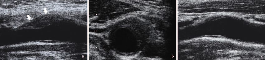
Continuing Education

                                                             b
Elektronischer Sonderdruck zur persönlichen Verwendung

                                                                      ▶ Fig. 2 A Typical location of the internal carotid artery dissection at the entrance to the petrous bone. ACI = internal carotid artery;
                                                                      ACC = common carotid artery Modified according to [9]. B Particularly common location of arterial vertebral dissection: Section V3
                                                                      above and below the 1st cervical vertebra (C1). Modified according to [9].

                                                                      ▶ Abb. 2 a Typische Lokalisation der Arteria-carotis-interna-Dissektion am Eintritt in das Felsenbein. ACI = Arteria carotis interna;
                                                                      ACC = Arteria carotis communis. Aus [9], modifiziert. b Besonders häufige Lokalisation der Arteria-vertebralis-Dissektion: V3-Ab-
                                                                      schnitt ober- und unterhalb des 1. Halswirbels (C1). Aus [9], modifiziert.

                                                                   Dissection Locations                                                        artery is documented in ▶ Fig. 3 and the „Segments of
                                                                                                                                               the vertebral artery“ information box. Dissections also
                                                                   Typical sites of spontaneous dissections are characterized                  occur in segment V1 before entering the spine at C6, in
                                                                   by the transition of vessels from a movable section to a                    segment V2 between C6 and C2, and when passing
                                                                   bony fixed section, where they are subjected to particular                  through the dura in the foramen magnum [6]. The dis-
                                                                   mechanical stress. In the internal carotid artery, this is the              section can proceed intracranially from the V3 segment
                                                                   segment before it enters the petrous bone (▶ Fig. 2A);                      to the basilar artery and also extend extracranially over a
                                                                   the dissection may extend a long distance caudally from                     long distance. In rare cases, other intracranial vessels can
                                                                   the petrous bone to a few centimeters above the extra-                      be affected [6].
                                                                   cranial bifurcation [8].

                                                                   In the vertebral artery, section V3 above and below the
                                                                   first cervical vertebra (C1) is particularly frequently affec-
                                                                   ted (▶ Fig. 2B) [9]; the segmental division of the vertebral

                                                                                                                                    Arning C. Ultrasound Criteria for… Ultraschall in Med | © 2023. Thieme. All rights reserved.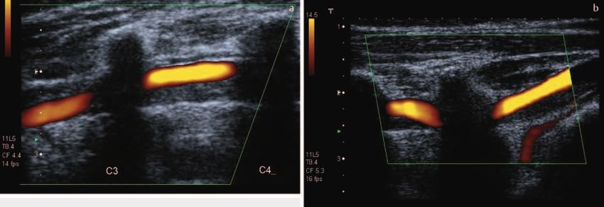
b
                                                             I N F O B OX : S E G M E N T E D E R A R T E R I A V E R T E-
                                                             BR ALI S ( S. ▶ ABB. 3)
                                                             V0: Abgang aus der Arteria subclavia
                                                             V1: Anfangsabschnitt, von distal des Abgangs bis
                                                             zum Eintritt in die Wirbelsäule bei C6
                                                             V2: Intravertebrales Segment zwischen C6 und C2
                                                             V3: Untere und obere Atlasschleife von C2 bis zum
                                                             Foramen magnum
                                                             V4: Intrakranielles Segment vom Foramen magnum
                                                             bis zur Einmündung in die Arteria basilaris

                                                         Diagnose der Halsarterien-Dissektion
                                                         mit Ultraschall
                                                         Mit der klinischen Verdachtsdiagnose erfolgt eine Bildge-
                                                         bung für die definitive Diagnose „Dissektion“. Nach inter-
Elektronischer Sonderdruck zur persönlichen Verwendung

                                                         nationalem Expertenkonsensus sind folgende Befunde
                                                         typisch für eine Dissektion der Halsarterien: Wandhäma-
                                                         tom; Pseudoaneurysma; langstreckige, spitz zulaufende
                                                         Stenose; Intimalefze; Doppellumen; Verschluss mehr als
                                                         2 cm oberhalb der Karotisbifurkation mit Nachweis eines
                                                         Pseudoaneurysmas [10].

                                                         Die Duplexsonografie weist Dissektionen der Arteria carotis
                                                         interna und der Arteria vertebralis mit unterschiedlicher
                                                         Sensitivität nach. Für Dissektionen der Arteria vertebralis
                                                         wird eine Sensitivität von 92 % angegeben [11], wobei we-
                                                         niger erfahrene Untersucher diese Ergebnisse wahrschein-
                                                         lich nicht erreichen. Die Arteria carotis interna ist unmittel-
                                                         bar unterhalb der Schädelbasis im Ultraschallbild nicht                                        ▶ Fig. 3 Segments V0 to V4 of the vertebral artery. From:
                                                         direkt darstellbar, sodass kurzstreckige, gering stenosieren-                                  Arning C. Farbkodierte Duplexsonografie der hirnversor-
                                                         de Dissektionen der Arteria carotis interna dem sonografi-                                     genden Arterien. 3rd edition. Thieme, Stuttgart, 2002,
                                                                                                                                                        modified.
                                                         schen Nachweis entgehen. Für Dissektionen der Arteria ca-
                                                         rotis interna, die nur zu lokalen klinischen Symptomen                                         ▶ Abb. 3 Segmente V0 bis V4 der Arteria vertebralis. Aus:
                                                         geführt haben, wird die Sensitivität mit 69 % angegeben.                                       Arning C.: Farbkodierte Duplexsonografie der hirnversor-
                                                         Hochgradig stenosierende Dissektionen, die mit einem er-                                       genden Arterien, 3. Auflage. Thieme, Stuttgart, 2002,
                                                                                                                                                        modifiziert.
                                                         höhten Schlaganfallrisiko verbunden sind, können jedoch
                                                         mittels hämodynamischer Kriterien erkannt werden [12].
                                                         Bei Dissektionen der Arteria carotis interna, die zu einer ze-                            interna ist eine langstreckige, spitz zulaufende Stenose mit
                                                         rebralen Ischämie geführt haben, wird die Sensitivität der                                Nachweis eines exzentrischen echoarmen Wandhämatoms
                                                         Duplexsonografie im Vergleich mit der Katheterangiografie                                 im distalen extrakraniellen Abschnitt der Arterie
                                                         mit 96 % und die Spezifität mit 94 % angegeben [12].                                      (▶ Abb. 4a–b und Fallbeispiel 1). Da der Gefäßabschnitt
                                                                                                                                                   der Arteria carotis interna unmittelbar unter der Schädelba-
                                                                                                                                                   sis sonografisch nicht direkt beurteilt werden kann, lässt
                                                         Ultraschallbefunde bei Dissektion der                                                     sich ein Stenosebefund bei Dissektion, auch wenn das
                                                         Arteria carotis interna                                                                   Wandhämatom im Ultraschallbild erkennbar ist, nicht voll-
                                                                                                                                                   ständig abbilden; deshalb kann der Stenosegrad nicht
                                                         Die Ultraschalluntersuchung der Arteria carotis interna soll-
                                                                                                                                                   durch direkte Dopplermessung bestimmt werden. Hoch-
                                                         te das Gefäß immer so weit wie möglich nach kranial dar-
                                                                                                                                                   gradig stenosierende Dissektionen können mit hämodyna-
                                                         stellen. Wichtig ist dabei eine gute Geräteeinstellung mit
                                                                                                                                                   mischen Kriterien erkannt werden: durch den Vergleich der
                                                         möglichst geringer Farbfensterkippung, niedriger Pulsrepe-
                                                                                                                                                   Strompulskurven in der Arteria carotis communis, der Arte-
                                                         titionsfrequenz und hoher Farbverstärkung. Der zusätzliche
                                                                                                                                                   ria carotis interna (▶ Abb. 5a–b), der A. cerebri media oder
                                                         Einsatz einer niederfrequenten Sektorsonde kann sinnvoll
                                                                                                                                                   durch den Nachweis von Kollateralen [12].
                                                         sein. Typischer Befund einer Dissektion der Arteria carotis

                                                         Arning C. Ultrasound Criteria for… Ultraschall in Med | © 2023. Thieme. All rights reserved.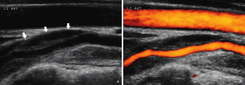
Continuing Education

                                                             b
                                                                      ▶ Fig. 4 a Dissection of the internal carotid artery with a elongated, tapering stenosis beginning 3 cm above the origin. Stenosis
                                                                      and eccentric hypoechoic mural hematoma (arrows) in longitudinal section. b Eccentric hypoechoic mural hematoma (arrows) in
                                                                      transverse section.

                                                                      ▶ Abb. 4 a Dissektion der Arteria carotis interna mit langstreckiger, spitz zulaufender Stenose, 3 cm oberhalb des Abgangs begin-
                                                                      nend. Stenose und exzentrisches echoarmes Wandhämatom (Pfeile) im Longitudinalschnitt. b Exzentrisches echoarmes Wandhä-
                                                                      matom (Pfeile) im Transversalschnitt.
Elektronischer Sonderdruck zur persönlichen Verwendung

                                                                                                                                             that have only led to local clinical symptoms. However,
                                                                      INFORMAT ION B OX: S EGM ENTS O F T HE V ER-                           highly stenosing dissections associated with an increased
                                                                      T E B R A L A R T E RY ( S E E ▶ FIG. 3)                               risk of stroke can be detected using hemodynamic crite-
                                                                      V0: Exit from the subclavian artery                                    ria [12]. In cases of internal carotid artery dissection lead-
                                                                      V1: Initial section, from distal to the exit to the entry              ing to cerebral ischemia, the sensitivity of duplex sono-
                                                                      into the spine at C6                                                   graphy compared with catheter angiography is reported
                                                                      V2: Intravertebral segment between C6 and C2                           to be 96 % and the specificity 94 % [12].
                                                                      V3: Lower and upper atlas loop from C2 to foramen
                                                                      magnum
                                                                      V4: Intracranial segment from the foramen magnum                       Ultrasound Findings in Dissection of the
                                                                      to the junction with the basilar artery                                Internal Carotid Artery
                                                                                                                                             An ultrasound examination of the internal carotid artery
                                                                                                                                             should always show the vessel as far cranially as possible.
                                                                                                                                             A good device setting with the weakest possible color box
                                                                   Diagnosis of Cervical Artery Dissection
                                                                                                                                             tilting, low pulse repetition frequency and high color gain
                                                                   using Ultrasound                                                          is important. The additional use of a low-frequency sec-
                                                                   With the suspected clinical diagnosis, imaging is per-                    tor probe may be useful. A typical finding of dissection
                                                                   formed for a definitive diagnosis of dissection. According                of the internal carotid artery is an elongated, tapering
                                                                   to international expert consensus, the following findings                 stenosis with evidence of an eccentric low-echo mural
                                                                   are typical of cervical artery dissection: mural hematoma;                hematoma in the distal extracranial portion of the artery
                                                                   pseudoaneurysm; elongated, tapered stenosis; intimal                      (▶ Fig. 4a–b and Case Report 1). Because the vascular
                                                                   fovea; double lumen; occlusion more than 2 cm above                       segment of the internal carotid artery immediately below
                                                                   the carotid bifurcation with evidence of pseudoaneurysm                   the skull base cannot be directly assessed sonographi-
                                                                   [10].                                                                     cally, a stenosis finding during dissection cannot be fully
                                                                                                                                             visualized, even if the mural hematoma is visible in the
                                                                   Duplex sonography detects dissections of the internal                     ultrasound image; therefore the degree of stenosis can-
                                                                   carotid artery and vertebral artery with varying sensitiv-                not be determined by direct Doppler measurement.
                                                                   ity. A sensitivity of 92 % has been reported for vertebral                Highly stenosing dissections can be detected with hemo-
                                                                   artery dissections [11], although less experienced exami-                 dynamic criteria by comparing the flow pulse curves in
                                                                   ners are unlikely to achieve these results. The internal car-             the common carotid artery, internal carotid artery
                                                                   otid artery cannot be directly visualized on ultrasound                   (▶ Fig. 5a–b), middle cerebral artery, or by detecting
                                                                   immediately below the skull base; thus short-segment,                     collaterals [12].
                                                                   low-grade stenosing dissections of the internal carotid
                                                                   artery escape sonographic detection. Sensitivity of 69 %
                                                                   is indicated for dissections of the internal carotid artery

                                                                                                                                  Arning C. Ultrasound Criteria for… Ultraschall in Med | © 2023. Thieme. All rights reserved.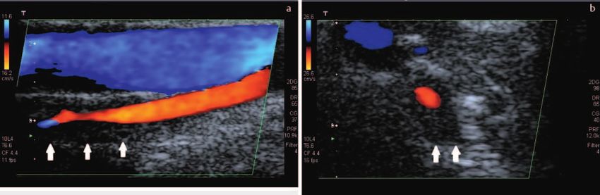
b
                                                             ▶ Fig. 5 a Indirect hemodynamic signs of a high-grade stenosing dissection of the right internal carotid artery. Doppler sonogra-
                                                             phy shows slowing of flow and increased pulsatility in the initial section of the right internal carotid artery compared to the left side
                                                             (b). b Normal finding of the contralateral internal carotid artery.

                                                             ▶ Abb. 5 a Indirekte hämodynamische Zeichen einer hochgradig stenosierenden Dissektion der rechten Arteria carotis interna. Mit
                                                             Dopplersonografie Nachweis einer Strömungsverlangsamung und erhöhten Pulsatilität im Anfangsabschnitt der rechten Arteria
                                                             carotis interna im Vergleich zur linken Seite (b). b Normalbefund der Arteria carotis interna kontralateral.
Elektronischer Sonderdruck zur persönlichen Verwendung

                                                                                                                                                   Ultraschallbefunde bei Dissektion
                                                             FA L L B E I S P I E L 1 : D I S S E K T I O N , Z U N Ä C H S T
                                                                                                                                                   der Arteria vertebralis
                                                             ÜB ERS EH EN
                                                             Ein 67-jähriger Mann mit anamnestisch bekannter                                       Für den Nachweis von Dissektionen der Arteria vertebralis
                                                             Migräne klagt über Schmerzen in der linken Augen-                                     ist die Duplexsonografie besonders vorteilhaft, da das
                                                             höhle mit Ausstrahlung in die linke Kopfseite. An Be-                                 Gefäß vom Anfangssegment bis zur Einmündung in die
                                                             gleitsymptomen lassen sich Schluckstörungen erfra-                                    Arteria basilaris, mit kurzen Unterbrechungen im Verlauf
                                                             gen und ein diskret hängendes Augenlid links. Ein                                     durch knöcherne Strukturen, dargestellt werden kann
                                                             Kopf-MRT am 6. Schmerztag zeigt 2 kleine kortikale                                    (▶ Abb. 7a–b) [9]. Von besonderer Bedeutung ist die Un-
                                                             Mediainfarkte links, die Diagnostik auf der Stroke-                                   tersuchung oberhalb und unterhalb von C1 (▶ Abb. 8a–
                                                             Unit ergibt Normalbefunde, auch die Duplexsono-                                       b). Eine weitere wichtige Lokalisation ist der Eintritt der
                                                             grafie der Gefäße ist als unauffällig beschrieben. Der                                Arteria vertebralis in den Querfortsatz des Halswirbels
                                                             Patient wird am 9. Schmerztag entlassen mit der Di-                                   C6 (▶ Abb. 9). Dissektionen können auch innerhalb des
                                                             agnose „Bekannte Migräne, nebenbefundlich am                                          V2-Segments zwischen C6 und C2 auftreten, deshalb
                                                             ehesten klinisch inapparente Hirninfarkte“.                                           sollte auch dieses Segment mit Ultraschall vollständig un-
                                                             Wegen fortbestehender Schmerzen erfolgt eine zwei-                                    tersucht werden. Häufigster Befund bei Dissektion der
                                                             te Gefäßsonografie, jetzt mit der klinischen Verdachts-                               Arteria vertebralis ist ein Wandhämatom (▶ Abb. 7a und
                                                             diagnose „Dissektion“: Sie zeigt ein Wandhämatom an                                   Fallbeispiele 2 und 3), selten findet sich kurzstreckig ein
                                                             der Arteria carotis interna (▶ Abb. 6a) und als indirek-                              doppeltes Lumen (▶ Abb. 7b).
                                                             tes Stenosezeichen eine Strömungsverlangsamung der
                                                             A. cerebri media links. Auch die mit der Frage nach ei-
                                                             ner Dissektion erneut durchgeführte MRT weist an der                                       FALLBEISPIEL 2: DISSEK TION NUR MIT ULTR A-
                                                             Arteria carotis interna links nun eindeutig einen Dis-                                     SCHALL NACHWEISBAR, MRT UNAUFFÄLLIG
                                                             sektionsbefund nach. Bei Ultraschall-Kontrolle nach 3                                      Eine 32-jährige Frau wird durch Schläge an den Kopf
                                                             Monaten ist der Befund partiell gebessert und nach                                         verletzt, Nacken-Hinterkopf-Schmerzen lassen an
                                                             weiteren 6 Monaten normalisiert (▶ Abb. 6b).                                               eine Arteria-vertebralis-Dissektion denken. Zunächst
                                                             Fazit: Ohne die klinische Verdachtsdiagnose wird die                                       wird ein MRT durchgeführt, das einen vollständig un-
                                                             Dissektion mit MRT und Ultraschall zunächst überse-                                        auffälligen Befund zeigt (auch bei späterer Überprü-
                                                             hen, mit der richtigen Verdachtsdiagnose ist sie mit                                       fung in Kenntnis des Ultraschallbefunds). Mit Ultra-
                                                             beiden Methoden erkennbar.                                                                 schall wird an beiden Vertebralarterien ein typischer
                                                                                                                                                        Dissektionsbefund mit exzentrischem Wandhäma-
                                                                                                                                                        tom nachgewiesen (▶ Abb. 10a–b).
                                                                    Merke
                                                         Gezielte Diagnostik unter der klinischen Verdachtsdi-
                                                         agnose „Dissektion“ verbessert die Sensitivität der
                                                         Bildgebung.

                                                         Arning C. Ultrasound Criteria for… Ultraschall in Med | © 2023. Thieme. All rights reserved.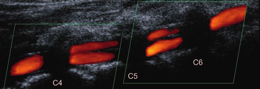
Continuing Education
b
▶ Fig. 6 a Eccentric wall thickening of the distal-extracranial internal carotid artery during dissection. b Follow-up examination of
Elektronischer Sonderdruck zur persönlichen Verwendung
the finding from a after 9 months: finding has normalized.
▶ Abb. 6 a Exzentrische Wandverdickung der Arteria carotis interna, distal-extrakraniell bei Dissektion. b Kontrolluntersuchung des
Befunds aus a nach 9 Monaten: Befund hat sich normalisiert.
Note
C A SE STUDY 1: D I SS EC T I O N I NITI ALLY OV ER- Targeted diagnostics under the suspected clinical diag-
LO O K E D nosis of dissection improves the sensitivity of the ima-
A 67-year-old male with a history of migraine com- ging.
plains of pain in the left orbit with radiation to the
left side of the head. Accompanying symptoms in-
clude dysphagia and a discrete drooping left eyelid. Ultrasound Findings in Dissection
A head MRI on the 6th day of pain shows two small of the Vertebral Artery
cortical medial infarcts on the left side; diagnostics
Duplex sonography is particularly advantageous for the de-
on the stroke unit reveal normal findings; duplex ul-
tection of vertebral artery dissections because the vessel
trasound of the vessels is also described as unre-
can be visualized from the initial segment to its junction
markable. The patient is discharged on the 9th day
with the basilar artery, with brief interruptions in its course
of pain with the diagnosis „Known migraine, second-
by bony structures (▶ Fig. 7a–b) [9]. Of particular impor-
ary findings most likely clinically inapparent cerebral
tance is an examination above and below C1 (▶ Fig. 8a–b).
infarction“.
Another important location is the entrance of the vertebral
Because of the persistent pain, a second vascular ul-
artery into the transverse process of the C6 cervical verte-
trasound is performed, now with the suspected clin-
bra (▶ Fig. 9). Dissections can also occur within the V2 seg-
ical diagnosis of dissection. The examination reveals
ment between C6 and C2, therefore this segment should
a mural hematoma on the internal carotid artery
also be fully examined with ultrasound. The most common
(▶ Fig. 6a) and, as an indirect sign of stenosis, slow-
finding in vertebral artery dissection is a mural hematoma
ing of the flow of the left middle cerebral artery.
(▶ Fig. 7a and Case Studies 2 and 3); rarely, a short-seg-
The MRI, which was performed again with the ques-
ment double lumen is found (▶ Fig. 7b).
tion of dissection, now clearly shows a dissection
finding on the left internal carotid artery. At ultra-
sound follow-up after 3 months, the findings are par-
tially improved and normalized after another 6 CASE STUDY 2: DISSECTION DE TECTABLE
months (▶ Fig. 6b). O N LY W I T H U LT R A S OU N D , M R I UN R E M A R K -
Bottom line: without the suspected clinical diagno- AB LE
sis, the dissection with MRI and ultrasound is initially A 32-year-old female is injured by blows to the head,
overlooked; with the correct suspected diagnosis it pain in the back of the neck suggests a dissection of
can be detected with both methods. the vertebral artery. First, an MRI is performed, which
shows completely unremarkable findings (even when
reviewed later with knowledge of the ultrasound
Arning C. Ultrasound Criteria for… Ultraschall in Med | © 2023. Thieme. All rights reserved.b
▶ Fig. 7 a Eccentric hypoechoic wall hematoma (arrows) in vertebral artery dissection in the V2 segment between C4 and C6. b Another
finding of a vertebral artery dissection between C4 and C6 with evidence of stenosis (aliasing) in the color Doppler image (arrowhead),
short-segment double lumen and eccentric wall thickening (arrows).
▶ Abb. 7 a Exzentrisches echoarmes Wandhämatom (Pfeile) bei Dissektion der Arteria vertebralis im V2-Segment zwischen C4 und
C6. b Ein weiterer Befund einer A.-vertebralis-Dissektion zwischen C4 und C6 mit Stenosebefund (Aliasing) im Farbdopplerbild
(Pfeilspitze), mit kurzsegmentigem Doppellumen und exzentrischer Wandverdickung (Pfeile).
Elektronischer Sonderdruck zur persönlichen Verwendung
Merke dickung der Arteria carotis an der Bifurkation oder an der
Falsch-negative Befunde kommen bei allen bildgeben- Arteria carotis communis unmittelbar proximal der Bifur-
den Verfahren vor. Bei der MRT ist der charakteristische kation; die Arterie ist genau an dieser Stelle druckemp-
Befund, die exzentrische Signalanhebung in den fett- findlich, der Befund lässt sich durch leichten Druck mit
supprimierten T1-Sequenzen, in den ersten 72 Stunden dem Schallkopf lokalisieren. Die Wandverdickung führt
nach Eintritt der Dissektion oft noch nicht nachweisbar. zu einer geringen Lumeneinengung und einer deutlichen
Erweiterung der Gefäßwand nach außen (▶ Abb. 12a–b).
In einigen Fällen zeigt sich die Wandverdickung in 2 ver-
FA L L B E I S P I E L 3 : A K U T E R N A C K E N - H I N T E R - schiedenen Schichten der Arterienwand (▶ Abb. 12b).
KO P F -S C HM ER Z Bei der Ultraschall-Nachuntersuchung nach 3–5 Wochen
sind die pathologischen Befunde deutlich geringer aus-
Eine 24-jährige Frau klagt über spontan aufgetretene
geprägt (Fallbeispiel 4), gleichzeitig bilden sich die
Nacken-Hinterkopf-Schmerzen links. Vor Behandlung
Schmerzen spontan zurück. Ursache für diese Erkrankung
durch den Orthopäden wird eine Duplexsonografie
ist eine unspezifische Entzündung der Gefäßwand [13],
durchgeführt, die eine Dissektion der Arteria verte-
dazu passend zeigt die MRT eine abnorme Kontrastmit-
bralis unter- und oberhalb des 6. Halswirbels zeigt
telanreicherung der Arterienwand [13].
(▶ Abb. 11a–b). Bei diesem Befund darf keine Manu-
altherapie erfolgen!
Nach eigener Erfahrung entsteht bei idiopathischer Karo-
tidynie besonders häufig der Fehlbefund einer Dissektion,
da die Ultraschallbefunde sehr ähnlich aussehen. Folgen-
de Kriterien ermöglichen die Unterscheidung: 1.) Die Ka-
Differenzialdiagnosen der Dissektion im
rotisbifurkation, typischer Ort der Karotidynie, wäre eine
Ultraschallbild ganz ungewöhnliche Lokalisation für eine spontane Dis-
Einige Ultraschallbefunde sehen einer Dissektion sehr sektion (s. o.: „Lokalisation von Dissektionen“). 2.) Eine
ähnlich, obwohl sie auf eine andere Gefäßpathologie Wandverdickung in 2 Gefäßwandschichten, typisch für
oder eine Normvariante zurückzuführen sind. Die Krite- eine Karotidynie, ist untypisch für eine Dissektion. 3.) Pa-
rien für die Unterscheidung werden im Folgenden be- tienten mit einer spontanen Karotisdissektion klagen
schrieben. über Kopfschmerzen, Patienten mit Karotidynie über
Schmerzen seitlich am Hals. 4.) In unklaren Fällen zeigt
Idiopathische Karotidynie die ergänzend durchgeführte MRT eine Kontrastmittelan-
reicherung in der Gefäßwand, während die MRT bei Dis-
Die Ultraschallbefunde der idiopathischen Karotidynie sektion eine Signalanhebung ohne Kontrastmittel zeigt.
wurden erstmals 2004 beschrieben [13, 14]. Die Patien- Die Differenzierung von Dissektion und Karotidynie ist
ten leiden unter akuten einseitigen Schmerzen seitlich wichtig, da bei einer Karotidynie, die das Lumen nur mini-
am Hals, verbunden mit einer lokalen Druckschmerzhaf- mal einengt, keine Gerinnungstherapie indiziert ist.
tigkeit. Das Ultraschallbild zeigt eine echoarme Wandver-
Arning C. Ultrasound Criteria for… Ultraschall in Med | © 2023. Thieme. All rights reserved.Continuing Education
b
▶ Fig. 8 a Dissection of the vertebral artery above C1. Eccentric wall thickening (arrows) at the upper atlas loop with stenosis found
in the color Doppler image. b Dissection of the vertebral artery. Eccentric wall thickening (arrows) at the inferior atlas loop between
C1 and C2.
▶ Abb. 8 a Dissektion der Arteria vertebralis oberhalb C1. Exzentrische Wandverdickung (Pfeile) an der oberen Atlasschleife mit
Stenosebefund im Farbdopplerbild. b Dissektion der Arteria vertebralis. Exzentrische Wandverdickung (Pfeile) an der unteren
Atlasschleife zwischen C1 und C2.
Elektronischer Sonderdruck zur persönlichen Verwendung
findings). Ultrasound discloses typical dissection
with eccentric mural hematoma in both vertebral ar-
teries (▶ Fig. 10a–b).
Note
False-negative findings occur with all imaging modal-
ities. On MRI, the characteristic finding, eccentric signal
enhancement in the fat-suppressed T1 sequences, is
often not yet detectable in the first 72 hours after dis-
section onset.
▶ Fig. 9 Dissection of the vertebral artery with mural
C A S E S T U DY 3: AC U T E N EC K O CC I P I T A L PA I N hematoma directly below C6.
A 24-year-old female complains of spontaneous onset ▶ Abb. 9 Dissektion der Arteria vertebralis mit Wand-
of left occipital neck pain. Prior to treatment by the or- hämatom unmittelbar unterhalb von C6.
thopedist, duplex sonography is performed, demon-
strating dissection of the vertebral artery below and
above the 6th cervical vertebra (▶ Fig. 11a–b). Man- cal tenderness. The ultrasound image shows a hypoecho-
ual therapy must not be performed with this finding! ic wall thickening of the carotid artery at the bifurcation
or at the common carotid artery just proximal to the bi-
furcation; the artery is sensitive to pressure at this exact
location, and the finding can be localized by gentle pres-
Differential Diagnoses of Dissection on sure with the transducer. Wall thickening results in minor
lumen narrowing and marked outward dilation of the
the Ultrasound Image vessel wall (▶ Fig. 12a–b). In some cases, wall thickening
Some ultrasound findings look very similar to a dissec- is evident in two different layers of the arterial wall
tion, although they may be due to a different vascular pa- (▶ Fig. 12b). At the follow-up ultrasound examination
thology or a norm variant. The criteria for differentiation after 3–5 weeks, the pathological findings are significant-
are described below. ly less pronounced (Case Study 4), and at the same time
the pain regresses spontaneously. The cause of this con-
Idiopathic carotidynia dition is nonspecific inflammation of the vessel wall [13];
appropriately, MRI shows abnormal contrast enhance-
Ultrasound findings of idiopathic carotidynia were first
ment of the arterial wall [13].
described in 2004 [13, 14]. Patients suffer from acute
unilateral pain on the side of the neck associated with lo-
Arning C. Ultrasound Criteria for… Ultraschall in Med | © 2023. Thieme. All rights reserved.b
▶ Fig. 10 a Dissection of the right vertebral artery. Eccentric wall thickening between C2 and C4. b Dissection of the left vertebral
artery. Eccentric wall thickening above and below C6.
▶ Abb. 10 a Dissektion der Arteria vertebralis rechts: Exzentrische Wandverdickung zwischen C2 und C4. b Dissektion der Arteria
vertebralis links: Exzentrische Wandverdickung ober- und unterhalb von C6.
Elektronischer Sonderdruck zur persönlichen Verwendung
den, s. Fallbeispiel 5 [15]. Die Analyse der Gefäßwand er-
FA L L B E I S P I E L 4 : F E H L D I AG N OS E D I S S E K T I O N möglicht jedoch die Unterscheidung von einer Dissekti-
BEI K AROTIDYNIE on: Die Arterienwand ist nicht verdickt und das Gefäß ist
Eine 41-jährige Frau klagt seit 5 Tagen über spontan nicht nach außen erweitert. Die Lokalisation des Befunds
aufgetretene Schmerzen an der rechten Halsseite, – nahe der Bifurkation – ist ein weiteres Kriterium für die
verbunden mit lokaler Druckempfindlichkeit und ver- Unterscheidung von einer spontanen Dissektion.
mehrter Pulsation. Bei auswärtiger Sonografie wird
eine Dissektion diagnostiziert.
Das Ultraschallbild zeigt eine echoarme exzentrische FA L L B E I S P I E L 5 : F E H L D I AG N OS E D I S S E K T I O N
Verdickung der Gefäßwand unmittelbar an der Karo- BE I C AROTID WEB
tisbifurkation mit geringer Lumeneinengung und Ein 42-jähriger Mann erhält im Rahmen einer Vorsor-
deutlicher Auftreibung des Gefäßes nach außen: geuntersuchung eine Duplexsonografie der Halsarte-
den typischen Befund einer idiopathischen Karotidy- rien. Dabei wird an der Arteria carotis communis
nie (▶ Abb. 13a–b). Gegen eine spontane Dissektion rechts eine Dissektion festgestellt, der Patient wird
sprechen die Lokalisation an der Bifurkation, die Ver- notfallmäßig in die Klinik eingewiesen. Er ist asymp-
dickung in 2 Gefäßwandschichten und die Lokalisati- tomatisch; Kopfschmerzen oder Symptome einer ze-
on der Schmerzen (seitlich am Hals, nicht am Kopf). rebrovaskulären Störung sind nicht zu erfragen. Er
Die Kontrolluntersuchung nach 6 Wochen zeigt den hat keine Verletzung am Hals erlitten.
typischen Verlauf der Karotidynie: Die vorbeschrie- Das Ultraschallbild zeigt im Endabschnitt der A. caro-
bene echoarme Wandverdickung ist nur noch sehr tis communis rechts eine lamellenartige Septierung
gering ausgeprägt (▶ Abb. 13c) mit 2 Lumina (▶ Abb. 15a–b); kurzstreckig sind so-
gar 3 Lumina nachweisbar. Die Gefäßwand erscheint
sonst unauffällig, weist kein Wandhämatom und kei-
Merke ne Erweiterung nach außen hin auf. Im Farbdoppler-
Bei Idiopathischer Karotidynie entsteht nicht selten der und Spektraldoppler-Modus sind keine Stenosezei-
Fehlbefund einer spontanen Dissektion. Wichtigstes chen erkennbar, die Strompulskurven sind normal.
Kriterium für die Unterscheidung ist die Lokalisation Es handelt sich um ein Carotid Web und nicht um eine
des Befunds an oder nahe der Karotisbifurkation. spontane Dissektion, da die Arterienwand nicht ver-
dickt und das Gefäß nicht nach außen erweitert ist.
Carotid Web
Das Carotid Web ist eine seltene Form der intimalen fi-
bromuskulären Dysplasie, die aus dünnen Gewebesträn- Merke
gen besteht – ausgehend von der Arterienwand in der Bei bindegewebiger Septierung des Gefäßlumens durch
Nähe der Karotisbifurkation. In seltenen Fällen bildet sie ein Carotid Web kann der Fehlbefund einer Dissektion
ein Netzwerk, das das Gefäßlumen verengt (▶ Abb. 14a– entstehen. Wichtigstes Kriterium für die Unterschei-
c). Der Befund kann als Dissektion fehlinterpretiert wer- dung ist die unauffällige Gefäßwand.
Arning C. Ultrasound Criteria for… Ultraschall in Med | © 2023. Thieme. All rights reserved.Continuing Education
b
▶ Fig. 11 a Vertebral artery dissection showing eccentric wall thickening below C6 in B-mode. b Vertebral artery dissection showing
eccentric wall thickening below C6. Finding from a in color Doppler image.
▶ Abb. 11 a Dissektion der Arteria vertebralis mit exzentrischer Wandverdickung unterhalb von C6 im B-Bild. b Dissektion der Arteria
vertebralis mit exzentrischer Wandverdickung unterhalb von C6. Befund aus a im Farbdoppler-Bild.
Elektronischer Sonderdruck zur persönlichen Verwendung
▶ Fig. 12 a Idiopathic carotidynia in the distal section of the common carotid artery, below the bifurcation. In the longitudinal section,
pronounced hypoechoic thickening of the vessel wall with slight narrowing of the lumen and clear expansion to the outside. b Idiopathic
carotidynia. Finding from a in transverse section with wall thickening in 2 vessel wall layers.
▶ Abb. 12 a Idiopathische Karotidynie im distalen Abschnitt der Arteria carotis communis, unterhalb der Bifurkation. Im Longitu-
dinalschnitt ausgeprägte echoarme Verdickung der Gefäßwand mit geringer Lumeneinengung und deutlicher Erweiterung nach
außen. b Idiopathische Karotidynie. Befund aus a im Transversalschnitt mit Wandverdickung in 2 Gefäßwandschichten.
In our own experience, the misdiagnosis of dissection is important, because no anticoagulation therapy is indicated
particularly common in idiopathic carotidynia since the for this condition that minimally constricts the lumen.
ultrasound findings look very similar. The following criteria
support the differentiation: 1. The carotid bifurcation, typ-
ical site of carotidynia, would be a very unusual site for a C ASE STUDY 4: MISDIAGNOSIS OF DISSECTION
spontaneous dissection (see above: „Location of Dissec- IN THE CASE OF C AROTIDYNIA
tions“). 2. Wall thickening in 2 vessel wall layers, typical of
A 41-year-old woman has complained for 5 days of
carotidynia, is atypical of dissection. 3. Patients with spon-
spontaneously occurring pain on the right side of
taneous carotid dissection complain of headache, and pa-
the neck associated with local pressure tenderness
tients with carotidynia report pain in the side of the neck.
and increased pulsation. Off-site ultrasound has diag-
4. In ambiguous cases, supplemental MRI shows contrast
nosed dissection.
enhancement in the vessel wall, whereas MRI in the case
The ultrasound image shows a hypoechoic, eccentric
of dissection shows a signal enhancement without contrast
thickening of the vessel wall directly at the carotid bi-
medium. Differentiation of dissection from carotidynia is
furcation with slight narrowing of the lumen and
Arning C. Ultrasound Criteria for… Ultraschall in Med | © 2023. Thieme. All rights reserved.b
▶ Fig. 13 a Idiopathic carotidynia. Pronounced hypoechoic thickening of the vessel wall directly at the carotid bifurcation with slight narrowing of
the lumen and clear expansion to the outside. b The same finding as in a in transverse section. Two wall layers are affected. c The same finding as in
a–b at the follow-up after 6 weeks: the wall thickening is only very slightly pronounced.
▶ Abb. 13 a Idiopathische Karotidynie: Ausgeprägte echoarme Verdickung der Gefäßwand unmittelbar an der Karotisbifurkation mit geringer
Lumeneinengung und deutlicher Erweiterung nach außen. b Derselbe Befund wie in a im Transversalschnitt. Zwei Wandschichten sind betroffen.
c Derselbe Befund wie in a–b bei Kontrolluntersuchung nach 6 Wochen: Die Wandverdickung ist nur noch sehr gering ausgeprägt.
Elektronischer Sonderdruck zur persönlichen Verwendung
▶ Fig. 14 a Carotid web in the cranial section of the common carotid artery, just below the bifurcation. Septation of the vessel lumen by connective
tissue strands with formation of several lumina. The vessel wall appears unremarkable, unlike in dissection. b Finding from a in color Doppler mode
showing multiple lumina. c Finding from a in transverse section. Septation of the vessel lumen by tissue strands with formation of several lumina.
▶ Abb. 14 a Carotid Web im kranialen Abschnitt der Arteria carotis communis, unmittelbar unterhalb der Bifurkation. Septierung des Gefäßlumens
durch Bindegewebsstränge mit Ausbildung mehrerer Lumina. Die Gefäßwand erscheint unauffällig – anders als bei Dissektion. b Befund aus a im
Farbdoppler-Modus mit Darstellung mehrerer Lumina. c Befund aus a im Transversalschnitt. Septierung des Gefäßlumens durch Gewebebänder mit
Ausbildung mehrerer Lumina.
Großgefäß-Vaskulitis terung nach außen ist im Falle einer Vaskulitis gering und
bei einer Dissektion deutlich ausgeprägt.
Zwei Formen der Immunvaskulitis können sich an den gro-
ßen extrakraniellen Gefäßen manifestieren und mit Ultra-
Fensterung
schall nachgewiesen werden: Takayasu-Arteriitis und Rie-
senzellarteriitis. In den typischen Fällen zeigt das Gefäß Die Fensterung der Arteria vertebralis, eine segmentale
eine lange, homogen echoarme, konzentrische Wandver- Verdopplung der Arterie in einem kurzen Gefäßabschnitt,
dickung, die zu einer Lumeneinengung führt [9]. Das Gefäß ist eine seltene angeborene Anomalie, die keine klinische
ist auch geringgradig nach außen erweitert. Im Farbdopp- Bedeutung hat. Allerdings kann der Ultraschallbefund einer
lerbild erscheint die verdickte, echoarme Wand als dunkler Fensterung zur Fehlinterpretation als arterielle Dissektion
Halo, der das farbige Lumen umgibt (▶ Abb. 16a–b). mit Doppellumen führen (▶ Abb. 17). Die Unterscheidung
zwischen extrakranieller Dissektion und dieser Normvaria-
Vaskulitis und Dissektion stellen sich im Ultraschallbild ähn- nte ist möglich, da im Falle einer Dissektion mit 2 Lumina
lich dar, und mit der Sonografie kann nicht zwischen Häma- eine pathologische Strompulskurve in mindestens einem
tom und Entzündung als Ursache der echoarmen Wandver- Lumen zu erwarten ist [16]. Bei der Normvariante ist die
dickung differenziert werden. Die Unterscheidung ist aber Form des Dopplerspektrums in allen Gefäßabschnitten nor-
nach der Form des Befunds in der Gefäßwand möglich, ins- mal (▶ Abb. 18a–b), sodass eine Dissektion ausgeschlos-
besondere bei Untersuchung im Transversalschnitt [9]: Die sen und die Diagnose einer Fensterung gestellt werden
Vaskulitis zeigt typischerweise eine konzentrische Verdi- kann. Bei einem derartigen Befund ist ein Restzustand
ckung der Gefäßwand (▶ Abb. 16b) – bei einer Dissektion nach vorangegangener Dissektion mit einem persistieren-
ist die Wandverdickung exzentrisch (▶ Abb. 4b). Die Erwei- den kurzen Doppellumen zwar nicht völlig auszuschließen.
Arning C. Ultrasound Criteria for… Ultraschall in Med | © 2023. Thieme. All rights reserved.Continuing Education
b
▶ Fig. 15 a Carotid web. Lamellar septation in the end section of the common carotid artery with two lumina. b Carotid web. The
same finding as in a in color Doppler mode.
▶ Abb. 15 a Carotid Web: Lamellenartige Septierung im Endabschnitt der Arteria carotis communis mit 2 Lumina. b Carotid Web:
Derselbe Befund wie in a im Farbdoppler-Modus.
Elektronischer Sonderdruck zur persönlichen Verwendung
clear outward expansion of the vessel, the typical
finding of idiopathic carotidynia (▶ Fig. 13a–b). The C A S E S T U DY 5 : M I S D I A G N O S I S O F D I S S E C -
location at the bifurcation, thickening in 2 vessel T I O N I N T H E C A S E O F C A ROT I D W E B
wall layers, and the localization of the pain (lateral A 42-year-old male undergoes duplex sonography of
to the neck, not to the head) argue against a sponta- the cervical arteries as part of a screening examina-
neous dissection. tion. A dissection is found in the right common caro-
The follow-up examination after 6 weeks shows the tid artery, and the patient is admitted to the hospital
typical course of carotidynia: the previously de- as an emergency case. The patient is asymptomatic,
scribed low echo wall thickening is only very slightly with no headache or symptoms of cerebrovascular
pronounced (▶ Fig. 13c) disorder. He did not suffer any injury to his neck.
The ultrasound image shows a lamellar septation with
two lumina in the terminal segment of the right com-
Note mon carotid artery (▶ Fig. 15a–b), and three lumina
A false finding of a spontaneous dissection is not un- are detectable in the short segment. The vessel wall
common in the case of idiopathic carotidynia. The most otherwise appears unremarkable, showing no mural
important criterion for differentiation is the location of hematoma and no extension to the outside. Color
the finding at or near the carotid bifurcation. and spectral Doppler modes do not disclose signs of
stenosis, and the flow pulse curves are normal.
Carotid web This is a carotid web and not a spontaneous dissec-
tion because the arterial wall is not thickened and
Carotid web is a rare form of intimal fibromuscular dys- the vessel is not dilated outward.
plasia consisting of thin strands of tissue, originating
from the arterial wall near the carotid bifurcation. In rare
cases, it forms a network that constricts the vessel lumen
Note
(▶ Fig. 14a–c). The finding may be misinterpreted as a
An erroneous finding of dissection may be the result of
dissection, see Case Study 5 [15]. However, analysis of
connective tissue septation of the vessel lumen by a
the vessel wall allows differentiation from dissection:
carotid web. An unremarkable vascular wall is the most
The artery wall is not thickened and the vessel is not dila-
important criterion for differentiation.
ted outward. The location of the finding – proximity to
the bifurcation – is another criterion for distinguishing it
Large-vessel vasculitis
from spontaneous dissection.
Two forms of immune vasculitis may manifest in the large
extracranial vessels and be detected by ultrasound: Ta-
kayasu arteritis and giant cell arteritis. In typical cases, the
vessel shows a long, homogeneously hypoechoic, concen-
tric wall thickening leading to lumen narrowing [9]. The
Arning C. Ultrasound Criteria for… Ultraschall in Med | © 2023. Thieme. All rights reserved.You can also read