Uterine Prolapse: The Other Exceptional Complication of Spina Bifida in Newborns
←
→
Page content transcription
If your browser does not render page correctly, please read the page content below
Open Journal of Pediatrics, 2021, 11, 50-54
                                                                                               https://www.scirp.org/journal/ojped
                                                                                                            ISSN Online: 2160-8776
                                                                                                              ISSN Print: 2160-8741

Uterine Prolapse: The Other Exceptional
Complication of Spina Bifida in Newborns

T. Mukenge1, F. B. Balde1*, Z. Benmassaoud1, I. Oualili1, O. Alaoui1,2, A. Mahmoudi1,2,
K. Khattala1,2, Y. Bouabdallah1,2
1
    Department of Pediatric Surgery, Hassan II University Hospital, Fez, Morocco
2
    Sidi Mohamed Ben Abdellah University, Fez, Morocco

How to cite this paper: Mukenge, T.,            Abstract
Balde, F.B., Benmassaoud, Z., Oualili, I.,
Alaoui, O., Mahmoudi, A., Khattala, K. and      Introduction: Spina bifida is the most common neural tube defect. Uterine
Bouabdallah, Y. (2021) Uterine Prolapse:        prolapse is an exceptional presentation of its complications. We aim to de-
The Other Exceptional Complication of
                                                scribe the clinical and progressive features of uterine prolapse in a newborn
Spina Bifida in Newborns. Open Journal of
Pediatrics, 11, 50-54.                          baby with spina bifida. Observation: 19-day-old newborn. Admitted for spi-
https://doi.org/10.4236/ojped.2021.111005       na bifida. The clinical examination showed an anal gaping, the presence of
                                                uterovaginal prolapse and bilateral equine varus clubfoot. The particularity
Received: January 9, 2021
                                                was that this prolapse had a spontaneous resolution but appears with screams.
Accepted: March 5, 2021
Published: March 8, 2021
                                                We made a compression bandage at the time of resolution. After a 6-month
                                                of follow-up, the baby is in good general condition and, there is no recurrence
Copyright © 2021 by author(s) and               of the prolapse. Conclusion: Uterine prolapse is a rare complication of spina
Scientific Research Publishing Inc.
                                                bifida. The main therapeutic component remains the prevention of spina bi-
This work is licensed under the Creative
Commons Attribution International
                                                fida.
License (CC BY 4.0).
http://creativecommons.org/licenses/by/4.0/     Keywords
                 Open Access
                                                Uterovaginal Prolapse, Congenital, Spina Bifida

                                              1. Introduction
                                              Spina bifida is a neural tube closure defect occurring during the fourth week of
                                              gestation. It is the most common neural tube defect [1]. It has consequences
                                              whose physiopathological evolution is often severe and requires long and evolu-
                                              tionary management. These are hydrocephalus, sensory and motor deficits, or-
                                              thopaedic and urogenital abnormalities. The uterine prolapse is an exceptional
                                              presentation of the long list of its complications. In the neonatal period, uterine
                                              prolapse is associated with spina bifida in more than 80% [2] [3]. Management
                                              ranges from manual reduction to surgical treatment whose radicality has evolved

DOI: 10.4236/ojped.2021.111005        Mar. 8, 2021                 50                                    Open Journal of Pediatrics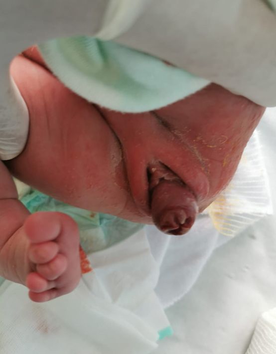
T. Mukenge et al.
over the years [4]. The purpose of this study is to describe the clinical and evolu-
tionary characteristics of the case of a newborn with uterine prolapse in spina bi-
fida.
2. Observation
Infant, 19 days old, female, from a non-consanguine marriage, mother, 18 years
old G1P1 no folic acid intake by the mother, irregular follow-up pregnancy. No
antenatal imaging. Born by natural way, to term according to the date of the last
menses with good adaptation to the extra-uterine life. Admitted for a lumbosa-
cral swelling seen since birth. She was in good general condition, stable and apy-
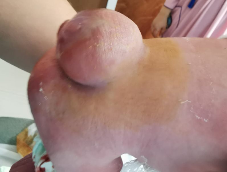
retic. There was a 4 × 4 cm lumbosacral myelomeningocele (Figure 1). An anal
gaping, a prolapsed mass between the two large lips (uterine prolapsus) (Figure
2). According to the mother, this mass is spontaneously resolute but reappears
after an episode of crying. The urethral orifice was normal. The baby also had a
bilateral equine varus clubfoot (EVCF). The weight was 3 kg 200, and the head
perimeter was 34 cm. We performed a compression dressing at the time of
spontaneous resolution of the prolapse for 24 hours. We noted a complete dis-
appearance of the prolapse, even at the scrams. We cure the myelomeningocele
by closing the neural plaque, closure of the dura mater and cutaneous closure.
We noted an increase of 2 cm of the head perimeter 6-days after surgery of spina
bifida. Postoperative trans-fontanelle ultrasound at J + 6 found hydrocephalus.
We performed a ventriculoperitoneal shunt (VPS) as indicated by our protocol
for all active hydrocephalus. Ponsetti’s protocol was started for the bilateral
EVCF.
After 6-months of follow-up, the baby is in good general condition, no recur-
rence of the prolapse (Figure 3). However, there is a delay in motor vehicle ac-
quisitions: head holding and sitting position.
Figure 1. Profile image of the baby’s back, a 4 × 4 cm lumbosacral
spina bifida covered at the periphery by the skin, an unbroken
translucent membrane un the centre.
DOI: 10.4236/ojped.2021.111005 51 Open Journal of Pediatrics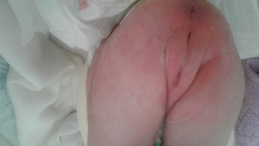
T. Mukenge et al.

                                           Figure 2. Clinical image showing uterine prolapse of the newborn.

                                           Figure 3. The clinical image of the 3-month follow-up showing an
                                           absence of uterine prolapse.

                                 3. Discussion
                                 Uterine prolapse is an unfolding circumferential protrusion to a varying degree
                                 of the uterus through the vaginal opening. Caused by the absence of innervation
                                 of the muscular structures of the perineum. In particular, the levator anus, which
                                 is the first support for the pelvic organs [2].
                                    The neonatal forms are exceptional. The first description was in 1723, and
                                 since less than 100 cases have been reported in the literature [5]. This case is the
                                 second encountered in our training in 10 years of experience. In the neonatal
                                 period, uterine prolapse is more than 80% associated with spina bifida [2].
                                    It is essential to formally eliminate the other diagnostic possibilities, in front
                                 of an inter labial mass of the newborn: such as the vaginal polyp, urethral pro-
                                 lapse, a paraurethral cyst, or rhabdomyosarcoma [2]. In severe forms, there it is
                                 associated with rectal prolapse [5].
                                    Uterine prolapse is a therapeutic emergency to prevent complications. Such as

DOI: 10.4236/ojped.2021.111005                         52                                    Open Journal of Pediatrics
T. Mukenge et al.
urinary obstruction, which can lead to hydronephrosis, or endometrial metapla-
sia [6] [7]. No consensus management protocol has been established. The au-
thors make proposals for support through the sharing of experiences. It ranges
from conservative treatment to surgical treatment. Manual reduction under
anaesthesia has been strongly reported [1] [6].
This reduction is held in place either by an intravaginal wick with a compres-
sion bandage for 72 hours on average [6]. Using a Folley catheter with the in-
flated balloon held in place for 2-weeks helps keep the reduction [2] [3].
Frequently the procedure is repeated because of recurrence of the prolapse, or
require surgical treatment. Surgical treatment ranges from a simple labial suture
to amputation of the cervix or even hysterectomy. Transabdominal uteropexy
has also been reported [1] [3].
Cases of spontaneous resolution are reported with isolated uterine prolapse of
the newborn [3].
The peculiarity is that our patient presented a spontaneous reduction in pro-
lapse, which is why we took this advantage to apply a compression bandage held
in place for 24 hours. It saved the baby from the risks of iterative anaesthesia.
The other possibility was to cure the myelomeningocele and reduce the pro-
lapse at the same time. But the prolapse had completely disappeared.
Hydrocephalus can depend or not to the spina bifida. Its can appear sponta-
neously or after cure of myelomeningocele. Anywhere discussion is open about
when to conduced a ventriculoperitoneal shunt. From 1998 to 2014, hydroce-
phalus treatment has become more delayed. A meta-analysis found that there is
not differ between delayed and simultaneous hydrocephalus treatment [8].
After a follow-up of 6 months, no recurrence of the prolapse was reported
even in the literature, no case of recurrence is reported
4. Conclusion
Uterine prolapse is an exceptional complication of spina bifida. The treatment
ranges from manual reduction to actual surgery, the radical nature of which has
changed over the years. In all cases, the therapeutic component remains the pre-
vention of Spina bifid by taking folic acid before and during the first months of
pregnancy.
Ethical Aspect
Parental consent has obtained for the use of patient data, and no image allows
identification.
Conflicts of Interest
The authors declare no conflicts of interest regarding the publication of this paper.
References
[1] Aykanat, A., Solakoglu, E., Bilginer, B., Celik, H.T. and Yigit, S. (2020) Uterovaginal
DOI: 10.4236/ojped.2021.111005 53 Open Journal of PediatricsT. Mukenge et al.
Prolapse in a Newborn with Meningomyelocele: Case Report. Journal of Pediatric
and Adolescent Gynecology, 33, e607-e609.
https://doi.org/10.1016/j.jpag.2020.04.001
[2] Abdelsalam, S.E.A., Desouki, N.M. and Alaal, N.A.A. (2006) Use of Foley Catheter
for Management of Neonatal Genital Prolapse: Case Report and Review of the Lite-
rature. Journal of Pediatric Surgery, 41, 449-452.
https://doi.org/10.1016/j.jpedsurg.2005.11.031
[3] Baskaran, D. and Mohan Nazeeb, P. (2012) Purse String Suturing in a Neonatal Pro-
lapsed Uterus. Indian Journal of Surgery, 74, 143-145.
https://doi.org/10.1007/s12262-011-0361-z
[4] Ajabor, L.N. and Okojie, S.E. (1976) Genital Prolapse in the Newborn. International
Surgery, 61, 496-497.
[5] Yıldızdaş, H.Y., Ece, Ü., Yurdakul, G.B. and Kolağasıgil, E. (2019) Spontaneously
Resolved Uterine Prolapse in a Neonate with Spina Bifida. The Turkish Journal of
Pediatrics, 61, 979-981. https://doi.org/10.24953/turkjped.2019.06.026
[6] Jijo, Z.W., Betele, M.T. and Ali, A.S. (2018) Congenital Uterovaginal Prolapse in a
Newborn. Case Reports in Obstetrics and Gynecology, 2018, Article ID: 1425953.
https://doi.org/10.1155/2018/1425953
[7] Lockwood, G., Durkee, C. and Groth, T. (2012) Genital Prolapse Causing Urinary
Obstruction and Hydronephrosis in a Neonate: A Case and Review of the Litera-
ture. Journal of Neonatal Surgery, 1, 39.
[8] McCarty, D.J., Sheinberg, D.L., Luther, E. and McCrea, H.J. (2019) Myelomeningo-
cele Associated Hydrocephalus: Nationwide Analysis and Systematic Review. Jour-
nal of Neurosurgery, 47, E5. https://doi.org/10.3171/2019.7.FOCUS19469
DOI: 10.4236/ojped.2021.111005 54 Open Journal of PediatricsYou can also read


