The information on this cover page is based on the most recent submission data from the authors. It may vary from the final published article. Any ...
←
→
Page content transcription
If your browser does not render page correctly, please read the page content below
Kidney360 Publish Ahead of Print, published on September 9, 2021 as doi:10.34067/KID.0002642021

                                                         American Society of Nephrology
                                                         1401 H St NW , Suite 900
                                                         W ashington, DC 20005
                                                         Phone: 202-640-4660 | Fax 202-637-9793
                                                         vramsey@kidney360.org

   The information on this cover page is based on the most recent submission
  data from the authors. It may vary from the final published article. Any fields
             remaining blank are not applicable for this manuscript.

Article Type: Original Investigation

Discovery of Novel Proteomic Biomarkers for the Prediction of Kidney Recovery from Dialysis-
Dependent AKI Patients
DOI: 10.34067/KID.0002642021

Jaclyn Daniels, Jennie Ma, Zhijun Cao, Richard Beger, Jinchun Sun, Laura Schnackenberg, Lisa Pence, Devasmita Choudhury, Paul Palevsky,
Didier Portilla, and Li-Rong Yu

Key Points:
*High throughput proteomics detected serum protein levels in AKI-D patients who recovered kidney function

*Novel predictive biomarkers of kidney recovery from AKI-D patients were discovered

*Potential biological pathways associated with kidney remodeling, repair, and regeneration were suggested

Abstract:
Background: Acute kidney injury requiring dialysis (AKI-D) is associated with prolonged hospitalization, mortality, and progressive chronic kidney
disease (CKD) among survivors. Previous studies have examined only select urine or serum biomarkers for predicting kidney recovery from
AKI. Methods: Serum samples collected on day 8 of randomized renal replacement therapy from 72 patients enrolled in the Veteran's
Affairs/National Institutes of Health Acute Renal Failure Trial Network study were analyzed by the SOMAscan proteomic platform to profile 1305
proteins in each sample. Of these patients, 38 recovered kidney function and dialysis was discontinued, while another 34 patients remained on
dialysis by day 28. Results: Differential serum levels of 119 proteins, with 53 higher and 66 lower, were detected in samples from patients who
discontinued dialysis compared to patients who remained on dialysis by day 28. Patients were classified into tertiles based on SOMAscan protein
measurements for the 25 proteins most differentially expressed. The association of serum levels of each protein with kidney recovery was
further evaluated using logistic regression analysis. Higher serum levels of CXCL11, CXCL2/CXCL3, CD86, Wnt-7a, BTK, c-Myc, TIMP-3, CCL5,
ghrelin, PDGF-C, survivin, CA2, IL-9, EGF, and neuregulin-1, and lower levels of soluble CXCL16, IL1RL1, stanniocalcin-1, IL-6, and FGF23 when
classified in tertiles, were significantly associated with better kidney recovery. This significant association persisted for each of these proteins
after adjusting for potential confounding risk factors including age, gender, cardiovascular SOFA score, congestive heart failure, diabetes,
modality of intensive dialysis treatment, cause of AKI, baseline serum creatinine, day-8 urine volume, and estimated 60-day mortality risk.
Conclusions: These results suggest concerted changes between survival-related proteins and immune-regulatory chemokines in regulating
angiogenesis, endothelial and epithelial remodeling, and kidney cell regeneration, illustrating potential mechanisms of kidney recovery. Thus, this
study identifies potential novel predictive biomarkers of kidney recovery in AKI-D patients.

Disclosures: J. Ma reports the following: Scientific Advisor or Membership: Statistician on the Editorial Board for Frontiers in Molecular
Psychiatry, Editorial Board of American Journal of Physiology - Renal Physiology, DSMB Member for the Clinical Trials network, the National
Institute on Drug Abuse. R. Beger reports the following: Scientific Advisor or Membership: Editor for Metabolomics Society and Scientific Reports.
D. Choudhury reports the following: Research Funding: Janssen Pharma, AstraZeneca, Abbvie, Boehringer Ingelheim, CorMedix, GSK, Hope,
Duke Clinical Research Institute, Minolock, Bayer; Honoraria: Virginia Tech Carilion School of Medicine; Scientific Advisor or Membership: Salem
Research Institute-VA nonprofit research corporation - advisory board member; Other Interests/Relationships: Board member Salem Research
Institute, Salem VA Medical Center. P. Palevsky reports the following: Consultancy Agreements: Janssen Research & Development, LLC;
Scientific Advisor or Membership: National Kidney Foundation: President, Member, Scientific Advisory Board; Renal Physicians Association:
Member, Quality, Safety and Accountability Committee; Quality Insights Renal Network 4: Chair, Medical Review Board; UpToDate: Section
Editor, Renal Failure; Journal of Intensive Care Medicine: Member, Editorial Board. D. Portilla reports the following: Scientific Advisor or
Membership: Kidney360. L. Yu reports the following: Scientific Advisor or Membership: Member, Editorial Board of Journal of Proteomics. The
remaining authors have nothing to disclose.

                                        Copyright 2021 by American Society of Nephrology.
Funding: HHS | U.S. Food and Drug Administration (FDA): Li-Rong Yu, E07571.11; HHS | National Institutes of Health (NIH): Didier Portilla,
DK75976

Author Contributions: Jaclyn Daniels: Data curation; Methodology; Writing - review and editing Jennie Ma: Formal analysis; Writing - review
and editing Zhijun Cao: Formal analysis; Visualization; Writing - review and editing Richard Beger: Conceptualization; Supervision; Writing - review
and editing Jinchun Sun: Funding acquisition; Writing - review and editing Laura Schnackenberg: Writing - review and editing Lisa Pence: Writing -
review and editing Devasmita Choudhury: Resources; Writing - review and editing Paul Palevsky: Investigation; Resources; Writing - review and
editing Didier Portilla: Conceptualization; Funding acquisition; Investigation; Resources; Writing - review and editing Li-Rong Yu: Conceptualization;
Data curation; Formal analysis; Funding acquisition; Investigation; Methodology; Validation; Visualization; Writing - original draft

Clinical Trials Registration: No

Registration Number:

Registration Date:

How to Cite this article: Jaclyn Daniels, Jennie Ma, Zhijun Cao, Richard Beger, Jinchun Sun, Laura Schnackenberg, Lisa Pence, Devasmita
Choudhury, Paul Palevsky, Didier Portilla, and Li-Rong Yu, Discovery of Novel Proteomic Biomarkers for the Prediction of Kidney Recovery from
Dialysis-Dependent AKI Patients, Kidney360, Publish Ahead of Print, 10.34067/KID.0002642021

Discovery of Novel Proteomic Biomarkers for the Prediction of Kidney Recovery from Dialysis-

Dependent AKI Patients

Jaclyn R. Daniels1#, Jennie Z. Ma2, 3#, Zhijun Cao1, Richard D. Beger1, Jinchun Sun1, Laura

Schnackenberg1, Lisa Pence1, Devasmita Choudhury3,4, Paul M. Palevsky5, Didier Portilla3, Li-

Rong Yu1*

1
    Division of Systems Biology, National Center for Toxicological Research, U.S. Food and Drug

Administration, Jefferson, Arkansas, 2Division of Biostatistics, Department of Public Health

Sciences, University of Virginia, Charlottesville, Virginia, 3Division of Nephrology, Center for

Immunity, Inflammation and Regenerative Medicine, University of Virginia, Charlottesville,

Virginia, 4Salem Veterans Affairs Medical Center, Salem, Virginia, 5VA Pittsburgh Healthcare

System, University of Pittsburgh, Pennsylvania

#
These authors contributed equally.

*Address correspondence to: Dr. Li-Rong Yu, Division of Systems Biology, National Center for

Toxicological Research, U.S. Food and Drug Administration, 3900 NCTR Rd., Jefferson, AR

72079, USA, Email: Lirong.Yu@fda.hhs.gov

Abbreviations: FYN, tyrosine-protein kinase Fyn; CXCL11, C-X-C motif chemokine 11 (I-TAC);
CXCL2/CXCL3, C-X-C motif chemokine 2/C-X-C motif chemokine 3 (Gro-beta/gamma); CD86, T-
lymphocyte activation antigen CD86 (activation B7-2 antigen); WNT7A, protein Wnt-7a; BTK,
tyrosine-protein kinase BTK; c-Myc, Myc proto-oncogene protein; TIMP-3, metalloproteinase
inhibitor 3; CCL5, C-C motif chemokine 5 (T-cell-specific protein RANTES); PDGF-C, platelet-
derived growth factor C; CA2, carbonic anhydrase 2; IL-9, interleukin-9; EGF, epidermal growth
factor; YKL-40, chitinase-3-like protein 1; CXCL16, C-X-C motif chemokine 16; NGAL, neutrophil
gelatinase-associated lipocalin; IL1RL1, interleukin-1 receptor-like 1; IL-6, interleukin-6; CA3,
carbonic anhydrase 3; FGF23, fibroblast growth factor 23; CK-MB, creatine kinase-MB.

                                                  1
Key Points
High throughput proteomics detected serum protein levels in AKI-D patients who recovered
kidney function
Novel predictive biomarkers of kidney recovery from AKI-D patients were discovered
Potential biological pathways associated with kidney remodeling, repair, and regeneration
were suggested
ABSTRACT
Background: Acute kidney injury requiring dialysis (AKI-D) is associated with prolonged
hospitalization, mortality, and progressive chronic kidney disease (CKD) among survivors.
Previous studies have examined only select urine or serum biomarkers for predicting kidney
recovery from AKI.
Methods: Serum samples collected on day 8 of randomized renal replacement therapy from 72
patients enrolled in the Veteran's Affairs/National Institutes of Health Acute Renal Failure Trial
Network study were analyzed by the SOMAscan proteomic platform to profile 1305 proteins in
each sample. Of these patients, 38 recovered kidney function and dialysis was discontinued,
while another 34 patients remained on dialysis by day 28.
Results: Differential serum levels of 119 proteins, with 53 higher and 66 lower, were detected
in samples from patients who discontinued dialysis compared to patients who remained on
dialysis by day 28. Patients were classified into tertiles based on SOMAscan protein
measurements for the 25 proteins most differentially expressed. The association of serum
levels of each protein with kidney recovery was further evaluated using logistic regression
analysis. Higher serum levels of CXCL11, CXCL2/CXCL3, CD86, Wnt-7a, BTK, c-Myc, TIMP-3,
CCL5, ghrelin, PDGF-C, survivin, CA2, IL-9, EGF, and neuregulin-1, and lower levels of soluble
2CXCL16, IL1RL1, stanniocalcin-1, IL-6, and FGF23 when classified in tertiles, were significantly
associated with better kidney recovery. This significant association persisted for each of these
proteins after adjusting for potential confounding risk factors including age, gender,
cardiovascular SOFA score, congestive heart failure, diabetes, modality of intensive dialysis
treatment, cause of AKI, baseline serum creatinine, day-8 urine volume, and estimated 60-day
mortality risk.
Conclusions: These results suggest concerted changes between survival-related proteins and
immune-regulatory chemokines in regulating angiogenesis, endothelial and epithelial
remodeling, and kidney cell regeneration, illustrating potential mechanisms of kidney recovery.
Thus, this study identifies potential novel predictive biomarkers of kidney recovery in AKI-D
patients.
3INTRODUCTION
Acute kidney injury requiring dialysis (AKI-D) is associated with high mortality in hospitalized
patients. Of those that survive, a fraction recovers kidney function but most progress to chronic
kidney disease (1-3). Current assessment of kidney recovery primarily relies on urine output
and measurement of serum creatinine to estimate glomerular filtration rate; however, these
parameters are inconsistent and limited in predicting kidney recovery (4).
Previous studies have examined the potential of decreased levels of select kidney injury
biomarkers (5-7) or inflammatory biomarkers (8, 9) for the prediction of kidney recovery from
AKI, including urine hepatocyte growth factor (uHGF), urine neutrophil gelatinase-associated
lipocalin (uNGAL), and plasma NGAL (pNGAL). Studies also found that urine insulin-like growth
factor-binding protein 7 (uIGFBP-7) and tissue inhibitor of metalloproteinase 2 (uTIMP-2)
provided more accurate prediction of kidney recovery than other investigated urine AKI
biomarkers (10, 11). Recent studies showed that post-AKI proteinuria was associated with
kidney disease progression (12) and pre-admission proteinuria was an independent risk factor
for non-recovery for AKI-D (13). The Translational Research Investigating Biomarker Endpoints
for AKI (TRIBE-AKI) study found that high postoperative plasma levels of vascular endothelial
growth factor A isoform (VEGF) and placental growth factor (PGF) were independently
associated with reduced risk for AKI, prolonged AKI, and mortality, while high levels of the anti-
angiogenic marker, soluble VEGF receptor 1 (sVEGFR1) were associated with increased risk for
these outcomes in patients after cardiac surgery (14). These studies suggest that
serum/plasma/urine AKI or angiogenic biomarkers with use of appropriate mathematical
models could provide prognostic information for the prediction of kidney function recovery.
However, the performance of these biomarkers/models has not been well established or
validated.
Using the slow off-rate modified aptamers scan (SOMAscan) proteomics platform, we
recently identified fibroblast growth factor-23 (FGF23), tissue plasminogen activator (tPA),
4neutrophil collagenase (matrix metalloproteinase-8), soluble urokinase plasminogen activator
receptor (suPAR), and interleukin 6 (IL-6) as potential mortality-associated biomarkers in AKI-D
patients (15). In this study, we aimed to identify biomarkers for prediction of kidney function
recovery by analysis of serum samples obtained on day 8 of randomized renal replacement
therapy (RRT) from 72 AKI-D patients enrolled in the Veteran's Affairs/National Institutes of
Health Acute Renal Failure Trial Network (ATN) study. While most previous studies
concentrated on kidney recovery within 60 or 90 days (5, 16), this study focused on kidney
recovery within 28 days. Our study not only confirmed that lower serum levels of AKI
biomarkers were associated with AKI recovery but also identified higher levels of survival-
related proteins as novel biomarkers of kidney recovery.
METHODS
Study Design
The ATN study was a prospective, multicenter clinical trial to evaluate strategies of intensive
versus conventional renal replacement therapy in critically ill patients with AKI-D; 1124 patients
were enrolled and randomly assigned to intensive or conventional RRT in 27 Veterans Affairs
and 12 academic medical centers across the United States. Outcomes included 60-day
mortality, recovery of kidney function, and intensive care unit (ICU) and hospital lengths of stay.
Details of the study protocol including inclusion and exclusion criteria have been previously
published (17, 18). Patients enrolled in the ATN study were critically ill adults (18 years or older)
who had AKI clinically consistent with acute tubular necrosis and failure of one or more non-
renal organ(s) [defined as a non-renal sequential organ failure assessment (SOFA) score of ≥2]
or sepsis. Patient consent for serum sample collection and at least one sample were obtained
from 827 of the 1124 subjects who participated in the ATN study. A total of 819 patients
provided samples on day 1 and 573 patients on day 8, with 565 patients contributing samples
5on both day 1 and day 8 of randomized RRT. Among the 626 patients who survived to day 28,
343 patients were dialysis independent on day 28 while the remaining 283 patients remained
on dialysis. Selection of the samples used for this study was random and the decision was made
by the ATN Study coordinating center/biorepository with constraints regarding survival status
that were imposed. This post-hoc proteomic biomarker study of kidney recovery included day 8
serum samples from 72 randomly selected patients who either survived independent of dialysis
(N=38) or survived dependent on dialysis (N=34) by day 28. We did not analyze urine samples
since urine was not stored in the ATN study. Power analysis indicated that differentially
expressed proteins could be identified with a power of >0.8. In this study, kidney function
recovery was defined as alive and free of dialysis on day 28. Clinical data available through the
NIDDK data repository via a crosswalk file was then linked to the de-identified samples analyzed
in this study. This post hoc analysis was approved by the Salem VAMC and FDA Institutional
Review Boards.
SOMAscan Proteomic Profiling
Quantitative proteome profiling of AKI samples was performed by SOMAscan assay (version
1.3k) developed by SomaLogic Inc. (Boulder, CO) as described previously (15, 19). The assay
quantified 1,305 low, middle, and high abundance proteins using single-stranded DNA slow off-
rate modified aptamers (SOMAmers) with 5 calibration samples and 2 quality control samples
for each assay. Briefly, serum samples (50 µL) were incubated with pre-immobilized
SOMAmers, which were subjected to a series of washes to remove non-specific bindings.
SOMAmers specifically bound to their cognate proteins were released and hybridized to custom
DNA microarrays. The microarrays were scanned using an Agilent C scanner (G2505C, Agilent
Technologies, Palo Alto, CA). The raw data were processed as described previously (15).
Mainly, hybridization control normalization was applied to relative fluorescence units (RFUs) to
remove variation introduced during the hybridization and scanning processes, followed by
6median signal normalization to eliminate intra-run bias, and finally calibration to account for
inter-run differences.
Olink Assay of Inflammatory Proteins
The Olink inflammation panel consisting of 92 proteins was measured by Olink Proteomics
(Boston, MA) using the Proximity Extension Assay technology with consumption of 1 µL of each
serum sample, as previously described (20). Quality control was performed by adding four
internal controls into all samples and running external controls in every assay plate. Assay
results were reported in arbitrary, relative units as Normalized Protein eXpression (NPX) on a
log2 scale. More information regarding the Olink platform, NPX, assay validation data, and the
full list of proteins in the panel is available at Olink’s website (www.olink.com). Correlation
between SOMAscan and Olink data was assessed using multivariate correlation analysis in JMP
(version 12.1.0, SAS Institute Inc., Cary, NC) to calculate the correlation coefficient and p-value.
Identification of Differential Proteins
Day 8 samples were stratified based on dialysis status on day 28. Welch’s t-test was performed
for log-transformed SOMAscan RFUs to find significantly changed proteins between the dialysis
dependent and independent groups. A fold change of ≥1.2 and p < 0.05 were set as significant
changes as the criteria were evaluated and validated (15). The t-test p-values were also
adjusted for this multiplex assay to calculate false discovery rate (FDR) using the Benjamini and
Hochberg method (21). For protein expression mean, standard deviation (SD), and fold changes,
RFUs were used for SOMAscan data while log scale NPX was converted to a linear scale NPX
(i.e., non‐log transformed, linear NPX) for Olink data.
Ingenuity Pathway Analyses (IPA)
Ingenuity Pathway Analysis software was used for gene ontology, pathways, and core analysis
comparison of proteins with significant fold changes in patients who discontinued dialysis
versus those who continued dialysis on day 28. A p-value of < 0.05 was considered significant.
7Statistical Analysis of Proteins for Kidney Recovery
The top proteins with altered abundance (either higher or lower serum levels) were of interest
in this study. Since the distributions of these proteins were relatively skewed, patients were
classified into tertiles for each of these proteins, and the associations of categorized proteins
with the kidney recovery response were further evaluated and the effects of elevated protein
levels were quantified. Continuous variables were expressed as mean with standard deviation
or median (25th, 75th percentile) and categorical variables as frequencies and percentages. The
differences between patients with and without kidney recovery were evaluated using t-test for
continuous variables and Chi-square test (or Fisher’s exact test) for categorical variables.
Tertile-specific recovery rate (% of recovery) was calculated for each protein, and Chi-square
test or Fisher’s exact test was performed to test the difference in recovery rate among tertiles.
The association of serum levels of each protein with the probability of kidney recovery was
evaluated in the univariate logistic regression initially, and then in the multivariable logistic
regression with adjustment for age, gender, congestive heart failure (CHF), cardiovascular SOFA
(CV-SOFA) score, diabetes, intensive dialysis treatment, cause of AKI, baseline serum creatinine,
day-8 urine volume, and estimated 60-day mortality risk. The lower tertile level for every
protein was considered as the reference group. Reported p-values were not adjusted for
multiple comparisons. All statistical analyses were performed using the SAS software version
9.4 (SAS Institute, Cary, NC).
RESULTS
Patient Characteristics
We analyzed a total of 72 randomly selected samples obtained on day 8 of randomized RRT as
part of the ATN study. Table 1 shows the characteristics of the patients stratified by the status
of kidney function recovery by day 28 (i.e., dialysis on or off). Kidney function recovered (i.e.,
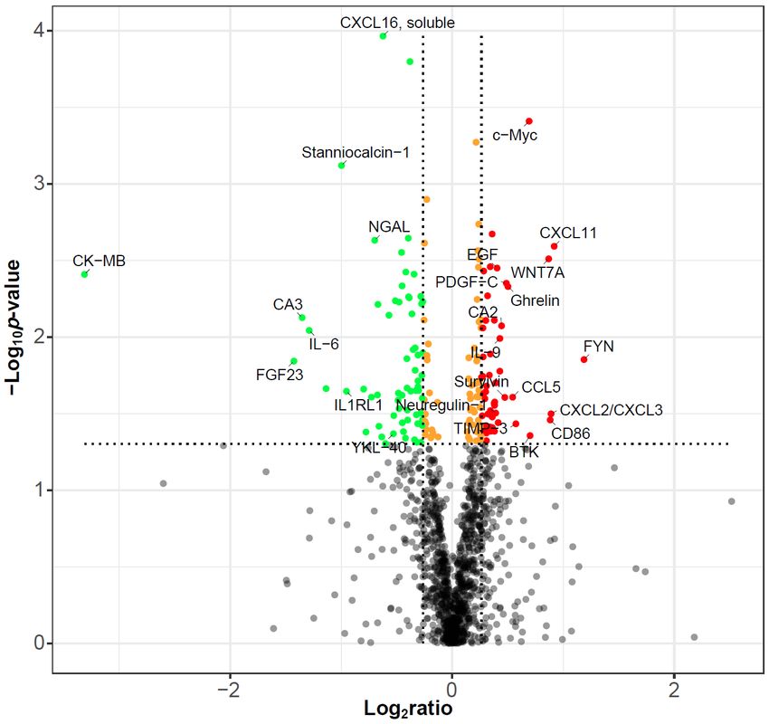
8survived without dialysis) in 38 (52.8%) patients while the remaining 34 (47.2%) patients survived with dialysis by day 28. Thus, recovery rate of kidney function in this subset of patients that we studied was similar to that in the overall ATN study cohort in which 343 of 626 patients (54.8%) surviving to day 28 were dialysis independent on day 28. The number (percentage) of patients with or without recovered kidney function by day 28 were also similar in diabetes, CV- SOFA score ≥2, intensive dialysis treatment arm, and cause of AKI, with no statistically significant differences (p >0.05). Higher baseline SOFA scores (both overall and CV-component) were associated with mortality; however, they were not associated with recovery of kidney function among survivors. The median baseline serum creatinine levels and estimated 60-day mortality risk scores were the same between the two groups. In addition, the median urine volume on day 8 was not significantly different (p=0.0597) between the kidney-recovered group (81.5, 60-478 mL/day) and the non-recovered group (59.5, 10-207 mL/day). These urine volumes were far less than that expected for kidney function recovery (>500 mL/day). SOMAScan Proteomic Profile of Day 8 AKI-D Serum Samples A volcano plot of SOMAscan analysis of 1305 serum proteins (Figure 1) demonstrates fold changes (FC cutoff=1.2) and statistical significance (p
discontinued dialysis by day 28, such as tyrosine-protein kinase Fyn (FC=2.28, p=0.014) and BTK

(FC=1.63, p=0.044), protein Wnt-7a (FC=1.83, p=0.0031), Myc proto-oncogene protein (c-Myc,

FC=1.62, p=0.0004), ghrelin (FC=1.42, p=0.0047), platelet-derived growth factor C (PDGF-C,

FC=1.40, p=0.0045), survivin (FC=1.39, p=0.0248), epidermal growth factor (EGF, FC=1.32,

p=0.0035), and neuregulin-1 (FC=1.31, p=0.0314). The SOMAscan measurements of all 1305

proteins are presented in Supplemental Table 2.

IPA Analysis of Biological Function Changes Related to Tissue Repair

IPA core analysis of proteins with significant changes in abundance revealed significant

alterations in diseases and biological functions on day 8 (Figure 2, Supplemental Table 3). The

analysis indicated increased functions in cell-to-cell signaling and interaction, cellular growth

and proliferation, cellular development, embryonic development, tissue morphology, tissue

development, organismal development, molecular transport, and post-translational

modification. The analysis also showed decreased function in cell death, connective tissue

disorders, organismal injury and abnormalities, and inflammatory response and disease. The

results suggest that molecular pathways related to these biological functions were already

activated or suppressed on day 8, which potentially affected kidney recovery by day 28.

Olink Verification of Top-changed Proteins

Of the top 25 proteins with significant changes identified from the SOMAscan assay (Table 2),

CXCL11, IL-6, and FGF23 were also included on the Olink inflammation panel (Supplemental

Table 4). Consistent results were obtained for all three proteins between the SOMAscan and

Olink assays. Strong positive correlations between the two assays were observed for CXCL11

and IL-6 with correlation coefficients of 0.86 (p
To establish a direct association between the serum levels of proteins that were significantly

different in the two groups and the end points of recovered or non-recovered kidney function,

we stratified their serum levels by tertiles of top-changed proteins (i.e., Fyn, CXCL11,

CXCL2/CXCL3, CD86, Wnt-7a, BTK, c-Myc, TIMP-3, CCL5, ghrelin, PDGF-C, survivin, CA2, IL-9,
EGF, neuregulin-1, YKL-40, soluble CXCL16, NGAL, IL1RL1, stanniocalcin-1, IL-6, CA3, FGF23, and

CK-MB) in day 8 samples. Higher serum levels for each of CXCL11, CXCL2/CXCL3, CD86, Wnt-7a,

BTK, c-Myc, TIMP-3, ghrelin, CA2, IL-9, EGF, and neuregulin-1 on day 8 were associated with

increased recovery rate of kidney function by day 28, with a statistical significance p
Myc levels were significantly associated with kidney recovery, with a ~56-fold increase in the

chance of recovery compared to lower c-Myc levels (odds ratio [OR]= 55.79, 95% confidence

interval [CI] (5.53, 562.99), p=0.0007). In contrast, for the proteins with lower serum levels on

day 8, patients with the upper serum levels for each of soluble CXCL16, IL1RL1, stanniocalcin-1,
IL-6, and FGF23 were unlikely to show kidney recovery by day 28 compared to those with the

lower corresponding protein levels, and these associations remained significant (p
who survived through day 8 had some degree of kidney recovery, thus we focused on the

identification of potential biomarkers in day 8 samples to predict kidney recovery by day 28 in

this study. Here, kidney function recovery was defined for the AKI patients who survived and

discontinued dialysis by day 28.

       Of the proteins identified from the SOMAscan assay that showed the greatest change,

CXCL11, IL-6, and FGF23 were also verified by the Olink assay. While strong correlation between

the two assays was observed for CXCL11 and IL-6 (r>0.85), FGF23 showed moderate correlation

(r=0.47). In our previous analysis, the correlation between SOMAscan and ELISA for FGF23 was

also moderate (r=0.61, p
that AKI biomarkers have the potential to be predictive biomarkers of kidney recovery.
However, these biomarkers need to be verified in larger clinical studies.
Inflammatory response is associated with phases of acute kidney injury and repair (25,
26). Reduced levels of the inflammatory biomarker IL-18 were found to be indicative of kidney
recovery (27). In this study, we found that lower serum levels of inflammation-related proteins
(FGF23, soluble CXCL16, IL1RL1, and IL-6) on day 8 were associated with increased kidney
recovery by day 28 and demonstrated that lower levels of these proteins were more
significantly associated with kidney recovery than AKI biomarkers YKL-40 and NGAL.
Furthermore, we also found that higher serum levels of CXCL11, CXCL2/CXCL3, CD86, CCL5, and
IL-9 were significantly associated with kidney recovery. Chemokine CXCL11, CXCL2/CXCL3, and
CCL5 target T-lymphocytes, endothelial cells, monocytes, and macrophages during wound
healing through their receptors on these cells (28). CXCL11 is a ligand of CXCR3, which has been
shown to play roles in protecting the kidney from ischemia reperfusion injury by recruiting
CXCR3+ natural killer T (NKT) cells (29, 30) and by promoting re-epithelialization in wound tissue
repair (31). CXCL2 and CXCL3 are ligands of CXCR2, which also play important roles in wound
healing (32). It has been shown that CXCL2/3-driven macrophage-myofibroblast crosstalk
promotes intestinal repair (33). Studies have shown that CD86 is required for a robust
regulatory T cell (Treg) response during the recovery phase for lung tissue repair after Influenza
A virus clearance (34). Furthermore, IL-9 is required to promote tissue repair in the recovery
phase of inflammatory lung (35) and inhibit early podocyte injury and progressive
glomerulosclerosis (36). These findings suggest that reduced inflammation and increased
chemokines/cytokines responsible for recruiting tissue repair immune cells or protecting kidney
cells from injury, may be useful predictive biomarkers of kidney recovery, underlining potential
mechanism(s) of renal regeneration and repair.
This study indicates that a large fraction of the proteins associated with kidney recovery
in AKI-D patients are related to cellular growth, survival, or proliferation. Higher serum levels of
14Wnt-7a and c-Myc on day 8 were associated with kidney recovery by day 28. Protein Wnt-7a is
a ligand in the canonical Wnt/beta-catenin signaling pathway, which has been shown to be
involved in renal tubular protection after AKI (37) and repair and regeneration after kidney
injury (38). Wnt/β-catenin signaling regulates the expression of the key driver of angiogenesis,
VEGF (39-41). The recent TRIBE-AKI study found that high postoperative plasma levels of VEGF
A isoform and PGF were associated with reduced risk for AKI and mortality (14). The present
finding of high levels of Wnt-7a could coincide with VEGF effects on angiogenesis and
endothelial remodeling, thus biomarkers associated with vascular regeneration could be
potential biomarkers of kidney recovery (42). C-Myc is a transcription factor that activates the
expression of growth-related genes (43). It is also well established that c-Myc is the major
transcription factor promoting VEGFA gene expression (44), illustrating its role in cell growth
and angiogenesis.
Coincidentally, higher serum levels of BTK, ghrelin, PDGF-C, survivin, EGF, and
neuregulin-1 were associated with kidney recovery by day 28. BTK is a tyrosine-protein kinase
and plays roles in B-cell development. PDGF-C, EGF, and neuregulin-1 are growth factors, and
both EGF and neuregulin-1 belong to the epidermal growth factor family. Delayed recovery
from AKI was found in mice with specific deletion of the EGF receptor in renal proximal tubule
epithelial cells (45). Ghrelin is expressed in tissues other than kidney (46); however, ghrelin
improves kidney function in mice with ischemic acute kidney failure (47) and reduces kidney
tissue damage in rats (48), probably due to its anti-inflammatory and antioxidant effects (49).
Increased expression of survivin in kidney epithelial cells was induced by AKI and kidney
recovery was markedly delayed in mice with renal proximal tubule-specific deletion of survivin
(50), suggesting that survivin plays a direct role in kidney recovery. Taken together, low levels of
inflammation-associated proteins (e.g., FGF23, CXCL6), high levels of chemokines/cytokines for
inflammatory tissue repair, and high levels of Wnt-7a, BTK, c-Myc, ghrelin, PDGF-C, survivin,
EGF, and neuregulin-1 are likely to play concerted roles in the coordination and regulation of
15angiogenesis, endothelial and epithelial remodeling, kidney cell regeneration in the transition
from inflammation to kidney recovery, and thus may be used as potential biomarkers for the
prediction of kidney recovery.
This study had several limitations. First, the availability of randomized day 8 serum
samples with secondary data analyses from the clinical trial study population was limited. This
restricted the development of a predictive model. However, we addressed these shortcomings
by using multiple statistical methods to attenuate potential biases. Second, both SOMAscan
and Olink assays measured relative protein level changes but not absolute protein
concentrations in serum, and not all the proteins discovered in the SOMAscan assay could be
verified using Olink assays as many of them were not included on the Olink panel. Third, this is a
biomarker discovery study for kidney recovery and there is a lack of an independent cohort of
patients for validation of these novel biomarker candidates. The results from this study are
informative for further verification in a larger cohort of samples. Finally, we were unable to
follow the trajectory of protein changes over the course of kidney recovery due to lack of
samples collected at later time points. Further studies specifically designed for this purpose
may be able to address this issue. However, the novel biomarkers of kidney recovery
discovered in our study warrant further independent validation in AKI-D patients.
In conclusion, this study used a high throughput proteomic technology to identify
potential novel biomarkers of kidney recovery, and defined potential biological pathways
associated with kidney remodeling, repair, and regeneration after AKI-D. Biomarkers involved in
the progressive biological processes of reducing inflammation, recruiting tissue repair immune
cells, releasing growth factors, activating epithelial and endothelial remodeling, and increasing
kidney cell growth and regeneration may be important targets for future studies to evaluate
kidney function recovery.
16DISCLOSURES
J. Ma reports the following: Scientific Advisor or Membership: Statistician on the Editorial Board
for Frontiers in Molecular Psychiatry, Editorial Board of American Journal of Physiology - Renal
Physiology, DSMB Member for the Clinical Trials network, the National Institute on Drug Abuse.
R. Beger reports the following: Scientific Advisor or Membership: Editor for Metabolomics
Society and Scientific Reports. D. Choudhury reports the following: Research Funding: Janssen
Pharma, AstraZeneca, Abbvie, Boehringer Ingelheim, CorMedix, GSK, Hope, Duke Clinical
Research Institute, Minolock, Bayer; Honoraria: Virginia Tech Carilion School of Medicine;
Scientific Advisor or Membership: Salem Research Institute-VA nonprofit research corporation -
advisory board member; Other Interests/Relationships: Board member Salem Research
Institute, Salem VA Medical Center. P. Palevsky reports the following: Consultancy Agreements:
Janssen Research & Development, LLC; Scientific Advisor or Membership: National Kidney
Foundation: President, Member, Scientific Advisory Board; Renal Physicians Association:
Member, Quality, Safety and Accountability Committee; Quality Insights Renal Network 4:
Chair, Medical Review Board; UpToDate: Section Editor, Renal Failure; Journal of Intensive Care
Medicine: Member, Editorial Board. D. Portilla reports the following: Scientific Advisor or
Membership: Kidney360. L. Yu reports the following: Scientific Advisor or Membership:
Member, Editorial Board of Journal of Proteomics. The remaining authors have nothing to
disclose.
FUNDING
This study was supported with funds from the National Center for Toxicological Research, U.S.
Food and Drug Administration (NCTR/FDA), Jefferson, Arkansas (Protocol # E07571.11, LRY); the
National Institutes of Health Grant DK75976; and a Veterans Affairs Merit Award to DP. JRD
would like to acknowledge NCTR/FDA for postdoctoral support through the Oak Ridge Institute
for Science and Education (ORISE).
The Acute Renal Failure Trial Network (ATN) Study was conducted by the ATN Investigators and
supported by the Cooperative Studies program of the Department of Veterans Affairs (VA)
17Office of Research and Development as CSP #530 and by the National Institute of Diabetes and
Digestive and Kidney Diseases (NIDDK) under interagency agreement Y1-DK-3508-01.
ACKNOWLEDGEMENTS
This article reflects the views of the authors and does not necessarily reflect those of the U.S.
Food and Drug Administration. Any mention of commercial products is for clarification only and
is not intended as approval, endorsement, or recommendation.
This manuscript was not prepared in collaboration with the ATN Study Investigators and does
not necessarily reflect the opinions or views of the ATN Study, VA, or NIDDK.
AUTHOR CONTRIBUTIONS: Jaclyn Daniels: Data curation; Methodology; Writing - review and
editing
Jennie Ma: Formal analysis; Writing – review and editing
Zhijun Cao: Formal analysis; Visualization; Writing - review and editing
Richard Beger: Conceptualization; Supervision; Writing – review and editing
Jinchun Sun: Funding acquisition; Writing - review and editing
Laura Schnackenberg: Writing - review and editing
Lisa Pence: Writing - review and editing
Devasmita Choudhury: Resources; Writing - review and editing
Paul Palevsky: Investigation; Resources; Writing - review and editing
Didier Portilla: Conceptualization; Funding acquisition; Investigation; Resources; Writing -
review and editing
Li-Rong Yu: Conceptualization; Data curation; Formal analysis; Funding acquisition;
Investigation; Methodology; Validation; Visualization; Writing - original draft.
All authors reviewed and revised the manuscript.
18SUPPLEMENTAL MATERIALS Supplemental Table 1. Proteins with significant changes in serum levels (1.2-fold and p
REFERENCES
1.    Wald R, McArthur E, Adhikari NK, Bagshaw SM, Burns KE, Garg AX, Harel Z, Kitchlu A, Mazer CD,
      Nash DM, Scales DC, Silver SA, Ray JG, Friedrich JO: Changing incidence and outcomes following
      dialysis-requiring acute kidney injury among critically ill adults: a population-based cohort study.
      Am J Kidney Dis 65: 870-877, 2015
2.    Uchino S, Kellum JA, Bellomo R, Doig GS, Morimatsu H, Morgera S, Schetz M, Tan I, Bouman C,
      Macedo E, Gibney N, Tolwani A, Ronco C: Acute renal failure in critically ill patients: a
      multinational, multicenter study. JAMA 294: 813-818, 2005
3.    Bagshaw SM, Laupland KB, Doig CJ, Mortis G, Fick GH, Mucenski M, Godinez-Luna T, Svenson
      LW, Rosenal T: Prognosis for long-term survival and renal recovery in critically ill patients with
      severe acute renal failure: a population-based study. Crit Care 9: R700-709, 2005
4.    Gaiao SM, Paiva J: Biomarkers of renal recovery after acute kidney injury. Rev Bras Ter Intensiva
      29: 373-381, 2017
5.    Srisawat N, Wen X, Lee M, Kong L, Elder M, Carter M, Unruh M, Finkel K, Vijayan A, Ramkumar
      M, Paganini E, Singbartl K, Palevsky PM, Kellum JA: Urinary biomarkers and renal recovery in
      critically ill patients with renal support. Clin J Am Soc Nephrol 6: 1815-1823, 2011
6.    Srisawat N, Murugan R, Lee M, Kong L, Carter M, Angus DC, Kellum JA: Plasma neutrophil
      gelatinase-associated lipocalin predicts recovery from acute kidney injury following community-
      acquired pneumonia. Kidney Int 80: 545-552, 2011
7.    Dewitte A, Joannes-Boyau O, Sidobre C, Fleureau C, Bats ML, Derache P, Leuillet S, Ripoche J,
      Combe C, Ouattara A: Kinetic eGFR and Novel AKI Biomarkers to Predict Renal Recovery. Clin J
      Am Soc Nephrol 10: 1900-1910, 2015
8.    Pike F, Murugan R, Keener C, Palevsky PM, Vijayan A, Unruh M, Finkel K, Wen X, Kellum JA:
      Biomarker Enhanced Risk Prediction for Adverse Outcomes in Critically Ill Patients Receiving
      RRT. Clin J Am Soc Nephrol 10: 1332-1339, 2015
9.    Murugan R, Wen X, Shah N, Lee M, Kong L, Pike F, Keener C, Unruh M, Finkel K, Vijayan A,
      Palevsky PM, Paganini E, Carter M, Elder M, Kellum JA: Plasma inflammatory and apoptosis
      markers are associated with dialysis dependence and death among critically ill patients receiving
      renal replacement therapy. Nephrol Dial Transplant 29: 1854-1864, 2014
10.   Aregger F, Uehlinger DE, Witowski J, Brunisholz RA, Hunziker P, Frey FJ, Jorres A: Identification of
      IGFBP-7 by urinary proteomics as a novel prognostic marker in early acute kidney injury. Kidney
      Int 85: 909-919, 2014
11.   Meersch M, Schmidt C, Van Aken H, Martens S, Rossaint J, Singbartl K, Gorlich D, Kellum JA,
      Zarbock A: Urinary TIMP-2 and IGFBP7 as early biomarkers of acute kidney injury and renal
      recovery following cardiac surgery. PLoS One 9: e93460, 2014
12.   Hsu CY, Chinchilli VM, Coca S, Devarajan P, Ghahramani N, Go AS, Hsu RK, Ikizler TA, Kaufman J,
      Liu KD, Parikh CR, Reeves WB, Wurfel M, Zappitelli M, Kimmel PL, Siew ED: Post-Acute Kidney
      Injury Proteinuria and Subsequent Kidney Disease Progression: The Assessment, Serial
      Evaluation, and Subsequent Sequelae in Acute Kidney Injury (ASSESS-AKI) Study. JAMA Intern
      Med 180: 402-410, 2020
13.   Lee BJ, Go AS, Parikh R, Leong TK, Tan TC, Walia S, Hsu RK, Liu KD, Hsu CY: Pre-admission
      proteinuria impacts risk of non-recovery after dialysis-requiring acute kidney injury. Kidney Int
      93: 968-976, 2018
14.   Mansour SG, Zhang WR, Moledina DG, Coca SG, Jia Y, Thiessen-Philbrook H, McArthur E, Inoue
      K, Koyner JL, Shlipak MG, Wilson FP, Garg AX, Ishibe S, Parikh CR: The Association of

                                                  20
Angiogenesis Markers With Acute Kidney Injury and Mortality After Cardiac Surgery. Am J Kidney
Dis 74: 36-46, 2019
15. Yu LR, Sun J, Daniels JR, Cao Z, Schnackenberg L, Choudhury D, Palevsky PM, Ma JZ, Beger RD,
Portilla D: Aptamer-Based Proteomics Identifies Mortality-Associated Serum Biomarkers in
Dialysis-Dependent AKI Patients. Kidney Int Rep 3: 1202-1213, 2018
16. Lee BJ, Hsu CY, Parikh R, McCulloch CE, Tan TC, Liu KD, Hsu RK, Pravoverov L, Zheng S, Go AS:
Predicting Renal Recovery After Dialysis-Requiring Acute Kidney Injury. Kidney Int Rep 4: 571-
581, 2019
17. Palevsky PM, O'Connor T, Zhang JH, Star RA, Smith MW: Design of the VA/NIH Acute Renal
Failure Trial Network (ATN) Study: intensive versus conventional renal support in acute renal
failure. Clin Trials 2: 423-435, 2005
18. Palevsky PM, Zhang JH, O'Connor TZ, Chertow GM, Crowley ST, Choudhury D, Finkel K, Kellum
JA, Paganini E, Schein RM, Smith MW, Swanson KM, Thompson BT, Vijayan A, Watnick S, Star
RA, Peduzzi P: Intensity of renal support in critically ill patients with acute kidney injury. N Engl J
Med 359: 7-20, 2008
19. Gold L, Ayers D, Bertino J, Bock C, Bock A, Brody EN, Carter J, Dalby AB, Eaton BE, Fitzwater T,
Flather D, Forbes A, Foreman T, Fowler C, Gawande B, Goss M, Gunn M, Gupta S, Halladay D,
Heil J, Heilig J, Hicke B, Husar G, Janjic N, Jarvis T, Jennings S, Katilius E, Keeney TR, Kim N, Koch
TH, Kraemer S, Kroiss L, Le N, Levine D, Lindsey W, Lollo B, Mayfield W, Mehan M, Mehler R,
Nelson SK, Nelson M, Nieuwlandt D, Nikrad M, Ochsner U, Ostroff RM, Otis M, Parker T,
Pietrasiewicz S, Resnicow DI, Rohloff J, Sanders G, Sattin S, Schneider D, Singer B, Stanton M,
Sterkel A, Stewart A, Stratford S, Vaught JD, Vrkljan M, Walker JJ, Watrobka M, Waugh S, Weiss
A, Wilcox SK, Wolfson A, Wolk SK, Zhang C, Zichi D: Aptamer-based multiplexed proteomic
technology for biomarker discovery. PLoS One 5: e15004, 2010
20. Assarsson E, Lundberg M, Holmquist G, Bjorkesten J, Thorsen SB, Ekman D, Eriksson A, Rennel
Dickens E, Ohlsson S, Edfeldt G, Andersson AC, Lindstedt P, Stenvang J, Gullberg M, Fredriksson
S: Homogenous 96-plex PEA immunoassay exhibiting high sensitivity, specificity, and excellent
scalability. PLoS One 9: e95192, 2014
21. Benjamini Y, Hochberg Y: Controlling the False Discovery Rate - a Practical and Powerful
Approach to Multiple Testing. J R Stat Soc Ser B-Stat Methodol 57: 289-300, 1995
22. Zhang JH, Palevsky PM, Chertow GM, Hartigan J, O'Connor TZ, Guarino P, Zhou B: Piecewise
analysis of patient survival after onset of AKI. Clin J Am Soc Nephrol 8: 1679-1684, 2013
23. Daniels JR, Cao Z, Maisha M, Schnackenberg LK, Sun J, Pence L, Schmitt TC, Kamlage B, Rogstad
S, Beger RD, Yu LR: Stability of the Human Plasma Proteome to Pre-analytical Variability as
Assessed by an Aptamer-Based Approach. J Proteome Res 18: 3661-3670, 2019
24. Hebels DG, Georgiadis P, Keun HC, Athersuch TJ, Vineis P, Vermeulen R, Portengen L, Bergdahl
IA, Hallmans G, Palli D, Bendinelli B, Krogh V, Tumino R, Sacerdote C, Panico S, Kleinjans JC, de
Kok TM, Smith MT, Kyrtopoulos SA: Performance in omics analyses of blood samples in long-
term storage: opportunities for the exploitation of existing biobanks in environmental health
research. Environ Health Perspect 121: 480-487, 2013
25. Okusa MD, Chertow GM, Portilla D: The nexus of acute kidney injury, chronic kidney disease,
and World Kidney Day 2009. Clin J Am Soc Nephrol 4: 520-522, 2009
26. Mulay SR, Holderied A, Kumar SV, Anders HJ: Targeting Inflammation in So-Called Acute Kidney
Injury. Semin Nephrol 36: 17-30, 2016
27. Hall IE, Yarlagadda SG, Coca SG, Wang Z, Doshi M, Devarajan P, Han WK, Marcus RJ, Parikh CR:
IL-18 and urinary NGAL predict dialysis and graft recovery after kidney transplantation. J Am Soc
Nephrol 21: 189-197, 2010
2128. Balaji S, Watson CL, Ranjan R, King A, Bollyky PL, Keswani SG: Chemokine Involvement in Fetal
and Adult Wound Healing. Adv Wound Care (New Rochelle) 4: 660-672, 2015
29. Yang SH, Lee JP, Jang HR, Cha RH, Han SS, Jeon US, Kim DK, Song J, Lee DS, Kim YS: Sulfatide-
reactive natural killer T cells abrogate ischemia-reperfusion injury. J Am Soc Nephrol 22: 1305-
1314, 2011
30. Zhang C, Zheng L, Li L, Wang L, Li L, Huang S, Gu C, Zhang L, Yang C, Zhu T, Rong R: Rapamycin
protects kidney against ischemia reperfusion injury through recruitment of NKT cells. J Transl
Med 12: 224, 2014
31. Yates CC, Whaley D, A YC, Kulesekaran P, Hebda PA, Wells A: ELR-negative CXC chemokine
CXCL11 (IP-9/I-TAC) facilitates dermal and epidermal maturation during wound repair. Am J
Pathol 173: 643-652, 2008
32. Zaja-Milatovic S, Richmond A: CXC chemokines and their receptors: a case for a significant
biological role in cutaneous wound healing. Histol Histopathol 23: 1399-1407, 2008
33. Esser-von Bieren J, Volpe B, Sutherland DB, Bürgi J, Verbeek JS, Marsland BJ, Urban JF, Jr., Harris
NL: Immune antibodies and helminth products drive CXCR2-dependent macrophage-
myofibroblast crosstalk to promote intestinal repair. PLoS Pathog 11: e1004778, 2015
34. Moser EK, Hufford MM, Braciale TJ: Late engagement of CD86 after influenza virus clearance
promotes recovery in a FoxP3+ regulatory T cell dependent manner. PLoS Pathog 10: e1004315,
2014
35. Turner JE, Morrison PJ, Wilhelm C, Wilson M, Ahlfors H, Renauld JC, Panzer U, Helmby H,
Stockinger B: IL-9-mediated survival of type 2 innate lymphoid cells promotes damage control in
helminth-induced lung inflammation. J Exp Med 210: 2951-2965, 2013
36. Xiong T, Attar M, Gnirck AC, Wunderlich M, Becker M, Rickassel C, Puelles VG, Meyer-
Schwesinger C, Wiech T, Nies JF, Divivier M, Fuchs T, Schulze Zur Wiesch J, Taipaleenmäki H,
Hoxha E, Wirtz S, Huber TB, Panzer U, Turner JE: Interleukin-9 protects from early podocyte
injury and progressive glomerulosclerosis in Adriamycin-induced nephropathy. Kidney Int 98:
615-629, 2020
37. Zhou D, Li Y, Lin L, Zhou L, Igarashi P, Liu Y: Tubule-specific ablation of endogenous β-catenin
aggravates acute kidney injury in mice. Kidney Int 82: 537-547, 2012
38. Lin SL, Li B, Rao S, Yeo EJ, Hudson TE, Nowlin BT, Pei H, Chen L, Zheng JJ, Carroll TJ, Pollard JW,
McMahon AP, Lang RA, Duffield JS: Macrophage Wnt7b is critical for kidney repair and
regeneration. Proc Natl Acad Sci U S A 107: 4194-4199, 2010
39. Olsen JJ, Pohl SO, Deshmukh A, Visweswaran M, Ward NC, Arfuso F, Agostino M, Dharmarajan A:
The Role of Wnt Signalling in Angiogenesis. Clin Biochem Rev 38: 131-142, 2017
40. Levy L, Neuveut C, Renard CA, Charneau P, Branchereau S, Gauthier F, Van Nhieu JT, Cherqui D,
Petit-Bertron AF, Mathieu D, Buendia MA: Transcriptional activation of interleukin-8 by beta-
catenin-Tcf4. J Biol Chem 277: 42386-42393, 2002
41. Easwaran V, Lee SH, Inge L, Guo L, Goldbeck C, Garrett E, Wiesmann M, Garcia PD, Fuller JH,
Chan V, Randazzo F, Gundel R, Warren RS, Escobedo J, Aukerman SL, Taylor RN, Fantl WJ: beta-
Catenin regulates vascular endothelial growth factor expression in colon cancer. Cancer Res 63:
3145-3153, 2003
42. Bouchard J, Mehta RL: Angiogenesis Markers and Recovery From Acute Kidney Injury: A Piece of
the Puzzle? Am J Kidney Dis 74: 12-14, 2019
43. Pelengaris S, Khan M, Evan G: c-MYC: more than just a matter of life and death. Nat Rev Cancer
2: 764-776, 2002
44. Shi Y, Xu X, Zhang Q, Fu G, Mo Z, Wang GS, Kishi S, Yang XL: tRNA synthetase counteracts c-Myc
to develop functional vasculature. Elife 3: e02349, 2014
2245. Chen J, Chen JK, Harris RC: Deletion of the epidermal growth factor receptor in renal proximal
tubule epithelial cells delays recovery from acute kidney injury. Kidney Int 82: 45-52, 2012
46. Mori K, Yoshimoto A, Takaya K, Hosoda K, Ariyasu H, Yahata K, Mukoyama M, Sugawara A,
Hosoda H, Kojima M, Kangawa K, Nakao K: Kidney produces a novel acylated peptide, ghrelin.
FEBS Lett 486: 213-216, 2000
47. Takeda R, Nishimatsu H, Suzuki E, Satonaka H, Nagata D, Oba S, Sata M, Takahashi M, Yamamoto
Y, Terauchi Y, Kadowaki T, Kangawa K, Kitamura T, Nagai R, Hirata Y: Ghrelin improves renal
function in mice with ischemic acute renal failure. J Am Soc Nephrol 17: 113-121, 2006
48. Çimen S, Taşdemir C, Vardı N, Ateş B, Taşdemir S, Özaydoğdu Çimen A: Protective effects of
ghrelin on kidney tissue in rats with partial ureteral obstruction. Turk J Med Sci 49: 696-702,
2019
49. Baatar D, Patel K, Taub DD: The effects of ghrelin on inflammation and the immune system. Mol
Cell Endocrinol 340: 44-58, 2011
50. Chen J, Chen JK, Conway EM, Harris RC: Survivin mediates renal proximal tubule recovery from
AKI. J Am Soc Nephrol 24: 2023-2033, 2013
23Table 1. Characteristics of day 8 patient cohorts stratified by kidney recovery status by day 28
Characteristica Overall Not recovered Recovered p-valueb
(N=72) (N=34) (N=38)
Age (yrs.) 60.1 ± 15.1 62.8 ± 15.3 57.6 ± 14.8 0.1456
Female 27 (37.5%) 10 (29.4%) 17 (44.7%) 0.1799
Congestive heart failure 11 (15.7%) 3 (9.1%) 8 (21.6%) 0.1968
Diabetes mellitus 23 (32.4%) 10 (29.4%) 13 (35.1%) 0.6067
CV-SOFA ≥2 42 (58.3%) 21 (61.8%) 21 (55.3%) 0.5764
Intensive treatment arm 43 (59.7%) 19 (55.9%) 24 (63.2%) 0.5297
Ischemic 51 (70.8%) 25 (73.5%) 26 (68.4%) 0.634
Cause of AKI Nephrotoxic 18 (25.0%) 10 (29.4%) 8 (21.1%) 0.4135
Sepsis 40 (55.6%) 17 (50.0%) 23 (60.5%) 0.3695
Multifactorial 39 (54.2%) 19 (55.9%) 20 (52.6%) 0.7823
Baseline serum creatinine 1.3 (1.05, 1.8) 1.3 (1.0, 1.7) 1.3 (1.1, 1.8) 0.671
Urine volume on day 8 (mL/day) 81.5 (31, 255) 59.50 (10, 207) 81.5 (60, 478) 0.0597
Estimated 60-day mortality risk 0.55 (0.35, 0.81) 0.55 (0.33, 0.81) 0.55 (0.36, 0.81) 0.7736
Serum calcium (mg/dL) 7.8 ± 1.0 7.8 ± 1.0 7.8 ± 1.0 0.9333
Serum PO4 (mg/dL) 5.4 ± 1.9 5.5 ± 2.1 5.4 ± 1.8 0.7658
a th th
Continuous variables are expressed as mean ± SD or median (25 , 75 percentile), categorical variables
as %; CV-SOFA, cardiovascular sequential organ failure assessment; PO4, phosphate; bp-values
comparing patients who recovered kidney function versus those who did not recover were calculated
using Mann-Whitney non-parametric test for continuous variables and Chi-square test (or Fisher’s exact
test if the count isTable 2. Kidney recovery rates by tertiles of serum proteins
Protein Tertile 1a Tertile 2 a Tertile 3 a Chi-Square
(low levels) (intermediate levels) (high levels) p-value
Proteins with higher serum levels in the recovery group
FYN 11 (45.8) 11 (45.8) 16 (66.7) 0.2483
CXCL11 6 (25.0) 15 (62.5) 17 (70.8) 0.0032
CXCL2/CXCL3 8 (33.3) 13 (54.2) 17 (70.8) 0.0334
CD86 9 (37.5) 11 (45.8) 18 (75.0) 0.0239
WNT7A 10 (41.7) 9 (37.5) 19 (79.2) 0.0063
BTK 9 (37.5) 11 (45.8) 18 (75.0) 0.0239
c-Myc 9 (37.5) 8 (33.3) 21 (87.5) 0.0002
TIMP-3 9 (37.5) 10 (41.7) 19 (79.2) 0.0063
CCL5 9 (37.5) 12 (50.0) 17 (70.8) 0.0652
Ghrelin 9 (37.5) 10 (41.7) 19 (79.2) 0.0063
PDGF-C 9 (37.5) 13 (54.2) 16 (66.7) 0.1272
Survivin 9 (37.5) 12 (50.0) 17 (70.8) 0.0652
CA2 10 (41.7) 10 (41.7) 18 (75.0) 0.0283
IL-9 7 (29.2) 16 (66.7) 15 (62.5) 0.0171
EGF 9 (37.5) 11 (45.8) 18 (75.0) 0.0239
Neuregulin-1 9 (37.5) 11 (45.8) 18 (75.0) 0.0239
Proteins with lower serum levels in the recovery group
YKL-40 15 (62.5) 14 (58.3) 9 (37.5) 0.1777
CXCL16, soluble 18 (75.0) 13 (54.2) 7 (29.2) 0.0063
NGAL 17 (70.8) 11 (45.8) 10 (41.7) 0.0911
IL1RL1 17 (70.8) 12 (50.0) 9 (37.5) 0.0652
Stanniocalcin-1 19 (79.2) 12 (50.0) 7 (29.2) 0.0023
IL-6 16 (66.7) 14 (58.3) 8 (33.3) 0.0551
CA3 18 (75.0) 11 (45.8) 9 (37.5) 0.0239
FGF23 17 (70.8) 11 (45.8) 10 (41.7) 0.0911
CK-MB 15 (62.5) 15 (62.5) 8 (33.3) 0.0652
a
N=24 patients in each tertile for all the proteins. Data are presented in the number of patients with
recovered kidney function and corresponding percentage in parenthesis in the tertile of each protein.
FYN, tyrosine-protein kinase Fyn; CXCL11, C-X-C motif chemokine 11 (I-TAC); CXCL2/CXCL3, C-X-C motif
chemokine 2/C-X-C motif chemokine 3 (Gro-beta/gamma); CD86, T-lymphocyte activation antigen CD86
(activation B7-2 antigen); WNT7A, protein Wnt-7a; BTK, tyrosine-protein kinase BTK; c-Myc, Myc proto-
oncogene protein; TIMP-3, metalloproteinase inhibitor 3; CCL5, C-C motif chemokine 5 (T-cell-specific
protein RANTES); PDGF-C, platelet-derived growth factor C; CA2, carbonic anhydrase 2; IL-9, interleukin-
9; EGF, epidermal growth factor; YKL-40, chitinase-3-like protein 1; CXCL16, C-X-C motif chemokine 16;
NGAL, neutrophil gelatinase-associated lipocalin; IL1RL1, interleukin-1 receptor-like 1; IL-6, interleukin-6;
CA3, carbonic anhydrase 3; FGF23, fibroblast growth factor 23; CK-MB, creatine kinase-MB.
25Table 3. Odds ratios and 95% confidence intervals of day 8 serum proteins in tertiles significantly
associated with kidney function recovery by day 28 as analyzed by logistic regression
Univariate Analysis Multivariable Analysisd
th th
Median (25 , 75 P- P-
Protein Tertilea OR (95% CI)c OR (95% CI)
percentile)b value value
CXCL11 1st 2532 (2060, 3467) 1 1
2nd 8658 (7016, 10109) 5.00 (1.45, 17.27) 0.0109 11.51 (2.22, 59.63) 0.0036
3rd 18586 (14894, 37590) 7.29 (2.03, 26.10) 0.0023 11.31 (2.10, 60.78) 0.0047
CXCL2/CXCL3 1st 459 (406, 512) 1 1
2nd 695 (643, 766) 2.36 (0.74, 7.60) 0.1490 1.93 (0.40, 9.32) 0.4133
3rd 1198 (952, 1386) 4.86 (1.43, 16.50) 0.0113 8.27 (1.77, 38.67) 0.0073
CD86 1st 831 (762, 898) 1 1
2nd 1105 (1065, 1127) 1.41 (0.45, 4.46) 0.5587 1.45 (0.37, 5.78) 0.5957
3rd 1539 (1365, 1748) 5.00 (1.45, 17.27) 0.0109 6.74 (1.47, 30.80) 0.0139
WNT7A 1st 270 (246, 294) 1 1
2nd 376 (342, 412) 0.84 (0.26, 2.68) 0.7680 1.16 (0.25, 5.39) 0.8516
3rd 556 (500, 609) 5.32 (1.49, 19.06) 0.0103 13.10 (2.40, 71.52) 0.0030
BTK 1st 758 (673, 837) 1 1
2nd 1005 (958, 1106) 1.41 (0.45, 4.46) 0.5587 0.92 (0.20, 4.20) 0.9180
3rd 1544 (1233, 1838) 5.00 (1.45, 17.27) 0.0109 5.48 (1.19, 25.21) 0.0291
c-Myc 1st 638 (556, 683) 1 1
2nd 863 (771, 915) 0.83 (0.26, 2.72) 0.7629 2.45 (0.42, 14.40) 0.3202
3rd 1247 (1155, 1604) 11.67 (2.70, 50.49) 0.0010 55.79 (5.53, 562.99) 0.0007
TIMP-3 1st 1046 (680, 1381) 1 1
2nd 3017 (2377, 3486) 1.19 (0.37, 3.79) 0.7680 1.15 (0.21, 6.32) 0.8728
3rd 7306 (5328, 9175) 6.33 (1.75, 22.91) 0.0049 36.98 (4.31, 317.19) 0.0010
CCL5 1st 3697 (2197, 4689) 1 1
2nd 12782 (10444, 16950) 1.67 (0.53, 5.27) 0.3841 2.38 (0.56, 10.04) 0.2387
3rd 38218 (28916, 49428) 4.05 (1.21, 13.54) 0.0232 8.64 (1.64, 45.42) 0.0109
Ghrelin 1st 1519 (1401, 1707) 1 1
2nd 2120 (2001, 2216) 1.19 (0.37, 3.79) 0.7680 2.00 (0.47, 8.50) 0.3500
3rd 2852 (2506, 3311) 6.33 (1.75, 22.91) 0.0049 11.70 (2.21, 61.77) 0.0038
PDGF-C 1st 484 (411, 529) 1 1
2nd 648 (631, 723) 1.97 (0.62, 6.24) 0.2489 3.24 (0.73, 14.44) 0.1234
3rd 1001 (896, 1669) 3.33 (1.02, 10.90) 0.0464 5.12 (1.12, 23.41) 0.0354
Survivin 1st 1113 (1006, 1220) 1 1
2nd 1515 (1342, 1578) 1.67 (0.53, 5.27) 0.3841 1.33 (0.28, 6.39) 0.7226
3rd 2002 (1855, 2286) 4.05 (1.21, 13.54) 0.0232 10.71 (1.94, 59.27) 0.0066
CA2 1st 260 (247, 271) 1 1
2nd 380 (318, 431) 1.00 (0.32, 3.15) 1.0000 0.94 (0.24, 3.75) 0.9298
3rd 599 (559, 664) 4.20 (1.23, 14.37) 0.0222 5.11 (1.14, 22.98) 0.0333
IL-9 1st 388 (371, 415) 1 1
2nd 571 (515, 634) 4.86 (1.43, 16.50) 0.0113 14.59 (2.30, 92.51) 0.0044
3rd 798 (691, 979) 4.05 (1.21, 13.54) 0.0232 10.38 (1.83, 58.72) 0.0081
EGF 1st 747 (655, 866) 1 1
262nd 1114 (1035, 1346) 1.41 (0.45, 4.46) 0.5587 1.46 (0.30, 7.11) 0.6370
3rd 1899 (1577, 2356) 5.00 (1.45, 17.27) 0.0109 12.06 (2.20, 66.12) 0.0041
Neuregulin-1 1st 253 (223, 282) 1 1
2nd 360 (333, 408) 1.41 (0.45, 4.46) 0.5587 3.43 (0.77, 15.34) 0.1071
3rd 552 (488, 805) 5.00 (1.45, 17.27) 0.0109 7.92 (1.61, 38.90) 0.0108
CXCL16, soluble 1st 12884 (10813, 15447) 1 1
2nd 27219 (21844, 29434) 0.39 (0.12, 1.34) 0.1358 0.38 (0.09, 1.69) 0.2031
3rd 40953 (35772, 48512) 0.14 (0.04, 0.49) 0.0023 0.09 (0.02, 0.44) 0.0033
IL1RL1 1st 10425 (7549, 12137) 1 1
2nd 30420 (21322, 38962) 0.41 (0.13, 1.35) 0.1437 0.41 (0.09, 1.81) 0.2399
3rd 66622 (52872, 95325) 0.25 (0.07, 0.83) 0.0232 0.11 (0.02, 0.66) 0.0152
Stanniocalcin-1 1st 2594 (1831, 3082) 1 1
2nd 4166 (3851, 5157) 0.26 (0.07, 0.94) 0.0392 0.13 (0.02, 0.77) 0.0243
3rd 7557 (6282, 16587) 0.11 (0.03, 0.41) 0.0010 0.04 (0.01, 0.28) 0.0009
IL-6 1st 891 (764, 972) 1 1
2nd 1312 (1213, 1498) 0.70 (0.22, 2.27) 0.5517 0.59 (0.14, 2.49) 0.4712
3rd 3024 (2214, 4157) 0.25 (0.08, 0.83) 0.0236 0.22 (0.05, 0.99) 0.0490
FGF23 1st 1107 (1022, 1171) 1 1
2nd 1693 (1452, 1971) 0.35 (0.11, 1.15) 0.0828 0.32 (0.07, 1.49) 0.1464
3rd 5067 (2968, 9366) 0.29 (0.09, 0.97) 0.0451 0.16 (0.03, 0.79) 0.0250
a
N=24 patients in each tertile for all the proteins. bSOMAscan RFU. cOR, odds ratio; CI, confidence
interval. dage, gender, CHF, diabetes mellitus, CV-SOFA ≥2, intensive dialysis treatment, cause of AKI,
baseline serum creatinine, day-8 urine volume, and estimated 60-day mortality risk were adjusted in the
multivariable logistic regression analysis. The low level of each protein (Tertile 1) was considered as the
reference. CXCL11, C-X-C motif chemokine 11 (I-TAC); CXCL2/CXCL3, C-X-C motif chemokine 2/C-X-C
motif chemokine 3 (Gro-beta/gamma); CD86, T-lymphocyte activation antigen CD86 (activation B7-2
antigen); WNT7A, protein Wnt-7a; BTK, tyrosine-protein kinase BTK; c-Myc, Myc proto-oncogene
protein; TIMP-3, metalloproteinase inhibitor 3; CCL5, C-C motif chemokine 5 (T-cell-specific protein
RANTES); PDGF-C, platelet-derived growth factor C; CA2, carbonic anhydrase 2; IL-9, interleukin-9; EGF,
epidermal growth factor; CXCL16, C-X-C motif chemokine 16; IL1RL1, interleukin-1 receptor-like 1; IL-6,
interleukin-6; FGF23, fibroblast growth factor 23.
27You can also read