Giant chondroid lipoma of the breast: A case report and literature review
←
→
Page content transcription
If your browser does not render page correctly, please read the page content below
EXPERIMENTAL AND THERAPEUTIC MEDICINE 22: 1087, 2021

                             Giant chondroid lipoma of the breast:
                              A case report and literature review
               ADNAN AL ALOUL1,2, SILVIU SAVGA1, CAMELIA DIACONU3,4, CORNEL SAVU5,6,
           OVIDIU STIRU7,8, MIHAI DIMITRIU9,10, IRINA BALESCU11 and NICOLAE BACALBASA9,12,13

           1
            Department of Surgery, Ramnicu Sarat County Hospital, 125300 Ramnicu Sarat; 2Department of Surgery,
   Titu Maiorescu University, 040051 Bucharest; 3Department of Internal Medicine, ‘Carol Davila’ University of Medicine
        and Pharmacy, 020021 Bucharest; 4Department of Internal Medicine, Clinical Emergency Hospital of Bucharest,
           105402 Bucharest; 5Department of Thoracic Surgery, ‘Carol Davila’ University of Medicine and Pharmacy,
       020021 Bucharest; 6Department of Thoracic Surgery, ‘Marius Nasta’ Institute of Pneumology, 050159 Bucharest;
       7
        Department of Cardiovascular Surgery, ‘Carol Davila’ University of Medicine and Pharmacy, 020021 Bucharest;
       8
         Department of Cardiovascular Surgery, ‘Prof. Dr. C.C. Iliescu’ Emergency Institute for Cardiovascular Diseases,
     022322 Bucharest; 9Department of Obstetrics and Gynecology, ‘Carol Davila’ University of Medicine and Pharmacy,
         020021 Bucharest; 10Department of Obstetrics and Gynecology, ‘Sf. Pantelimon’ Emergency Clinical Hospital,
    021659 Bucharest; 11Department of Visceral Surgery, ‘Ponderas’ Academic Hospital, 021188 Bucharest; 12Department
      of Visceral Surgery, Center of Excellence in Translational Medicine, ‘Fundeni’ Clinical Institute, 022328 Bucharest;
          13
             Department of Obstetrics and Gynecology, ‘I. Cantacuzino’ Clinical Hospital, 030167 Bucharest, Romania

                                         Received April 20, 2021; Accepted May 20, 2021

                                                    DOI: 10.3892/etm.2021.10521

Abstract. Chondroid lipoma is a rare benign lesion affecting          Introduction
the breast, in which the diagnosis of malignancy is difficult
to be excluded during preoperative studies. In this respect,          Although lipoma is the most commonly encountered benign
a correct histopathological diagnosis is mandatory in order           mesenchymal tumor (1), chondroid lipoma is a rare entity
to avoid overdiagnosis and subsequently overtreatment.                originating from soft tissues that was initially described by
In the present study, the case of a 61‑year‑old patient who           Meis and Enzinger in 1993 (1). Due to the presence of certain
self‑referred for the development of a large tumor at the level       similarities with extra‑skeletal chondrosarcoma and round
of the left breast is reported. Biopsy raised the suspicion of a      cell liposarcoma, the diagnosis of this pathological entity
chondroid lipoma; thus, the patient was submitted to conserva‑        may become challenging especially in cases presenting large
tive surgery. The lesion was completely excised by performing         lesions (2,3). Histopathological and immunohistochemical
a total mastectomy. The histopathological studies confirmed           studies demonstrating the presence of an association between
the presence of chondroid lipoma, with no signs of malignant          adipose tissue, cartilaginous tissues, mature adipocytes,
transformation. At the 24‑month follow‑up interval no signs of        chondroblasts and hyaline matrix as well as the absence of
recurrence were detected. In conclusion, although it may reach        aberrant proliferation suggesting malignant transformation
significant dimensions, giant chondroid lipoma of the breast is       seems to play a crucial role in order to provide a differential
a benign lesion that may benefit from conservative treatment          diagnosis with malignant lesions such as chondrosarcoma
and does not recur.                                                   and liposarcoma (4). Providing a correct diagnosis is crucial
                                                                      in the prevention of the overtreatment of these lesions which
                                                                      otherwise may be treated as malignant lesions.
                                                                          In the present study, the case of a 61‑year‑old patient who
                                                                      self‑referred for the development of a large tumor at the level
Correspondence to: Dr Irina Balescu, Department of Visceral           of the left breast is reported.
Surgery, ‘Ponderas’ Academic Hospital, 85a Nicolae Caramfil
Street, 021188 Bucharest, Romania
E‑mail: irina.balescu@ponderas‑ah.ro                                  Case report

Abbreviations: BIRDAS, breast imaging‑reporting and data system       Patient data. A 61‑year‑old patient with no significant medical
                                                                      history self‑presented for the appearance of a large tumor
Key words: giant chondroid lipoma, differential diagnosis, surgery,   which was observed within the last eight months and which
breast surgery                                                        presented a rapid growth within the last three months.
                                                                          The clinical examination revealed the presence of a large
                                                                      tumoral mass having developed at the level of the left breast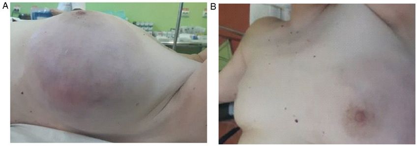
2 AL ALOUL et al: GIANT CHONDROID LIPOMA OF THE BREAST
Figure 1. (A) Initial preoperative procedure. The entire breast is deformed by the presence of a large tumoral mass inducing the apparition of collateral circulation.
(B) Initial preoperative procedure: The whole breast is deformed by the presence of a large tumoral mass while the nipple presents no significant modifications.
Figure 2. Mammography shows the presence of multiple calcifications.
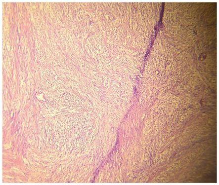
Figure 4. Histopathological studies included hematoxylin and eosin staining.
Magnification, x10. Mature adipose tissue and many cartilaginous islands
and mixoid cells are noted.
nipple presented no pathological aspects and no discharge.
Local examination of the left axilla failed to demonstrate
the presence of suspect adenopathies; furthermore, the
contralateral breast and axilla presented no pathological modi‑
fications (Fig. 1A and B).
Methods. The patient was further submitted to a mammo
graphy which confirmed the presence of a 22/18 cm lesion
with regular margins and intra‑tumoral calcifications. The
lesion was classified as a Breast Imaging‑Reporting and Data
System (BIRDAS) grade 2 tumor, and normal breast paren‑
chyma was almost absent (Fig. 2). The patient underwent breast
ultrasound which confirmed the presence of a heterogeneous
Figure 3. The specimen, obtained from total mastectomy, is a large, encap‑ mass measuring 20/18/15 cm with peripheral positive Doppler
sulated tumor. signal and minimal areas of normal breast parenchyma; the
ultrasound‑guided biopsy raised the suspicion of a chondroid
lipoma, with no signs of malignancy being encountered. The
measuring 20/15 cm, deforming the entire breast. Additionally, patient then underwent surgery with conservative intent;
signs of collateral circulation were observed at the level of the however, after excision of the encapsulated tumor, no remnant
adjacent skin (Fig. 1). The mass seemed to keep its mobility in breast parenchyma was found. Therefore, total mastectomy
the surrounding skin and to the prepectoral fascia while the was performed (Fig. 3). The histopathological studies, which
EXPERIMENTAL AND THERAPEUTIC MEDICINE 22: 1087, 2021                                                3

included hematoxylin and eosin staining, demonstrated the             liposarcoma (15‑28); in this respect, ultrasound‑guided biopsy
presence of a 20/18/10 cm encapsulated lesion presenting              is mandatory.
mesenchymal proliferation formed by chondroid myxoid                      Thus, appropriate diagnosis of benignity can be established
matrix with lipoblast areas and blood vessels and no mitotic          and therefore, the patient can be submitted to local excision of
activity (Fig. 4). In addition, a 1.5 cm fibroadenoma was also        the lesion, and overtreatment can be prevented (4,14,29‑36).
found in the close proximity of the giant chondroid lipoma.           However, in certain cases, due to the high volume of the lesion,
The postoperative outcome was uneventful. The patient then            retrieving the tumor can consist in fact in performing a total
underwent breast reconstruction with an implant two months            mastectomy (as presented in our case) although surgery is not
later. At 24 months, the patient is free of local or distant recur‑   intended to be a radical one as long as the biopsy has confirmed
rent disease.                                                         the absence of atypical mitoses (3). In order to prevent the risk
                                                                      of local recurrence, complete excision of the lesion en bloc
Discussion                                                            with the adjacent capsule is mandatory (4,15).
                                                                          Another important aspect which should be taken into
The structure of breast lipomas is usually modified by the            consideration when it comes to chondroid lipoma of the
presence of other mesenchymal elements leading to the                 breast is the one regarding the differential diagnostic with
development of fibrolipoma, angiolipoma, osteolipoma,                 malignant chondroid tumors such as primary chondroid
myxolipoma, or chondrolipoma (5). Most often cartilaginous            sarcoma (19‑24). Therefore, in cases in which malignant
transformation inside a lipomatous lesion leading to the              transformation is suspected, immunohistochemical studies
development of a chondrolipoma is related to a prolonged              aiming to investigate the presence of cytokeratin expression
evolution and to larger dimensions of a lipoma (6). Although          are mandatory (25‑28).
the exact process of chondrolipoma histogenesis is not                    In summary, giant breast chondroid lipoma represent scarce
well understood, there are three theories which have been             situations affecting women worldwide. Preoperative biopsy is
considered. The first one considers that in cases in which            mandatory in order to demonstrate the absence of any sign of
glandular components are found inside the tumor, they act             malignant disease and to further allow the surgeon to perform
like choristoma; the second theory considers that immature            tumoral enucleation without radical breast surgery. However,
mesenchymal cells develop towards both adipocytes and                 in certain cases the absence of normal breast parenchyma will
chondrocytes; while the third theory considers that these             transform the enucleation procedure into a total mastectomy
tumors originate from cartilaginous metaplasia of the adipose         followed by breast reconstructive surgery.
tissue in lipomas (7‑13).
    Chondroid lipomas are delimited, asymptomatic lesions             Acknowledgements
developed in the subcutaneous tissues or at the level of the
skeletal muscles, more commonly at the level of the arms, neck        Not applicable.
and head. Breast chondroid lipomas represent a scarce even‑
tuality, being more frequently encountered in young women             Funding
(during the third decade of life) and present as small volume
lesions (2). Their histopathological particularity is represented     No funding was received.
by the presence of cartilaginous tissue among mature fat
and glandular mammary parenchyma and their dimensions                 Availability of data and materials
usually range between 2 and 6 cm (4). Giant lesions are those
>5 cm in one dimension, with a weight of >500 g (14), but             Further information regarding the case study is available from
rarely surpassing 10 cm (15). The tumor is characterized by           the corresponding author on reasonable request.
the presence of peripheral compressed mammary parenchyma
which is transformed into a true capsule for the tumor while at       Authors' contributions
the level of the tumor ductal structures and mammary stroma
may not be present (4,16). In order to achieve proper diagnosis,      AAA and SS performed the surgical procedures. NB, MD and
imagistic studies such as breast ultrasound, mammography,             IB prepared the draft of the article in light of the literature data
and MRI followed by biopsy is mandatory. When it comes to             and case findings. NB was advisor of the surgical procedures.
the mammographic aspect of the lesion, it usually presents as         CD, CS and OS preoperatively investigated the patient. AAA
a radiolucent mass due to the presence of an increased amount         and NB revised the final draft of the manuscript. All authors
of fatty tissue in the absence of mammary stroma or ducts             read and approved the final manuscript.
in association with focal opacities induced by the presence
of islets of cartilaginous structures. However, the presence          Ethics approval and consent to participate
of calcifications is rather scarce, with few such cases being
reported thus far (5,7,17). Therefore, preoperatively, in the         The Ethics Committee of Ramnicu Sarat County Hospital
absence of a biopsy, the differential diagnosis with a malig‑         approved the study (no. 21/2018).
nant lesion is rather difficult to be established (5,18). The most
common entities which should be taken into consideration              Patient consent for publication
when performing a differential diagnosis are represented
by fat necrosis, giant fibroadenoma, supernumerary breast             Written informed consent was obtained from the patient on
as well as malignant lesions such as chondrosarcoma or                11.04.2018.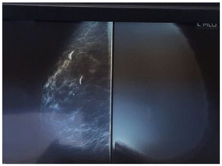
4                                    AL ALOUL et al: GIANT CHONDROID LIPOMA OF THE BREAST

Competing interests                                                    21. Montgomery E, Goldblum JR and Fisher C: Myofibrosarcoma:
                                                                           A clinicopathologic study. Am J Surg Pathol 25: 219‑228, 2001.
                                                                       22. Morgan PB, Chundra S, Hatch SS, Hawkins HK, Adegboyega PA
The authors declare that they have no competing interests.                 and Eltorky MA: Uncommon malignancies: Case 1. Low
                                                                           grade myofibroblastic sarcoma of the breast. J Clin Oncol 23:
                                                                           6249‑6251, 2005.
References                                                             23. Stark M, Hoffman A and Xiong Z: Mammary myofibrosarcoma:
                                                                           Case report and literature review. Breast J 17: 300‑304, 2011.
 1. Meis JM and Enzinger FM: Chondroid lipoma. A unique tumor          24. Brenn T and Fletcher CD: Radiation‑associated cutaneous
    simulating liposarcoma and myxoid chondrosarcoma. Am J Surg            atypical vascular lesions and angiosarcoma: Clinicopathologic
    Pathol 17: 1103‑1112, 1993.                                            analysis of 42 cases. Am J Surg Pathol 29: 983‑996, 2005.
 2. Vandeweyer E and Scagnol I: Axillary giant lipoma: A case          25. Cooper R, Rajak R, Valentine K and Bhargava V: Metaplastic
    report. Acta Chir Belg 105: 656‑657, 2005.                             carcinoma of the breast. Diagnostic Histopathol 24: 83‑85, 2018.
 3. Aljarrah A, Malik KA, Al Jarraha A, Sawhney S and Lakhtakia R:     26. Ginter PS, Mosquera JM, MacDonald TY, D'Alfonso TM,
    Chondroid lipoma of breast: A rare pathology. J Liaquat Univ           Rubin MA and Shin SJ: Diagnostic utility of MYC amplifica‑
    Med Health Sci 12: 131‑132, 2013.                                      tion and anti‑MYC immunohistochemistry in atypical vascular
 4. Banev SG and Filipovski VA: Chondrolipoma of the breast‑case           lesions, primar yor radiation induced mammary angiosarcoma
    report and a review of literature. Breast 15: 425‑426, 2006.           and primary angiosarcomas of other sites. Hum Pathol 45:
 5. Sudhamani S, Pandit AA and Kiri VM: Chondrolipoma of                   709‑716, 2014.
    breast: A case report with the review of the literature. J Sci     27. Cornejo KM, Deng A, Wu H, Cosar EF, Khan A, Cyr MS,
    Soc 39: 147‑148, 2012.                                                 Tomaszewicz K, Dresser K, O'Donnell P and Hutchinson L:
 6. Weiss SW and Goldblum JR: Cartilaginous soft tissue tumors.
    In: Enzinger and Weiss's Soft tissue tumors. 4th edition.              The utility of MYC and FLT4 in the diagnosis and treatment of
    Strauss M (ed).St. Louis, Mosby, p1361, 2001.                          post radiation atypical vascular lesion and angiosarcoma. Hum
 7. Kaplan L and Walts AE: Benign chondrolipomatous tumor of the           Pathol 46: 868‑875, 2015.
    human female breast. Arch Pathol Lab Med 101: 149‑151, 1977.       28. Maggiano F, Debiec‑Rychter M, Vanbockrijck M and Sciot R:
 8. Benisch B, Peison B and Sarno J: Benign mesenchymoma of the            Cellular angiofibroma: Another mesenchymal tumour with
    breast. Mt Sinai J Med 43: 530‑533, 1976.                              13q14 involvement, suggesting a link with spindle cell lipoma
 9. Fushimi H, Kotoh K, Nishihara K, Fujinaka H and Takao T:               and (extra)‑mammary myofibroblastoma. Histopathology 51:
    Chondrolipoma of the breast: A case report with cytological and        410‑412, 2007.
    histological examination. Histopathology 35: 478‑479, 1999.        29. Pasta V, Sottile D, Urciouli P, Del Vecchio L, Custureri F and
10. Fujimura N and Enomoto S: Lipoma of the tongue with cartilagi‑         D'Orazi V: Rare chondrosarcoma of the breast treated with
    nous change: A case report and review of the literature. J Oral        quadrantectomy instead of mastectomy: A case report. Oncol
    Maxillofac Surg 50: 1015‑1017, 1992.                                   Lett 9: 1116‑1120, 2015.
11. Lugo M, Reyes JM and Putong PB: Benign chondrolipomatous           30. Pasta V, Monti M, Cialini M, Vergine M, Urciuoli P, Iacovelli A,
    tumors of the breast. Arch Pathol Lab Med 106: 691‑692, 1982.          Rea S and D'Orazi V: Primitive sarcoma of the breast: New
12. Katzer B: Histopathology of rare chondroosteoblastic metaplasia        insight on the proper surgical management. J Exp Clin Cancer
    in benign lipomas. Pathol Res Pract 184: 437‑445, 1989.                Res 34: 72, 2015.
13. Shintaku M, Yamamoto Y, Kono F, Kitai T, Tsuji W, Yotsumoto F      31. Thilagavath G, Subramanian S, Samuel AV, Rani U and
    and Kushima R: Chondrolipoma of the breast as a rare variant of        Somasundaram C: Primary chondrosarcoma of the breast.
    myofibroblastoma: An immunohistochemical study of two cases.           J Indian Med Assoc 90: 16‑17, 1992.
    Virchows Arch 471: 531‑535, 2017.                                  32. Vandenhaute B, Validire P, Veillex C, Voelh P and Zafrani B:
14. Ricardo CR, Renato S, Luiz F and Espana Q: Breast reconstruc‑          Breast carcinoma with chondroid metaplasia. Ann Pathol 15:
    tion with parenchymal cross after giant lipoma removal. Aesth          53‑58, 1995.
    Plast Surg 32: 695‑697, 2008.                                      33. Pencavel TD and Hayes A: Breast sarcoma‑a review of diagnosis
15. Jorwekar GJ, Baviskar PK, Sathe PM and Dandekar KN: Giant              and management. Int J Surg 7: 20‑23, 2009.
    chondroid lipoma of breast. Indian J Surg 74: 342‑343, 2012.       34. Pasta V, Monti M, Antonucci D, Di Matteo FM, Boccaccini F
16. Marsh WL Jr, Lucas JG and Olsen J: Chondrolipoma of the                and Brescia A: Primary sarcoma of the breast: Criteria for radical
    breast. Arch Pathol Lab Med 113: 369‑371, 1989.                        surgery. G Chir 18: 703‑706, 1997.
17. Perez MT and Alexis JB: Chondrolipoma of the breast presenting     35. Stojadinovic A, Leung DH, Hoos A, Jaques DP, Lewis JJ and
    as calcifications in a routine mammogram. Histopathology 35:           Brennan MF: Analysis of the prognostic significance of micro‑
    189‑191, 1999.                                                         scopic margins in 2,084 localized primary adult soft tissue
18. Stark AM and Sunter JP: Benign chondrolipomatous tumour of             sarcomas. Ann Surg 235: 424‑434, 2002.
    the breast. Clin Radiol 39: 328‑329, 1988.                         36. Z elek L, Llomba r t‑ C ussac A, Ter r ier P, P ivot X,
19. Errarhay S, Fetohi M, Mahmoud S, Saadi H, Bouchikhi C and              Guinebretiere JM, Le Pechoux C, Tursz T, Rochard F,
    Banani A: Primary chondrosarcoma of the breast: A case presen‑         Spielmann M and Le Cesne A: Prognostic factors in primary
    tation and review of the literature. World J Surg Oncol 11: 208,       breast sarcomas: A series of patients with long‑term follow‑up.
    2013.                                                                  J Clin Oncol 21: 2583‑2588, 2003.
20. Nakanishi G, Lin SN, Asagoe K, Suzuki N, Matsuo A, Tanaka R,
    Makino E, Fujimoto W and Iwatsuki K: A novel fusion gene of
    collagen type 1 alpha 1 (exon 31) and platelet derived growth
    factor B‑chain( exon 2) in dermato fibrosarcome protuberans.
    Eur J Dermatol 17: 217‑219, 2007.
You can also read