Vanishing Non-immune Hydrops in Giant Chorioangioma of Placenta
←
→
Page content transcription
If your browser does not render page correctly, please read the page content below
DOI: 10.7860/JCDR/2021/46769.14583
Case Report
Vanishing Non-immune Hydrops in
Gynaecology Section
Obstetrics and
Giant Chorioangioma of Placenta
Sunita Dubey1, Aayushi Kaushal2, HN Pavithra3
ABSTRACT
Giant Chorioangioma of placenta is a rare nontrophoblastic tumour of placenta. It may lead to various maternal and foetal complications
like massive antepartum haemorrhage, sudden intrauterine foetal demise and non-immune hydrops, although in few cases mother
and the foetus remain unaffected. This report is of a 35-year-old G3P1L1A1, presented to hospital at 32 weeks gestation with pain
abdomen followed by watery discharge from vagina. Ultrasonography at 30 weeks revealed a huge mass on anterior wall with
placenta on posterior wall of uterus although her previous antenatal sonography did not reveal any abnormality either in the foetus
or in placenta. Diagnosis of preterm rupture of membranes was confirmed. Hence, she was kept on conservative management;
received antibiotics and steroids for foetal lung maturity. Subsequently, the foetus developed mild, steady non-immune hydrops
probably due to high output cardiac failure as Values of Middle Cerebral Artery’s Peak Systolic Velocity (MCA-PSV) were within
normal limits. Biophysical profile and nonstress test were normal. Guarded foetal prognosis was given due to non-immune hydrops
but she delivered a normal female baby with good Appearance, Pulse, Grimace, Activity and Respiration (APGAR) score with huge
chorioangioma of placenta. Although rare, chorioangiomas of placenta should be kept in differential diagnosis of non-immune
hydrops that needs regular foetal surveillance and timely intervention in affected foetuses to increase survival after birth.
Keywords: Nontrophoblastic tumour of placenta, Polyhydramnios, Preterm labour, Preterm rupture of membranes
CASE REPORT She received four doses of 6 mg dexamethasone given by
A 35-year-old G3P1L1A1 at 32+3 weeks gestation referred to the intramuscular route at six hourly intervals for foetal lung maturity
hospital with complaint of labour pains. She had one normal vaginal and was discharged following cessation of uterine contractions
delivery 17 years back, followed by spontaneous abortion one year with advise of follow-up in Outpatient Department. After one
later. Antenatal period was uneventful till admission. Her family and week, she presented with complaints of watery discharge per
medical history were not significant. vaginum that was confirmed by vaginal examination. Ultrasound
revealed; live foetus with breech presentation, reduced liquor
Ultrasonography of the foetus at 18 and 28 weeks gestation did not
reveal any abnormal mass within the placenta or uterine wall and there (Amniotic Fluid Index: 3 cm) along with the evidence of mild foetal
was no evidence of any malformations in the foetus too. Her vitals were scalp oedema and pericardial effusion. Thin streak of fluid was
stable, fundal height was of 32 weeks gestation. Ultrasound revealed seen around the left kidney suggestive of ascites. Abdominal
foetal parameters of 30 weeks with fundoposterior placenta and a wall muscles and overlying skin appeared thick and hypoechoic
well-defined multilobulated, hypoechoic mass measuring 98×75 mm suggestive of subcutaneous oedema with similar placental mass
with hyperechoic bands, arising from the chorionic plate of anterior as noticed earlier. Patient was admitted for evaluation of foetal
uterine wall was protruding inside the amniotic cavity [Table/Fig-1]. hydrops and management of preterm rupture of membranes along
with antibiotic cover as per our hospital protocol and monitored
for signs and symptoms of chorioamnionitis.
First abruptio placenta was excluded as she did not have any
episode of bleeding from vagina and there was no evidence
of increase in fundal height and uterine tenderness on clinical
examination. Differential diagnosis of succenturiate lobe and
submucosal fibroid uterus were made initially. After development
of mild foetal ascites and pericardial effusion, these diagnosis
were revised and diagnoses of placental tumour was made as a
cause of hydrops due to difference in echotexture of mass and
pattern of blood flow.
To rule out structural defects and arrhythmias as a cardiac cause
of foetal hydrops, foetal echocardiography was done that revealed
mild pericardial effusion and mild cardiomegaly. Foetal anaemia
was also excluded by Middle Cerebral Artery-Peak Systolic Velocity
(MCA-PSV) doppler study, value of which was 1.0 Multiple of Median
(MoM) for gestational age (40 cm/s at 32 weeks). Viral infections
were excluded by negative Toxoplasma, Rubella, Cytomegalovirus,
Herpes Simplex Virus (TORCH) profile. Maternal thalassaemia
[Table/Fig-1]: Transabdominal Ultrasonography revealed placenta (long arrow) screening and Indirect Coomb test were also done to exclude
with separate hypoechoic multilobulated mass (small arrow) with minimal blood flow inherited cause of foetal anaemia and possibility for alloantibodies
on colour doppler study. other than anti-Rh D, respectively. Possibility of aneuploidy was also
Journal of Clinical and Diagnostic Research, 2021 Mar, Vol-15(3): QD01-QD03 1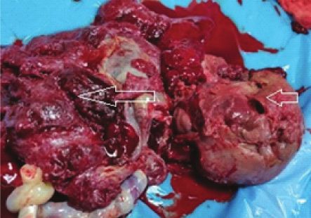
Sunita Dubey et al., Vanishing Non-immune Hydrops in Giant Chorioangioma of Placenta www.jcdr.net
excluded as echocardiography and level II scan did not reveal any Due to high perinatal mortality (30-40%) there is a need of timely
structural abnormality. interventions either in utero or in neonatal period particularly in cases
Subsequent monitoring by ultrasonography revealed normal liquor of foetal hydrops [5]. Very few case reports of giant chorioangioma
and features of hydrops were steady. MCA-PSV colour Doppler of placenta with intrauterine non-immune hydrops have resulted in
study was within normal limits with good biophysical profile. good neonatal outcome [6-9]. However, present case of mild foetal
hydrops was also born with good outcome without any intervention
Guarded foetal prognosis was explained to the relatives but they
that otherwise would have required in massive foetal hydrops,
opted for caesarean section. At 34 weeks she delivered a live born
girl of 1.7 kg birth weight with APGAR score of 9, 9 at 1 and 5 minute, merits it’s reporting.
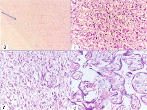
respectively. Following delivery of the placenta, a 10×8.5×6.2 cm Polyhydramnios, preterm labour are the most common maternal
fleshy vascularised mass was removed, histological examination complications, whereas maternal mirror syndrome and near miss
confirmed angiomatoid type of chorioangioma [Table/Fig-2,3]. mortality have also been noticed in patient with giant chorioangioma
of placenta [10-12]. In the present case, her amniotic fluid levels
were normal even after preterm rupture of membranes which might
be contributed to chorioangioma of placenta. So far, neonatal
outcome has been reported favourable in the few cases only
and minority of them may have foetal growth restriction only [4].
Conversely, spontaneous fetomaternal haemorrhage may lead to
sudden intrauterine deaths and foetal anaemia [13].
Foetal hydrops is a serious consequence of chorioangioma of
placenta that is defined as presence of fluid collection in any two of
the following cavities (abdominal, pericardial and pleural cavity or
skin oedema >5 mm). In placental chorioangioma, foetal hydrops
may be caused either by anaemia following sequestration of foetal
RBC within the tumour or due to increased cardiac fluid overload,
as assumed in present case. However, on doppler study; blood
flow within the tumour was not so profuse that may be the reason
why foetus continued to have mild foetal hydrops. Some authors
[Table/Fig-2]: Gross specimen of placenta (long arrow) with solid mass (small
have reported partial or complete resolution of hydrops in neonatal
arrow) suggestive of Placental tumour.
period similar to present case, reason being necrosis within the
placental tumour leading to decreased blood flow within the
tumour [8,13,14].
Disseminated intravascular coagulation-like picture have been
observed in neonates, secondary to formation of microthrombi
within the tumour [14]. Higher incidence of infantile haemangioma
and neonatal mortality due to purpura fulminance has been reported
with chorioangioma of placenta [15]. Thrombosis of umbillical
vein, cerebral infarction and cerebral ischemic stroke are various
complications reported in the neonatal period [16,17]. Malignant
potential in chorioangioma has also been reported with no evidence
of maternal and foetal metastasis [18]. Therefore, continuous
surveillance of baby is required even in neonatal period to detect
chorioangioma related complications.
Indomethacin can be used in polyhydramniose besides
amnioreduction to avoid preterm labour [19]. However, severe
constriction of the ductus arteriosus or tricuspid regurgitation needs
[Table/Fig-3]: H&E stain; 3a: 4X objective- Circumscribed lesion demarcated from discontinuation of this drug [20]. Intrauterine foetal transfusion can
the surrounding; 3b: 20x and 3c 40x objective- Proliferation of capillary sized blood be used as a supportive treatment for foetal anaemia whereas
vessels lined by plump endothelial cells; 3d: 40x objective- Proliferation of blood
vessels causing expansion of villi. laser ablation of the tumour is the definitive treatment for foetal
anaemia as well as polyhydramniose [21]. Non-availability of
On imaging by ultrasonography and chest X-ray, there was no laser ablation warrants ultrasound guided alcohol injection and
evidence of hydropes found in the baby. Her haemoglobin was microcoil or enbucrilate embolisation within or nearest to the tumour.
16 gm%, sepsis screen was negative. Baby is currently two-year- Hence, for these procedures patients need to be referred to higher
old with adequate psychomotor development and growth. centre. Whereas, unaffected foetus needs regular surveillance with
ultrasound only.
DISCUSSION
Chorioangioma of placenta is a rare but most frequent among CONCLUSION(S)
nontrophoblastic benign tumour of placenta which arise from the Prognosis of foetal hydops is usually guarded but in mild foetal
vessels of chorionic tissue. This tumour has three different histological hydrops of placental etiology can be unexpectedly good especially
types like angiomatous, cellular and degenerative type; out of them in third trimester with normal foetal MCA-PSV Doppler study.
angiomatous type is most commonly reported [1]. Most of them Coagulation of the feeding vessels by intrauterine endoscopic laser
are small in size (4 cm) has an
REFERENCES
incidence of 1 in 500 to 1 in 16000 pregnancies which may leads to [1] Marchetti AA. A consideration of certain types of benign tumours of the placenta.
various maternal and foetal complications [3,4]. Surg Gynecol Obstet. 1939;68:733-43.
2 Journal of Clinical and Diagnostic Research, 2021 Mar, Vol-15(3): QD01-QD03
www.jcdr.net                                                                       Sunita Dubey et al., Vanishing Non-immune Hydrops in Giant Chorioangioma of Placenta

 [2] Amer HZ, Heller DS. Chorangioma and related vascular lesions of the placenta-a      [12] Kawano R, Takemoto S, Shimamatsu K, Hori D, Kamura T. Fetomaternal
     review. Fetal Pediatr Pathol. 2010;29:199-206.                                           haemorrhage with intraplacental chorioangioma. J Obstet Gynaecol Res.
 [3] Zanardini C, Papageorghiou A, Bhide A, Thilaganathan B. Giant placental                  2013;39(2):583-87.
     chorioangioma: Natural history and pregnancy outcome. Ultrasound Obstet             [13] Willis C, Ferguson S, Soydemir F. Placental chorioangioma associated with
     Gynecol. 2010;35:332-36.                                                                 polyhydramnios and hydrops fetalis. BMJ Case Rep. 2019;29:12(1).
 [4] Wu Z, Hu W. Clinical analysis of 26 patients with histologically proven placental   [14] Abiramalatha T, Sherba B, Joseph R, Thomas N. Unusual complications of
     chorioangiomas. Eur J Obstet Gynecol Reprod Biol. 2016;199:156-63.                       placental chorioangioma: Consumption coagulopathy and hypertension in a
 [5] Batukan C, Holzgreve W, Danzer E, Bruder E, Hösli I, Tercanli S. Large placental         preterm newborn. BMJ Case Rep. 2016;2016:bcr2016215734.
     chorioangioma as a cause of sudden intrauterine fetal death. A case report. Fetal   [15] Selmin A, Foltran F, Chiarelli S, Ciullo R, Gregori D. An epidemiological study
     Diagn Ther. 2001;16(6):394-97. doi: 10.1159/000053946. PMID: 11694744.                   investigating the relationship between chorangioma and infantile hemangioma.
 [6] Caldas RT, Peixoto AB, Paschoini MC, Adad SJ, Souza ML, Araujo Júnior                    Pathol Res Pract. 2014;210(9):548-53.
     E. Giant placental chorioangioma with favorable outcome: A case report and          [16] Sivasli E, Tekşam O, Haliloğ  lu M, Güçer S, Orhan D, Gürgey A, Tekinalp G.
     literature review of literature. Ceska Gynekol. 2015;80(2):140-43.
                                                                                              Hydrops fetalis associated with chorioangioma and thrombosis of umbilical vein.
 [7] Fan M, Mootabar H. A rare giant placental chorioangioma with favorable
                                                                                              Turk J Pediatr. 2009;51(5):515-18.
     outcome: A case report and review of the literature. J Clin Ultrasound.
                                                                                         [17] Ghidini A, Locatelli A. Diffuse placental chorioangiomatosis causing multiple fetal
     2015;43(4):254-56.
                                                                                              cerebral embolism: a case report. J Reprod Med. 2006;51(4):321-24.
 [8] Chazotte C, Girz B, Koenigsberg M, Cohen WR. Spontaneous infarction of
                                                                                         [18] Ariel I, Boldes R, Weintraub A, Reinus C, Beller U, Arbel R.
     placental chorioangioma and associated regression of hydrops fetalis. Am J
     Obstet Gynecol. 1990;163(4 Pt 1):1180-81.                                                Chorangiocarcinoma: A case report and review of the literature. Int J Gynecol
 [9] Asokan S, Chad AK, Gard R. Prenatal diagnosis of placental tumour by                     Pathol. 2009;28(3):267-71.
     ultrasound. Journal of Clinical Ultrasound. 1978;6:180-81.                          [19] Moise KJ Jr. Indomethacin therapy in the treatment of symptomatic
[10] Yadav M, Maheshwari M, Sharma S, Godha Z, Garg P, Sharma G. Chorioangioma                polyhydramnios. Clin Obstet Gynecol. 1991;34(2):310-18.
     of placenta: A rare case of near-miss mortality. J Obstet Gynaecol India.           [20] Kriplani A, Abbi M, Banerjee N, Roy KK, Takkar D. Indomethacin therapy in the
     2017;67(3):224-26.                                                                       treatment of polyhydramnios due to placental chorioangioma. J Obstet Gynaecol
[11] García-Díaz L, Carreto P, Costa-Pereira S, Antiñolo G. Prenatal management and           Res. 2001;27(5):245-48.
     perinatal outcome in giant placental chorioangioma complicated with hydrops         [21] Hosseinzade P, Shamshirsaz AA, Javadian P, Espinoza J, Gandhi M, Ruano R,
     fetalis, fetal anemia and maternal mirror syndrome. BMC Pregnancy Childbirth.            et al. Prenatal therapy of large placental chorioangiomas: Case report and review
     2012;28:12-72.                                                                           of the literature. AJP Rep. 2015;5(2):e196-e202.

   PARTICULARS OF CONTRIBUTORS:
   1. Assistant Professor, Department of Obstetrics and Gynaecology, Government Medical College and Hospital, Sector 32, Chandigarh, India.
   2. Senior Resident, Department of Obstetrics and Gynaecology, Government Medical College and Hospital, Sector 32, Chandigarh, India.
   3. Demonstrator, Department of Pathology, Government Medical College and Hospital, Sector 32, Chandigarh, India.

   NAME, ADDRESS, E-MAIL ID OF THE CORRESPONDING AUTHOR:                                 PLAGIARISM CHECKING METHODS: [Jain H et al.]           Etymology: Author Origin
   Aayushi Kaushal,                                                                      • Plagiarism X-checker: Nov 17, 2020
   JK Hospital, Palm City, Attewali, Near Mata Sundri School, Fatehgarh Sahib,           • Manual Googling: Jan 09, 2021
   District Fatehgarh Sahib, Punjab, India.                                              • iThenticate Software: Jan 23, 2021 (5%)
   E-mail: kaushalaayushi@gmail.com

   Author declaration:                                                                                                                   Date of Submission: Nov 15, 2020
   • Financial or Other Competing Interests: None                                                                                        Date of Peer Review: Jan 01, 2021
   • Was informed consent obtained from the subjects involved in the study? Yes                                                          Date of Acceptance: Jan 09, 2021
   • For any images presented appropriate consent has been obtained from the subjects. Yes                                                Date of Publishing: Mar 01, 2021

Journal of Clinical and Diagnostic Research, 2021 Mar, Vol-15(3): QD01-QD03                                                                                                         3
You can also read