Palpebral Reconstruction in a Secondary Ophthalmology Center in Mali: About a Case
←
→
Page content transcription
If your browser does not render page correctly, please read the page content below
Surgical Science, 2023, 14, 97-103
                                                                                                   https://www.scirp.org/journal/ss
                                                                                                             ISSN Online: 2157-9415
                                                                                                               ISSN Print: 2157-9407

Palpebral Reconstruction in a Secondary
Ophthalmology Center in Mali:
About a Case

Dembélé Adama1*, Sidibe Moro1, Sidibe Oumar1, Dembele Ahmadou1, Poma Hachimi Amadou1,
Abdoulaye Konaté2, Oumar Diallo2, Djonny Jonas Dembele3, Mamadou Adama Togo2,
Kadiatou Ba Koita2, Abdoulaye Nouhoum Coulibaly2, Cheick Fantamady Tounkara2,
Nouhoum Touré2, Napo Abdoulaye2
1
  Régional Hospital of Sikasso, Sikasso, Mali
2
  Alliance for the Development of Community Ophthalmology (ADOC), Bamako, Mali
3
  Center Médical Jean Marie Cissé, Sikasso, Mali

How to cite this paper: Adama, D., Moro,       Abstract
S., Oumar, S., Ahmadou, D., Amadou,
P.H., Konaté, A., Diallo, O., Dembele, D.J.,   Introduction: Significant trauma to the periocular region can seriously dam-
Togo, M.A., Koita, K.B., Coulibaly, A.N.,
                                               age ocular structures and their adnexa. The eyelids can be damaged during
Tounkara, C.F., Touré, N. and Abdoulaye,
N. (2023) Palpebral Reconstruction in a        these traumas. The most frequent lesions are lacerations, the surgical treat-
Secondary Ophthalmology Center in Mali:        ment of which is generally simple. In some cases, there are complex traumas
About a Case. Surgical Science, 14, 97-103.    where there is a loss of tissue, which is difficult to treat. We report the case of
https://doi.org/10.4236/ss.2023.142013
                                               a 26-year-old young man, farmer with no medical and surgical history, re-
Received: December 16, 2022                    ferred by the odontostomatology and maxillofacial surgery department for
Accepted: February 14, 2023                    burns to the left hemi face by the exhaust pipe of his motorbike which oc-
Published: February 17, 2023                   curred following a public road accident (AVP). Observation: A 26-year-old
                                               young man with no medical-surgical history, visual acuity was 5/10 with good
Copyright © 2023 by author(s) and
Scientific Research Publishing Inc.            mobility of the globe. In collaboration with the maxillofacial surgeon, a graft
This work is licensed under the Creative       of the hemi face was performed using a flap from the inner side of the thigh
Commons Attribution International              first. In the second time we carried out a recovery by a flap which consisted in
License (CC BY 4.0).
                                               taking a supra-superciliary flap and suturing it at the level of the palpebral
http://creativecommons.org/licenses/by/4.0/
                                               edges which, in spite of a fragile vitality of the tissues. Conclusion: The re-
                Open Access
                                               construction of the eyelid is a real problem in our service because of the tech-
                                               nical platform and the availability of consumables, which limits us in the
                                               choice of the operating technique.

                                               Keywords
                                               Road Accident, Hemi-Face Burn, Eyelid Reconstruction

DOI: 10.4236/ss.2023.142013         Feb. 17, 2023                 97                                               Surgical Science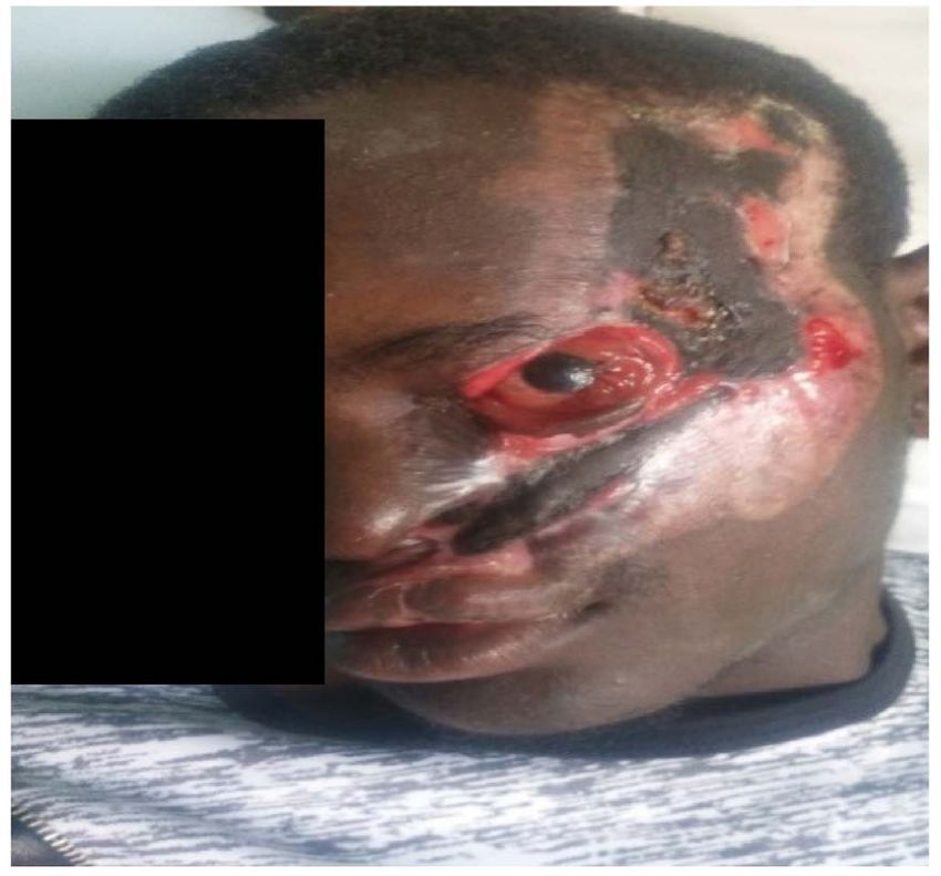
D. Adama et al.

                              1. Introduction
                              Injuries to the periorbital region are increasing in Sikasso Hospital due to the
                              evolution of the road infrastructure. Periorbital traumas are most often asso-
                              ciated with other lesions (maxillofacial, cranial and polytraumatized). These
                              traumas are most often due to public road accidents (AVP), burns, accidents
                              during work rural and ballistic or other brawls. Public road trauma accounted
                              for 78% of the causes of head trauma at Gabriel Touré Hospital from 2011 to
                              2012 and 80.3% of facial trauma at the CHU-CNOS (Centre Hospitalier Univer-
                              sitaire-Center National d’Odonto Stomatologie) in Bamako from January to De-
                              cember 2014 [1].
                                Significant trauma to the periocular region can seriously damage ocular
                              structures and their adnexa.
                                Palpebral involvement is often associated with lesions of the forehead, eye-
                              brows and cheeks. Burns affect the eyelids in 7.5% to 27% of cases in adults, and
                              they concern 71.7% of facial burns in children [1].
                                The eyelids can be damaged during these traumas. The most frequent lesions
                              are lacerations, the surgical treatment of which is generally simple. In some cas-
                              es, there are complex traumas where there is a loss of tissue, which is difficult to
                              treat.
                                At Sikasso Hospital, we do not have a study on periorbital trauma, despite the
                              high frequency of admissions to the emergency services for trauma related to
                              road traffic accidents.
                                We report the case of a 26-year-old young man, farmer with no medical and
                              surgical history, referred by the odontostomatology and maxillofacial surgery
                              department for burns to the left hemi face by the exhaust pipe of his motorbike
                              which occurred following a public road accident (AVP).

                              2. Observation
                              This was a 26-year-old young man with no particular history referred by the
                              maxillofacial surgery department for management of eyeball exposure, keratitis
                              and post-traumatic lagophthalmos after a motorcycle traffic accident.
                                The external examination found a wide wound of the hemiface about 20 cm
                              long on its long axis and 10 cm wide, with a significant loss of the upper left eye-
                              lid superior to three quarters of the eyelid and a burning of the epidermis of the
                              lower left eyelid.
                                Visual acuity was 10/10 in the right eye and 5/10 in the left eye with an intact
                              eyeball and good globe mobility. There was a total loss of the upper eyelid, mus-
                              cle and fat wasting resulting in lagophthalmos and serous secretions.
                                On biomicroscopic examination, conjunctival hyperaemia and superficial
                              punctate keratitis were observed in the left eye. In addition, the integrity of the
                              anterior chamber, pupil and iris was noted, with a good normal photomotor ref-
                              lex.

DOI: 10.4236/ss.2023.142013                         98                                            Surgical Science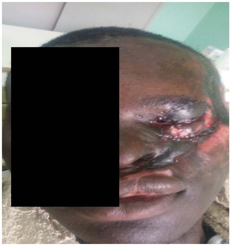
D. Adama et al.
The intraocular pressure taken in the air as well as the fundus taken with the
Volk 90 lens was normal in both eyes. The radiography of the facial mass
(face/profile) and computed tomography (CT scan) of the skull did not find any
anomaly or foreign body. The blood work was also unremarkable. Blood sugar
was 100 mg/dl, prothrombin Rate (PR) was 92%, creatinine was 10 mg/l, the
blood count formula was normal.
In collaboration with the maxillofacial surgeon, a graft of the hemi face was
performed using a flap from the inner side of the thigh first, combined with
medical treatment with antiseptic eye drops, wetting of the ocular surface and
corneal healing agents.
After three months of follow-up in the maxillofacial surgery department, the
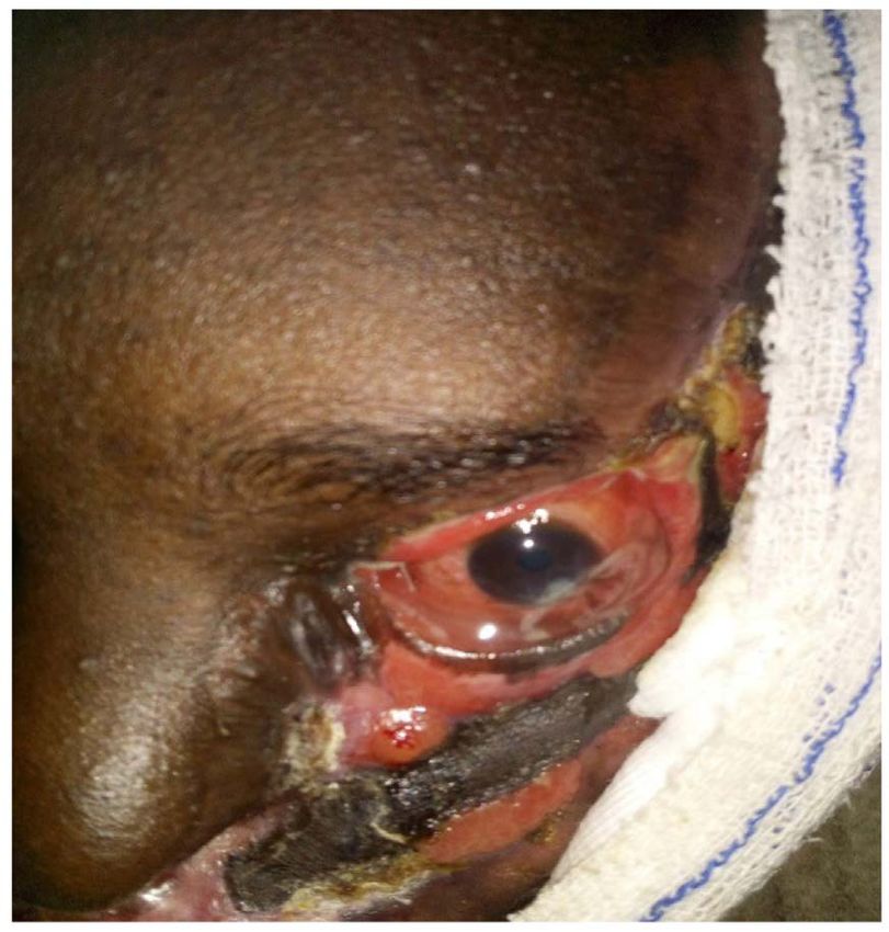
patient was referred to us for management of the palpebral inocclusion (lagoph-
thalmos) (Figure 1).
(a) (b)
2 Months
(c) (d)
3 Months
Figure 1. Image of patient (a): seven days after admission to the emergency room; (b):
One month after hemifacial graft break; (c): two months after break of the hemifacial
graft; (d): three months after placement of the hemifacial graft and 3 weeks after place-
ment of the flapsupra eyebrow.
DOI: 10.4236/ss.2023.142013 99 Surgical Science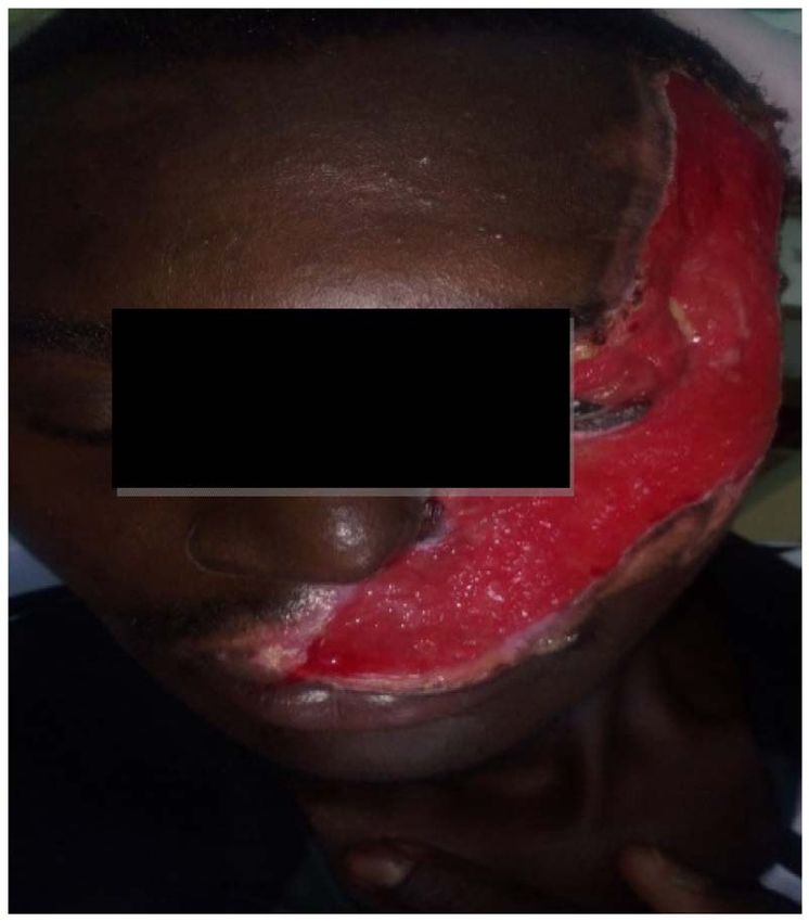
D. Adama et al.

                                 Faced with the evolution of the corneal ulceration and the risk of greater loss
                              of vision, recovery by a flap graft was performed, which consisted of removing a
                              myocutaneous flap above the eyebrow and suturing it with silk 6/0 at remaining
                              anterior eyelid edges and eyebrows with separated points followed by a progres-
                              sive ablation due to one point out of two from the seventh day. This recovery al-
                              lowed us, despite fragile tissue vitality, to have a considerable reduction in la-
                              gophthalmos and to obtain complete healing of the corneal ulceration. Four
                              weeks after placement of the myocutaneous flap, despite slight retraction of the
                              graft and excision of necrotic tissue. We suggested the need for a second inter-
                              vention at the level of the upper eyelid finally to carry out the positioning of the
                              posterior lamella by a tarso-conjunctival flap. Despite the explanations and the
                              advantages offered by this second intervention, especially on the functional level.
                              The patient already judged the aesthetic state of his face very satisfactory and
                              preferred to return to his village still refusing any other surgical intervention.

                              3. Comment and Discussion
                              The Sikasso region is considered the second largest city in Mali in terms of pop-
                              ulation but also with the highest number of traffic accidents after Bamako [2].
                              There are many reasons that cause accidents on our roads (lack of suitable infra-
                              structure, incivility, carelessness, etc.). At the Sikasso hospital, we do not have
                              statistics on road traffic accidents. Our patient was received in the context of a
                              public road accident with a burn of the hemi face by the exhaust pipes of the
                              motorcycle. Through a study, “Evaluation of road safety and associated trauma
                              in the city of Sikasso”, carried out by Emmanuel Bonnet [2], health geographer
                              and researcher at the IRD (Research Institute for Development) in 2019 showed
                              that more than 75% of accidents in the city of Sikasso involve pedestrians or
                              motorcyclists. Young people are the first victims’ road accidents with more than
                              53%. Our patient was 26 years old, close to the slice of E Bonnet who found that
                              young people between 18 and 24 years old constitute each year more than 25%
                              of the killed and 23% of the injured. The age of our patient was lower than the
                              average age found by AS Sangaré et al. which was 39 years (2 to 73 years) in
                              Mopti but the age group of 18 - 27 was the majority in this study. [3]. In Senegal
                              A. Lam et al. found an average age of 8.5 years much lower than the age of our
                              patient for an age group of 6 to 10 years most affected [4] probably linked to the
                              fact that this study only concerned children under 15 years of age. The eyelid
                              involvement was associated with other local lesions, namely keratitis, burning of
                              half of the left face, this association is known to several authors, especially when
                              they occur in the context of a public road accident. M Sissoko et al. found eyelid
                              lesions (78.25%) at CHU IOTA in 2020 following an accident that occurred
                              during the confinement of the COVID-19 pandemic [5]. Surgical management
                              of these lesions is often delayed because of the associated vital lesions. The pa-
                              tient most often passes through other departments before ophthalmology, such
                              was the case of our patient who spent a long time in the maxillofacial depart-
                              ment before being referred to ophthalmology.

DOI: 10.4236/ss.2023.142013                        100                                            Surgical Science
D. Adama et al.
Despite the delay in the surgical management on the ophthalmological level,
our patient benefited from medical treatments (Wetting agents of the ocular
surface and physiological saline) seven days after his admission to the emergency
department at the hospital, which allowed us to have a satisfactory visual acuity
of 5/10 in the affected eye. Especially since the quality of the functional and aes-
thetic result depends on the precocity of the assumption of responsibility.
The reconstruction of an upper eyelid being more complex than that of a low-
er eyelid because of its proximity to the eyeball and its function in all lacrimal
physiology, lid blinking and eye protection. Ideally, the ipsilateral or contralater-
al upper eyelid provides the best grafts for reconstruction. The pretarsal portion
of the retroauricular skin constitutes the second donor site, rather on the post-
erior surface of the pavilion than at the mastoid level, due to the difference in the
thickness of the skin [6].
We carried out a recovery using a frontal flap because of the importance of the
loss which was greater than three quarters of the eyelid and the urgency to pro-
tect the eyeball as well as the state of our technical platform at the Sikasso Hos-
pital. The choice of technique was mainly motivated by its mastery but also by
the quality of our technical platform. Because we do not have at the Sikasso
Hospital an adequate technical platform for eyelid and orbit surgery. Also our
patient had lesions such as scratches and micro wounds on the appropriate sam-
pling sites (micro wounds, scratches).
Other techniques, in particular that of the Tenzel flap reversed, are used espe-
cially in the losses of nasal substances or Cutler-Beard. These techniques being
more complex little mastered and require an adequate technical platform were
not chosen by our team.
The Abbot’s flap was not chosen because a transposition of the lower eyelid to
the upper one was difficult due to the importance of the burn of the hemiface
[7].
The difficulties of eyelid reconstruction are related to the anatomical and aes-
thetic particularities of the eyelids. These peculiarities have been reported by J.
Bouguilaand all [7]. They are mobile and fragile with a complex structure [8],
explaining the difficulties of the functional and anatomical management of these
lesions of palpebral burns [9]. This most often requires multiple surgical acts, as
in our case, where there was a need for a second surgery. Although the explana-
tions on the advantages of this second intervention were given, the patient
judged satisfied with the aesthetic state of his face and refused any other surgical
intervention.
As for aesthetics, it depends on the severity of the burn and the anatomical
and functional disorganization of the eyelids. It is recalled that the eyelid aes-
thetic extends to the orbital rim externally and towards the glabella internally,
going well beyond the external and internal canthus. [10]. The limits are esti-
mated at 0.5 cm inside the internal canthus and 1.5 cm outside the external can-
thus [9] [11]. The eyelids are held to the bony orbital frame by internal and ex-
ternal palpebral ligaments which attach them to the internal and external orbital
DOI: 10.4236/ss.2023.142013 101 Surgical ScienceD. Adama et al.
rims; the second means of attachment is represented by the orbicularis muscle.
The weakness of these attachments explains that these anatomical structures es-
sentially depend on retractions and scar tensions. [9]. The eyebrows also play an
important role in the aesthetic balance of the look. All it takes is a badly posi-
tioned eyebrow, or one whose hair grows in the wrong direction, to give the face
an incongruous and grotesque appearance. The bristles are vertical and oriented
slightly outwards in the inner part, laterally in the central part, outwards and
slightly downwards in the outer part [12].
Our decision to take charge of this patient was above all justified by the ur-
gency to protect the globe as well as the very limited means of the patient who
made a trip to the capital in Bamako.
4. Conclusion
Reconstruction of the eyelid after a road accident or a major burn is a real prob-
lem in our department and in our developing countries because of the slowness
of the treatment but especially because of the difficulty of a rapid multidiscipli-
nary treatment. The insufficiency of the technical platform and the availability of
consumables limit us in the choice of the operative technique. This reconstruc-
tion is also a problem of adherence for the patient, to multiple surgical proce-
dures and also the management of sequelae.
Conflicts of Interest
The authors declare no conflicts of interest regarding the publication of this
paper.
References
[1] Samaké, B., Togola, M., Maiga, H., Keïta, B., Mangané, Diallo, A. (2014) Le
traumatisme crânien au C.H.U. Gabriel Touré: Aspects cliniques et pronostiques.
Revue africaine d’anesthésiologie et de médecine d’urgence (RAMUR), 19, 33.
https://web-saraf.net/Le-traumatisme-cranien-au-C-H-U.html
[2] Bonnet, E. and Sanogo, M. (2020) Sikasso: Second City in Mali with the Highest
Number of Traffic Accidents According to a Study. JSTM.
https://www.jstm.org/sikasso-deuxieme-ville-au-mali-avec-le-plus-grand-nombre-d
accidents-de-circulation-selon-une-etude/
[3] Sangare, A.S., et al. (2020) Management of Road Traffic Accidents in the Mopti Re-
gion of Mali. Revue de Chirurgie Orthopédique et Traumatologique, 106, 429-433.
https://doi.org/10.1016/j.rcot.2020.02.005
[4] Lam, A., et al. (2007) 218 Eye Injuries in Children Aged 0 to 15 in Senegal. Journal
Français d’Ophtalmologie, 30, 2S212.
https://doi.org/10.1016/S0181-5512(07)80030-1
[5] Mr. Sissoko, et al. (2021) Ocular Trauma during the COVID-19 Health Crisis at the
Iota University Hospital. Journal Français d’Ophtalmologie, 44, 145-150.
https://doi.org/10.1016/j.jfo.2020.11.002
[6] Bardot, J., Casanova, D. and Malet, T. (2004) Chirurgie reconstructrice des paupières.
EMC - Chirurgie, 1, 365-390.
DOI: 10.4236/ss.2023.142013 102 Surgical ScienceD. Adama et al.
[7] Bouguila, J., et al. (2011) Particularities of the Management of Burnt Eyelids. Jour-
nal Français d’Ophtalmologie, 34, 655-662. https://doi.org/10.1016/j.jfo.2011.04.011
[8] Volpe, C.R. and Ramirez, O.M. (2005) The Beautiful Eye. Facial Plastic Surgery
Clinics of North America, 13, 493-504. https://doi.org/10.1016/j.fsc.2005.06.001
[9] Mandrekas, A.D., Zambacos, G.J. and Anastasopoulos, A. (2002) Treatment of Bi-
lateral Severe Eyelid Burns with Skin Grafts: An Odyssey. Burns, 28, 80-86.
https://doi.org/10.1016/S0305-4179(01)00072-9
[10] Bichet, J.C., Lakhel, A., Foyatier, J.L. and Cantaloube, D. (2001) Acute Stage Facial
Burns. Encycl Med Chir,” Stomatology, 22-088.
https://www.em-consulte.com/article/20334/brulures-de-la-face-au-stade-aigu
[11] Achauer, B.M. and Adair, S.R. (2000) Acute and Reconstructive Management of the
Burned Eyelid. Clinics in Plastic Surgery, 27, 87-96.
https://doi.org/10.1016/S0094-1298(20)32685-7
[12] Foyatier, J.-L., Voulliaume, D., Mojallal, A., Chekaroua, K. and Comparin, J.-P.
(2005) Treatment of Burn Sequelae: Facial Burns. EMC - Chirurgie, 2, 162-174.
https://doi.org/10.1016/j.emcchi.2005.01.003
DOI: 10.4236/ss.2023.142013 103 Surgical ScienceYou can also read