Development of a deep learning based software for calculating cleansing score in small bowel capsule endoscopy - Nature
←
→
Page content transcription
If your browser does not render page correctly, please read the page content below
www.nature.com/scientificreports
OPEN Development of a deep
learning‑based software
for calculating cleansing score
in small bowel capsule endoscopy
Ji Hyung Nam1,6, Youngbae Hwang2,6, Dong Jun Oh1, Junseok Park3, Ki Bae Kim4,
Min Kyu Jung5 & Yun Jeong Lim1*
A standardized small bowel (SB) cleansing scale is currently not available. The aim of this study was to
develop an automated calculation software for SB cleansing score using deep learning. Consecutively
performed capsule endoscopy cases were enrolled from three hospitals. A 5-step scoring system based
on mucosal visibility was trained for deep learning in the training set. Performance of the trained
software was evaluated in the validation set. Average cleansing score (1.0 to 5.0) by deep learning
was compared to clinical grading (A to C) reviewed by clinicians. Cleansing scores decreased as clinical
grading worsened (scores of 4.1, 3.5, and 2.9 for grades A, B, and C, respectively, P < 0.001). Adequate
preparation was achieved for 91.7% of validation cases. The average cleansing score was significantly
different between adequate and inadequate group (4.0 vs. 2.9, P < 0.001). ROC curve analysis revealed
that a cut-off value of cleansing score at 3.25 had an AUC of 0.977. Diagnostic yields for small, hard-
to-find lesions were associated with high cleansing scores (4.3 vs. 3.8, P < 0.001). We developed a
novel scoring software which calculates objective, automated cleansing scores for SB preparation. The
cut-off value we suggested provides a standard criterion for adequate bowel preparation as a quality
indicator.
Capsule endoscopy (CE) allows direct visualization of the entire small bowel (SB)1. It is also safe from sedation-
related complications with minimal i nvasiveness2. In current guidelines, CE is the first-line investigation method
for patients with obscure gastrointestinal bleeding or suspicious Crohn’s d isease3. It is also considered as an
initial diagnostic modality for various SB diseases including vascular or inflammatory diseases, SB tumors, and
polyposis syndrome3. With the expansion of CE indications and technological efforts, attempts have been made
to observe the entire SB in detail3–5. In addition, recently introduced deep learning method has shown excellent
performance for detecting SB lesions in C E6. It may overcome problems associated with time and effort needed
for CE interpretation. Despite these recent advances, CE has a limitation in that its quality is greatly influenced
by bowel preparation. In many cases, reading of CE videos is interrupted by air bubbles and residual materials.
Inadequate bowel preparation had led to repeat examination and cost increase7. As the diagnostic yield of CE
highly depends on the preparation quality of passively obtained images, effective bowel cleansing is essential for
qualified CE examination. Currently, the guideline recommends bowel preparation quality to be included in the
CE report, and the rate of adequate bowel preparation is considered one performance m easure3. Accordingly,
quality control of CE requires an objective scoring system to assess SB preparation. However, a standardized
and validated cleansing scale is currently unavailable, which is why the rate of bowel preparation is limited to
only minor performance measures.
Several grading scales to assess SB preparation quality have been reported7–9. Because the evaluation of bowel
preparation using these scales also depends on clinicians’ subjective judgment, validation does not guarantee
1
Division of Gastroenterology, Department of Internal Medicine, Dongguk University Ilsan Hospital, Dongguk
University College of Medicine, Goyang, Republic of Korea. 2Department of Electronics Engineering, Chungbuk
National University, Cheongju, Republic of Korea. 3Department of Internal Medicine, Digestive Disease
Center, Institute for Digestive Research, Soonchunhyang University College of Medicine, Seoul, Republic of
Korea. 4Department of Internal Medicine, Chungbuk National University College of Medicine, Cheongju, Republic
of Korea. 5Division of Gastroenterology and Hepatology, Department of Internal Medicine, Kyungpook National
University Hospital, Daegu, Republic of Korea. 6These authors contributed equally: Ji Hyung Nam and Youngbae
Hwang. *email: drlimyj@gmail.com
Scientific Reports | (2021) 11:4417 | https://doi.org/10.1038/s41598-021-81686-7 1
Vol.:(0123456789)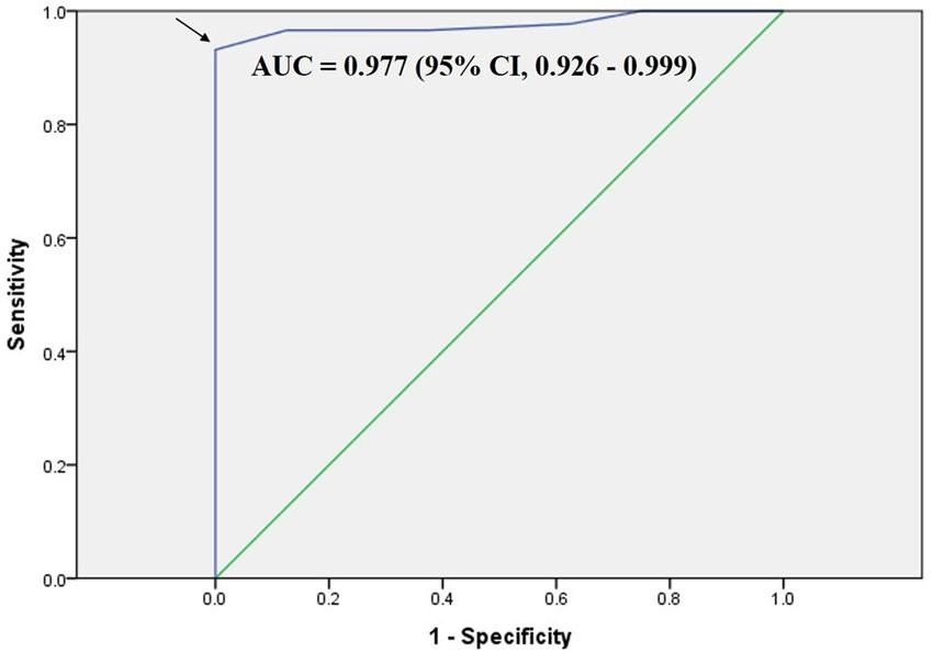
www.nature.com/scientificreports/
Figure 1. Data flow and deep learning process.
these scales’ objectivity. Computed cleansing scores using color intensities of tissue color bar (PillCam) or map
view (MiroCam) have also been d eveloped10,11. They can be integrated into their own CE reading programs to
provide objectively calculated scores. However, color intensities of condensed bands are insufficient to fully rep-
resent the cleanness of the entire CE image over tens of thousands. Also, if only the intensity of certain colors is
recognized, it can be difficult to distinguish between a color due to bleeding or ulcer and a color due to residual
materials. Besides, these integrated scales cannot be applied to other CE devices in general.
Thus, the aim of this study was to develop an automated calculation software for SB cleansing score that could
represent overall cleanness of the entire CE image to be actually read. This trial is expected to provide an objec-
tive cleansing scale for CE and suggest a standard criterion for adequate bowel preparation as a quality indicator.
Methods
Study design. Small bowel CE (PillCam SB3, GIVEN Imaging Ltd., Yoqneam, Israel) cases consecutively
performed at three University Hospital (Dongguk Univ., Chungbuk national Univ., and Kyungpook national
Univ.) of South Korea between 2016 and 2020 were screened. The SB3 cases with patients over 18 years of age
were enrolled. Reasons for CE examinations were obscure gastrointestinal bleeding, suspected or established
Crohn’s disease, and suspected small bowel tumor or polyposis. Overnight fasting was performed for all patients.
Bowel cleansing was achieved with 2 L polyethylene glycol (PEG) plus ascorbic acid (Coolprep; Taejoon Pharm.
Co., Seoul, Korea). Exclusion criteria were inaccessible videos due to mechanical error and incomplete cases
when the cecum was not reached due to capsule retention or power limitation. Among eligible CE cases, 72 cases
from Dongguk Univ. Hospital between Jan. 2016 and Dec. 2019 were selected for deep learning database (train-
ing set). In addition, a separate set of 96 CE cases from three Univ. Hospitals between December 2016 and May
2020 were selected for external validation (validation set) (Fig. 1). The study was conducted in accordance with
the guidelines of the Declaration of Helsinki and was approved by the Institutional Review Board of Dongguk
University Ilsan Hospital (IRB no. DUIH 2020-06-017). Because this is a retrospective study using CE images
that has already completed, informed consent was waived from IRB.
Data collection for training. As significant abnormalities such as bleeding and ulcer could confuse train-
ing of bowel cleansing state, video segments without any significant SB lesion were extracted from cases (n = 72)
of the training set (Fig. 1). In sequence, 71,191 frames (still-cut images) were separated from extracted video
segments using an OCR (optical character recognition) program. These separated frames were classified into
four categories: normal-clean mucosa, bubble-dominant mucosa, bile-dominant mucosa, and debris-dominant
mucosa. Two experienced CE readers (J.H.N. and D.J.O.) reviewed these frames and scored cleansing qualities
based on the proportion of visualized mucosa (Fig. 2). This scale used 5-step scores ranging from 5 (more than
90% of mucosa visible) to 1 (less than 25% of mucosa visible) depending on obscuration by bubble, bile, and
debris. If there were any discrepancies between the two readers, a final score was determined after re-evaluation
and discussion with a senior reader (Y.J.L.).
Deep learning process. To escape from data imbalance problem, 700 images per each cleansing score
were selected from the 5-step scored frames to develop a deep learning model that could classify SB cleansing
state. A total of 3500 images were randomly separated into 2500 and 1000 images for training and verification,
respectively. A deep learning network called InceptionResnetV2 was used for training due to its recent good per-
formance in ImageNet C hallenges12. Our dataset was not enough to train the deep network from scratch (empty
parameter). Therefore, training was started from a pre-trained model parameter using ImageNet dataset. First,
the last layer from the pre-trained parameter was trained with hyperparameters of 10,000 for the number of
steps, 24 for batch size, and 0.01 for learning rate. Top-1 and Top-2 accuracies of the trained network for the test
Scientific Reports | (2021) 11:4417 | https://doi.org/10.1038/s41598-021-81686-7 2
Vol:.(1234567890)www.nature.com/scientificreports/
Figure 2. Cleansing score used for deep learning: a 5-step scoring method based on the proportion of
visualized mucosa.
Segmental grading (mucosal invisibility of each segment)
< 5% of number of video imagea with > 50% invisible mucosa by bubbles,
Grade 1
bile, or debris
Grade 2 5–15%
Grade 3 15–25%
Grade 4 > 25%
Overall grading (overall cleansing quality)
Grade A Total grade 3–5
Grade B 6–8
Grade C 9–12
Clinically adequate preparation
Adequate Grade A or B
Inadequate Grade C
Table 1. A validated small bowel preparation scale using a quantitative parameter. a Still-cut image (frame).
were 50.4% and 74.5%, respectively. Full layers were then trained with hyperparameters of 220,000 for number
of steps, 24 for batch size, and 0.0001 for learning rate. Final Top-1 and Top-2 accuracies of the trained network
were 69.4% and 91.2%, respectively. Because the dataset was classified by clinicians subjectively, uncertainty
between two scores was allowed. Before a hard determination of the score, the probability for each score was
predicted applying softmax function. From the output of soft function, the final cleansing score was estimated
by computing expected value as:
5
Final_score(I) = i ∗ pi (I)
i=1
where i indicated the grade and pi (I) indicated the probability of i-th grade for an image I.
External validation. The trained scoring software was validated using 96 CE cases different from those
used in the training set. All video segments corresponding to SB sections of the validation set were separated into
frames using the OCR program. Extracted frames were divided into three equal number of segments according
to the time sequence of the video: segment 1 (seg1), proximal third; segment 2 (seg2), middle; and segment 3
(seg3), distal. Using the trained scoring software, a cleansing score was assigned to every frame of the valida-
tion set. Separately, two CE readers reviewed bowel preparation quality (clinical grading) of frames. They were
blinded to cleansing scores obtained from the trained software, clinical records, and original reports of the
validation CE cases. Clinical grading was assessed using a quantitative parameter of a previously validated grad-
ing system13 based on the proportion of non-prepped images in which bubble, bile, and debris disturbed more
than 50% of visualization (Table 1). Clinical grading of each segment (segmental grading, 1 to 4) was assessed
independently. Overall image quality (overall grading, A to C) was determined as the sum of segmental grading
Scientific Reports | (2021) 11:4417 | https://doi.org/10.1038/s41598-021-81686-7 3
Vol.:(0123456789)www.nature.com/scientificreports/
Segment 1 Segment 2 Segment 3
Grade n (%) Score, mean ± SD n (%) Score, mean ± SD n (%) Score, mean ± SD P-value
1 59 (61.5) 4.4 ± 0.3 62 (64.6) 4.3 ± 0.4 41 (42.7) 4.2 ± 0.4 0.006*
2 32 (33.3) 3.8 ± 0.4 26 (27.1) 3.5 ± 0.5 40 (41.7) 4.0 ± 0.5
3 5 (5.2) 3.3 ± 0.4 2 (2.1) 3.2 ± 0.0 7 (7.3) 3.3 ± 0.5
4 0 (0) – 6 (6.3) 2.3 ± 0.5 8 (8.3) 2.2 ± 0.5
Total 4.1 ± 0.5 4.0 ± 0.7 3.7 ± 0.7 < 0.001*
Table 2. Clinical grading and cleansing scores of each small bowel segment in the validation set (n = 96). SD,
standard deviation. *P-values for grade distribution and average cleansing scores per segment, respectively.
per CE case. Overall grading of A or B was classified as clinically adequate preparation while overall grading of
C was considered as inadequate. Any disagreement between the two readers was resolved after discussion with
the senior reader.
Capsule endoscopy studies of validation set were prospectively read using an analyzing software (Rapid reader
ver. RR83.24.14254.0) for PillCam SB3 by another CE reader (J.P.) who was blinded to results from cleansing
scores, clinical grading, and original CE findings. Diagnostic yield was defined as the detection of SB lesion
likely to provide diagnostic information such as erosion, ulcer, bleeding, hematin, vascular lesion, and mass.
Statistical analyses. The main outcome was the performance of deep learning for assessment of SB prepa-
ration quality. Average cleansing scores calculated by the deep learning-based software were compared with
clinical grading determined using a validated preparation scale.
In the validation set, average cleansing score (from 1.0 to 5.0) per segment and per case were calculated as
the sum of cleansing scores divided by the number of frames. ANOVA (analysis of variance) was performed to
compare average cleansing scores among different groups of segmental grading (1 to 4) and overall grading (A to
C). Post-hoc analysis was performed using Dunnett’s test. Average cleansing scores between clinically adequate
and inadequate preparation groups were compared using independent sample t-test. Sensitivity and specific-
ity for clinically adequate preparation were calculated for each average cleansing score (1.0 to 5.0). Receiver
operating characteristics (ROC) curve was generated to assess a cut-off value of cleansing score for clinically
adequate preparation. In addition, whether diagnostic yield differed according to bowel preparation quality was
analyzed. Two-sided P-values of less than 0.05 were considered statistically significant. All statistical analyses
were conducted using SPSS Statistics 19.0 (IBM, Armonk, NY, USA).
Results
Descriptive summary and deep learning recognition. Ninety-six CE cases were enrolled for the vali-
dation set. Their mean age was 58.1 ± 18.7 years (range, 18–92 years). There were 64 (66.7%) males. Mean SB
transit time was 6.1 ± 2.6 h (range, 1.7–13.7 h). Class Activation Map (CAM) applied to the recognition of cleans-
ing score of image frames using the deep learning software was confirmed (Supplementary Figure 1). Lower
cleansing score indicated higher weight for bubbles, bile, or debris, whereas higher cleansing score indicated
higher weight for clean mucosa and its folds.
Cleansing scores and clinical grading. The distribution of clinical grading and average cleansing scores
per segment is shown in Table 2. There was a tendency for clinical grading to get worse from seg1 to seg3
(P = 0.006). More than 60% of seg1 and seg2 had grade 1 whereas only 42.7% of seg3 had grade 1. Grade 4
accounted for 8.3% in seg3. However, it was absent in seg1. Average cleansing scores were also different among
segments, showing 4.1 ± 0.5 (range, 2.8–4.9), 4.0 ± 0.7 (range, 1.5–4.9), and 3.7 ± 0.7 (range, 1.4–4.9) for seg1,
seg2, and seg3, respectively.
Average cleansing scores and segmental grading per segment were analyzed (Table 2 and Fig. 3A). Average
cleansing scores tended to decrease from grade 1 to 4 for all segments (all P < 0.001). Numbers of cases with
overall image quality grades A, B, and C were 75 (78.1%), 13 (13.5%), and 8 (8.3%), respectively. Average cleans-
ing scores decreased when overall grading decreased from grade A to grade C, yielding 4.1 ± 0.4, 3.5 ± 0.5, and
2.9 ± 0.4 for grades A, B, and C, respectively (P < 0.001) (Fig. 3B, Supplementary Table 1). Grade A and grade B
showed significantly higher average cleansing scores than grade C (P < 0.001 and P = 0.001 respectively).
Clinically adequate preparation was achieved for 91.7% (88/96) of cases. The average cleansing score for the
adequate group was significantly higher than that for the inadequate group (4.0 vs. 2.9, P < 0.001). In ROC curve,
a cut-off value of cleansing score at 3.25 for clinically adequate preparation had a sensitivity of 93%, a specificity
of 100%, and an AUC (area under the curve) of 0.977 (95% CI: 0.926–0.999, P < 0.001) (Fig. 4).
Diagnostic yield. Main endoscopic findings of the validation set are shown in Table 3. The overall diag-
nostic yield was 62.5% (60/96). It was not significantly different between adequate and inadequate preparation
groups (61.4% vs. 75.0%, P = 0.446). The average cleansing score did not differ either according to the overall
diagnostic yield (4.0 vs. 3.8, P = 0.197). Excluding 36 cases of relatively easy-to-detect lesions such as bleeding,
large ulcers, diffuse inflammation or erosions, or mass, detection rate for small lesions such as tiny erosion, aph-
thous ulcer, hematin, and angioectasia accounted for 41.4% (24/58) in the clinically adequate preparation group.
Scientific Reports | (2021) 11:4417 | https://doi.org/10.1038/s41598-021-81686-7 4
Vol:.(1234567890)www.nature.com/scientificreports/
Figure 3. Average cleansing scores by (A) segmental grades and (B) overall grades. Scores decreased from
grade 1 to 4 and from A to C.
Figure 4. Receiver operating characteristic (ROC) curve of average cleansing score for clinically adequate
preparation. The curve estimated a cut-off value of 3.25 (arrow). AUC, area under the curve.
CE findingsa n (%)
Hematinsb 1 (1.0)
Erosionsb 15 (15.6)
Angioectasiab 1 (1.0)
Aphthous ulcerb 7 (7.3)
Ulcer 18 (18.8)
Bleeding 13 (13.5)
Mass 1 (1.0)
Diffuse inflammation or erosions 4 (4.2)
Total 60 (62.5)
Table 3. Main endoscopic findings of the validation set (n = 96). CE, capsule endoscopy. a Classified as a main
finding if CE result included various lesions. b These lesions were classified into difficult-to-detect small lesions.
No small lesion was detected in two inadequate cases. Average cleansing score was significantly higher when
small lesions were detected (4.3 vs. 3.8, P < 0.001). The AUC for cleansing score was 0.747 (95% CI: 0.622–0.871,
P = 0.001) for detecting the small lesions. The cut-off value of 3.25 of cleansing score for clinically adequate
preparation showed 100% sensitivity for diagnosing small lesions.
Discussion
We firstly developed a deep learning-based automation software for calculating SB cleansing score. It is of clinical
significance in that it demonstrated a performance of deep learning-based software using a previously validated
preparation scale. External validation of this deep learning-based software showed a good performance for
preparation quality assessment. In addition, a cut-off value was suggested for clinically adequate preparation.
A recently reported guideline recommends CE indications, cecal visualization, lesion detection, and the rate of
adequate bowel preparation as performance measures for qualified CE3. Based on 17-year data from the Korean
Scientific Reports | (2021) 11:4417 | https://doi.org/10.1038/s41598-021-81686-7 5
Vol.:(0123456789)www.nature.com/scientificreports/
Capsule Endoscopy Registry, one study has shown that inadequate bowel preparation is significantly associated
with capsule retention and incomplete e xamination14. Another recent study has shown that higher SB transit
time is associated with inadequate bowel p reparation15. However, the rate of adequate bowel preparation, despite
its importance to CE quality, is described as only a minor performance measure in the guideline3. The reason is
that there is no simplified objective criterion for assessing SB preparation yet. In addition, methods and proper
timing of SB preparation remain c ontroversial16–18. Unlike colonoscopy, it is not easy to assess the cleanness of
tens of thousands of SB images obtained over several hours. No matter how validated scales are used, the cur-
rent assessment of SB preparation quality by individual clinicians is inevitably subjective and time-consuming.
With the expansion of CE indications and recent increase in clinical use, an objective and automated calculation
system is essential. The calculating system should be based on clinically validated preparation scales and consist-
ent with experienced CE readers’ assessment. Meanwhile, newly introduced deep leaning-based computational
analysis of CE images allows more accurate detection of SB lesions with reduced reading time than conventional
CE reading19–22. However, as long as the CE subsequently analyze passively obtained images, the performance of
deep learning for lesion detection still depends on the quality of bowel preparation. Numerous grading scales with
different technical characteristics have been i ntroduced23. However, studies on the application of deep learning
for assessing SB preparation quality have not been reported yet. Accordingly, authors of this study developed
a deep learning-based objective and automated calculating system and showed its clinical usefulness through
external validation.
Usually, deep learning-based classification can be applied to explicit problems, for example, object classifica-
tion of images. Although the dataset in this work was built by CE readers subjectively, the output from the train-
ing was significant (Top-2 accuracy of 91.2%). The deep learning model was also validated by comparison with
clinical grading. Results of this study demonstrated that deep learning can be applied to subjective problems that
are usually determined by human specialists. Our model learned various images of cleanliness for each category
of bubble, bile, and debris, but the final cleansing score was derived regardless of the category. Since the dura-
tion or degree of mucosal obscuration can vary by category, an advanced model is needed that can differentiate
between categories and assign different cleansing scores.
We used a previously validated cleansing s cale8,13 for CE readers’ clinical grading. By calculating the percent-
age of frames with more than 50% not visible, it is considered a more detailed and less subjective method among
existing preparation s cales3,23. The original scale rated both mucosal invisibility and fluid transparency indepen-
dently. In our study, however, we did not rate the fluid transparency separately as the grading of transparency
seemed to be more subjective. Instead, we simply included fluid transparency in the grading of mucosal invis-
ibility. We regarded opaque fluid as ‘invisible’ portion, while images showing transparent fluid were considered
‘visible’. As transparent fluid is enough to detect underlying SB lesions, it is feasible to classify it as visible mucosa.
Compared to the grading scale used by CE readers, scoring for the training set required a more visually simpli-
fied scale capable of clearly recognizing the cleanliness of each image frame. The cleansing scale of colonoscopy
generally uses a 4-level scale based on the amount of fecal residue and turbid fluid, which is also applied to
colon CE24. For training of cleansing score, it may be common to use a 4-step score depending on the mucosal
visibility. However, score 5 (more than 90% mucosa are visible) was separately classified because we needed to
train completely cleaned mucosa. In addition, our 5-step scoring system for deep learning enhanced the aver-
age cleansing score. Meanwhile, the deep learning process did not train images containing SB lesions such as
bleeding or ulcer. Interestingly, the newly developed software calculated cleansing scores comparable to clinical
grading results even for CE cases involving SB lesions in the validation test. As proven by the CAM applied to
deep learning recognition, the software accurately recognized residual materials such as bubbles, bile, and debris
against clean mucosa and its folds.
The present study showed that cleansing score calculated by deep learning model was highly correlated with
clinical grading assessed by clinicians. We suggested a significant cut-off value for clinically adequate preparation
(AUC, 0.977). Based on the cut-off value of 3.25, it is possible to evaluate whether the CE was qualified and to
determine the need for repeat examination or additional diagnostic approach. There was no difference in bowel
preparation quality according to overall diagnostic yield. However, the detection of small, hard-to-find lesions
such as a few erosions, aphthous ulcers, and vascular lesions was significantly associated with a high average
cleansing score. It is conceivable that bleeding or large ulcers can be easily detected and diagnosed even for
inadequate bowel preparation cases. Contrary, small lesions are relatively difficult to be detected in an inadequate
preparation state. A cut-off value of 3.25 showed 100% sensitivity for diagnosing small lesions. This suggests that
such cut-off value for clinically adequate preparation is sufficient for the detection of small lesions.
Our deep learning model was validated with 100 CE images from 3 hospitals, and the clinical characteristics
of each case were not included in the analysis. Although reviewers who determined clinical grades were blinded
to the cleansing scores and CE findings of each case, a more independent assessment of bowel cleansing would
require more CE cases from more hospitals. In addition, this study is currently in a preliminary stage for devel-
oping a deep learning model and validating its performance before it is integrated into the CE reading system
and applied in real clinical practice. Further studies using prospectively enrolled CE cases should be warranted
to demonstrate the validity and reproducibility of our model with real CE cases in clinical practice.
In conclusion, our novel scoring software provides an objective and automated cleansing score for SB prepa-
ration in CE. The suggested cut-off value can be used as a criterion as to whether or not the bowel preparation
is appropriate to detect SB lesions in clinical practice. This study is expected to provide a standard for adequate
bowel preparation in the quality control of CE. The application of the deep learning model enables evaluation
of whether the CE examination was appropriate and its results reliable. Additional advances in the model are
expected with more CE case experiences in the future.
Scientific Reports | (2021) 11:4417 | https://doi.org/10.1038/s41598-021-81686-7 6
Vol:.(1234567890)www.nature.com/scientificreports/
Received: 15 August 2020; Accepted: 6 January 2021
References
1. Iddan, G., Meron, G., Glukhovsky, A. & Swain, P. Wireless capsule endoscopy. Nature 405(6785), 417 (2000).
2. Amornyotin, S. Sedation-related complications in gastrointestinal endoscopy. World J. Gastrointest. Endosc. 5(11), 527–533 (2013).
3. Spada, C. et al. Performance measures for small-bowel endoscopy: a European Society of Gastrointestinal Endoscopy (ESGE)
quality improvement initiative. Endoscopy 51(6), 574–598 (2019).
4. Ching, H. L. et al. Magnetically assisted capsule endoscopy in suspected acute upper GI bleeding versus esophagogastroduoden-
oscopy in detecting focal lesions. Gastrointest. Endosc. 90(3), 430–439 (2019).
5. Nam, S. J. et al. 3D reconstruction of small bowel lesions using stereo camera-based capsule endoscopy. Sci. Rep. 10(1), 6025 (2020).
6. Soffer, S. et al. Deep learning for wireless capsule endoscopy: a systematic review and meta-analysis. Gastrointest. Endosc. 92,
831–839 (2020).
7. Brotz, C. et al. A validation study of 3 grading systems to evaluate small-bowel cleansing for wireless capsule endoscopy: a quan-
titative index, a qualitative evaluation, and an overall adequacy assessment. Gastrointest. Endosc. 69(2), 262–270 (2009).
8. Goyal, J., Goel, A., McGwin, G. & Weber, F. Analysis of a grading system to assess the quality of small-bowel preparation for capsule
endoscopy: in search of the Holy Grail. Endosc. Int. Open 2(3), E183-186 (2014).
9. Park, S. C. et al. A novel cleansing score system for capsule endoscopy. World J. Gastroenterol. 16(7), 875–880 (2010).
10. Van Weyenberg, S. J., De Leest, H. T. & Mulder, C. J. Description of a novel grading system to assess the quality of bowel prepara-
tion in video capsule endoscopy. Endoscopy 43(5), 406–411 (2011).
11. Ponte, A. et al. Validation of the computed assessment of cleansing score with the Mirocam(R) system. Rev. Esp. Enferm. Dig.
Organo Oficial Soc. Esp. Patol. Dig. 108(11), 709–715 (2016).
12. Christian, S., Sergey, I. & Vincent, V.: Inception-v4, inception-resnet and the impact of residual connections on learning. In Thirty-
First AAAI Conference on Artificial Intelligence (2017).
13. Esaki, M. et al. Bowel preparations for capsule endoscopy: a comparison between simethicone and magnesium citrate. Gastrointest.
Endosc. 69(1), 94–101 (2009).
14. Kim, S. H. et al. Research Group for Capsule Endoscopy/Small Bowel E: Changes in performance of small bowel capsule endoscopy
based on nationwide data from a Korean Capsule Endoscopy Registry. Korean J. Intern. Med. 35, 889 (2019).
15. Ponte, A. et al. Predictive factors of an incomplete examination and inadequate small-bowel cleanliness during capsule endoscopy.
Rev. Esp. Enferm. Dig. Organo Oficial Soc. Esp. Patol. Dig. 110(10), 605–611 (2018).
16. Adler, S. N. et al. A novel purgative protocol for capsule endoscopy of the small bowel produces better quality of visibility than 2
l of PEG: Timing is of the essence. United Eur. Gastroenterol. J. 5(4), 485–490 (2017).
17. Shiotani, A., Opekun, A. R. & Graham, D. Y. Visualization of the small intestine using capsule endoscopy in healthy subjects. Dig.
Dis. Sci. 52(4), 1019–1025 (2007).
18. Gkolfakis, P., Tziatzios, G., Dimitriadis, G. D. & Triantafyllou, K. Meta-analysis of randomized controlled trials challenging the
usefulness of purgative preparation before small-bowel video capsule endoscopy. Endoscopy 50(7), 671–683 (2018).
19. Park, J. et al. Recent development of computer vision technology to improve capsule endoscopy. Clin. Endosc. 52(4), 328–333
(2019).
20. Aoki, T. et al. Clinical usefulness of a deep learning-based system as the first screening on small-bowel capsule endoscopy reading.
Dig. Endosc. Off. J. Jpn. Gastroenterol. Endosc. Soc. 32(4), 585–591 (2020).
21. Tsuboi, A. et al. Artificial intelligence using a convolutional neural network for automatic detection of small-bowel angioectasia
in capsule endoscopy images. Dig. Endosc. Off. J. Jpn. Gastroenterol. Endosc. Soc. 32(3), 382–390 (2020).
22. Ding, Z. et al. Gastroenterologist-level identification of small-bowel diseases and normal variants by capsule endoscopy using a
deep-learning model. Gastroenterology 157(4), 1044–1054 (2019).
23. Ponte, A., Pinho, R., Rodrigues, A. & Carvalho, J. Review of small-bowel cleansing scales in capsule endoscopy: a panoply of
choices. World J. Gastrointest. Endosc. 8(17), 600–609 (2016).
24. Leighton, J. A. & Rex, D. K. A grading scale to evaluate colon cleansing for the PillCam COLON capsule: a reliability study. Endos-
copy 43(2), 123–127 (2011).
Acknowledgements
This research was supported by a grant (Grant Number: HI19C0665) from the Korean Health Technology R & D
project through the Korean Health Industry Development Institute (KHIDI) funded by the Ministry of Health
& Welfare, Republic of Korea. We thank researchers Yejin Ha and Hee Kyoung Song for data anonymization,
image extraction, and image separation.
Author contributions
N.J.H. analyzed the data and wrote the paper. H.Y. created deep learning software and performed data inter-
pretation. O.D.J., P.J., K.K.B., and J.M.K. collected the data and performed critical revision. L.Y.J. conceived and
designed the study, and revised the manuscript. All authors reviewed the manuscript.
Funding
All authors have no financial relationships relevant to this article to disclose.
Competing interests
The authors declare no competing interests.
Additional information
Supplementary Information The online version contains supplementary material availlable at https://doi.
org/10.1038/s41598-021-81686-7.
Correspondence and requests for materials should be addressed to Y.J.L.
Reprints and permissions information is available at www.nature.com/reprints.
Publisher’s note Springer Nature remains neutral with regard to jurisdictional claims in published maps and
institutional affiliations.
Scientific Reports | (2021) 11:4417 | https://doi.org/10.1038/s41598-021-81686-7 7
Vol.:(0123456789)www.nature.com/scientificreports/
Open Access This article is licensed under a Creative Commons Attribution 4.0 International
License, which permits use, sharing, adaptation, distribution and reproduction in any medium or
format, as long as you give appropriate credit to the original author(s) and the source, provide a link to the
Creative Commons licence, and indicate if changes were made. The images or other third party material in this
article are included in the article’s Creative Commons licence, unless indicated otherwise in a credit line to the
material. If material is not included in the article’s Creative Commons licence and your intended use is not
permitted by statutory regulation or exceeds the permitted use, you will need to obtain permission directly from
the copyright holder. To view a copy of this licence, visit http://creativecommons.org/licenses/by/4.0/.
© The Author(s) 2021
Scientific Reports | (2021) 11:4417 | https://doi.org/10.1038/s41598-021-81686-7 8
Vol:.(1234567890)You can also read