Diagnosis and Treatment of Snake Envenomation in Dogs in Queensland, Australia - MDPI
←
→
Page content transcription
If your browser does not render page correctly, please read the page content below
veterinary
              sciences

Communication
Diagnosis and Treatment of Snake Envenomation in Dogs in
Queensland, Australia
Ludovica Valenza, Rachel Allavena                   , Mark Haworth         , Jonathon Cochrane and Joerg Henning *

                                          School of Veterinary Science, University of Queensland, Gatton, QLD 4343, Australia;
                                          ludovalenza@hotmail.com (L.V.); r.allavena@uq.edu.au (R.A.); m.haworth@uq.edu.au (M.H.);
                                          jonno_vet@hotmail.com (J.C.)
                                          * Correspondence: j.henning@uq.edu.au

                                          Abstract: Australia has some of the most venous snakes in the world, and envenomations of do-
                                          mestic dogs are common, but clinical signs as well as the diagnostic procedures and treatments of
                                          snake envenomations are poorly described. Therefore, we invited veterinary clinics in the state of
                                          Queensland, Australia, to provide detailed data on snake envenomation cases in dogs. A total of
                                          230 cases were reported from 19 veterinary hospitals, with an average of 12.1 dogs per clinic, per
                                          year. Detailed case data were provided from 20 dogs—of these, 65.0% (13/20) were envenomated
                                          during the daytime, with collapse and paresis being the most common signs reported by owners.
                                          The median time between the onset of clinical signs and admission to the veterinary hospital was
                                          60 min. Clinical signs were the sole diagnostic modality utilised by veterinarians in 30.0% (6/20) of
                                          cases. Activated clotting time was the most common diagnostic procedure conducted, while snake
                                          venom detection kits (SVDK) were only used in 15.0% (3/20) of cases. Of the dogs that received
                                          antivenom (85.0%, 17/20), the tiger/multibrown combination (3000 units tiger/4000 units brown)
                                was predominately (13/17) provided. Three of the 17 dogs that received antivenom (17.6%) died or

                                          were euthanised. About 82.4% (14/17) of the dogs treated with antivenom, but only 33.3% (1/3) of
Citation: Valenza, L.; Allavena, R.;      the dogs not treated with antivenom, recovered (p = 0.140). Overall, veterinarians relied frequently
Haworth, M.; Cochrane, J.; Henning,       on medical history, clinical signs, and diagnostic tests other than the SVDK and, thus, most likely,
J. Diagnosis and Treatment of Snake       administered snake envenomation treatment based on their clinical experience.
Envenomation in Dogs in
Queensland, Australia. Vet. Sci. 2021,
                                          Keywords: snake; envenomation; dogs; Australia
8, 14. https://doi.org/10.3390/
vetsci8020014

Academic Editor: Patrick Butaye
                                          1. Introduction
Received: 8 January 2021
Accepted: 15 January 2021                       Australia is home to some of the world’s deadliest terrestrial snakes. Of the estimated
Published: 20 January 2021                140 snake species recognised, 92 possess venom glands [1]. With the exception of the brown
                                          tree snake, the venom-producing snakes belong to the family Elapidae, which deliver their
Publisher’s Note: MDPI stays neutral      venom through two hollow, fixed, front fangs. The Australian elapids of clinical importance
with regard to jurisdictional claims in   are grouped into five major categories: brown snakes (Pseudonaja), tiger snakes (Notechis),
published maps and institutional affil-   black snakes (Pseudechis), death adders (Acanthophis), and taipans (Oxyuranus) [2].
iations.                                        It had been estimated that between 500 and 1000 humans are bitten by snakes in
                                          Australia every year [3,4]. Of 718 confirmed human snake bite cases reported between
                                          2005 and 2015, 41% were caused by eastern brown snakes, 17% by tiger snakes, and 16% by
                                          red-bellied black snakes [5]. Reliable companion animal statistics are harder to acquire due
Copyright: © 2021 by the authors.         to a lack of confirmed diagnosis. Research conducted more than 20 years ago estimated
Licensee MDPI, Basel, Switzerland.        6200 cases of snake envenomation in domestic species in Australia per year with 61% and
This article is an open access article    35% of these cases occurring in dogs and cats, respectively [6]. Although reports on snake
distributed under the terms and           envenomation in small animals have been published [7–9], the clinical signs reported by
conditions of the Creative Commons        owners and treating veterinarians as well as the diagnostic procedures and treatments of
Attribution (CC BY) license (https://     elapid snake envenomation conducted by veterinarians in small animal practices are poorly
creativecommons.org/licenses/by/          described. Therefore, the objective of this research was to describe the frequency, clinical
4.0/).

Vet. Sci. 2021, 8, 14. https://doi.org/10.3390/vetsci8020014                                                https://www.mdpi.com/journal/vetsci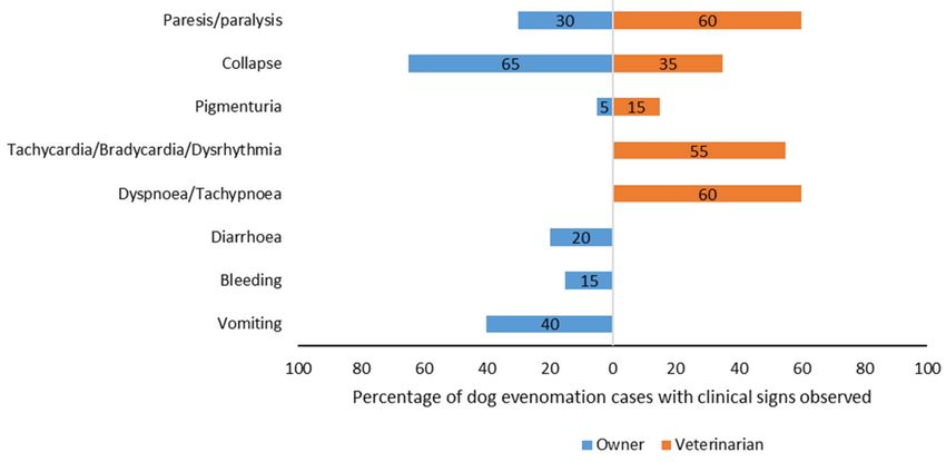
Vet. Sci. 2021, 8, 14                                                                                            2 of 8

                        characteristics, diagnosis, treatments, and outcomes of dogs admitted with assumed elapid
                        snake envenomation to veterinary clinics in Queensland, Australia.

                        2. Materials and Methods
                             Small and mixed animal veterinary clinics in Queensland, Australia, were targeted
                        to collect data on snake envenomations in dogs. The platform “Survey Monkey” [10]
                        was used to develop an electronic data collection form (a copy of the form is provided
                        as Supplementary Figure S1). The form had two sections. Firstly, the frequency of snake
                        envenomations over the period of one year was explored (January until December 2016),
                        while in the second part, information on individual patients was collected, including the
                        environment where the envenomation occurred, the clinical signs as observed by owners
                        and recorded by veterinarians (including the time between onset of clinical signs and
                        admission). Diagnostic tests and treatments performed by the veterinarian, the number of
                        days of hospitalisation, and the outcome of hospitalisation were also recorded.
                             As no register of veterinary clinics was publicly available, a sampling frame was
                        created by identifying 145 postal addresses of veterinary clinics in Queensland through
                        Google searches. A postcard describing the project and providing the address to the project
                        website [11], which contained a link to the data collection form, was sent out to these
                        veterinary clinics. In addition, the Australian Veterinary Association [12] published the
                        link to the data collection form in their monthly e-newsletter and on their Facebook site.
                        Finally, a link to the data collection was posted on a professional Facebook group webpage
                        for veterinarians and veterinary nurses in Queensland. Data collection was conducted
                        between 1 April and 30 August 2017.
                             Data were summarised using descriptive statistics and cross-tabulations. The outcome
                        of hospitalisation was compared between usage and non-usage of antivenom using the
                        Fisher’s exact test. Results were considered significantly different at a p-value < 0.05.

                        3. Results
                              A total of 19 veterinary clinics completed the data collection form, reporting 230
                        envenomations in dogs over the 12-month period, with an average (range) of 12.1 (1–51)
                        and a median of 11 dog envenomations reported per clinic, per year in Queensland,
                        Australia, in 2016. The locations of the veterinary clinics that responded providing data
                        are shown in Supplementary Figure S2. The peak months for snake envenomation were
                        between September and March, corresponding with the warmer weather in Australia
                        (Figure 1).
                              A complete history was provided for 20 dogs with snake envenomation, and 12 were
                        females and eight were males. The majority of dog breeds affected were terriers (40.0%,
                        8/20, including four Jack Russell, two Fox, two Staffordshire Bull, one Boston, and one
                        West Highland Terrier) and cattle dogs (25.0%, 5/20), followed by crossbreed dogs (2/20)
                        and range of single dogs of other breeds (Border Collie, Dachshund, Labradors, Pointer,
                        Shih Tzu). Forty percent (8/20) of dogs were between one and two years old, and 30.0%
                        (6/20) were between three and five or between six and ten years old, respectively.
                              Recorded owner observations indicated that signs of the envenomation were mostly
                        noted after 8 a.m. and before 6 p.m. (65.0%, 13/20). Envenomation occurred in 80.0%
                        (16/20) of dogs in the backyard of houses, followed by pastures and bushland (10.0%,
                        2/20), respectively. A snake was sighted in two cases. The most common signs of snake
                        envenomation observed by owners were collapse and paresis/paralysis (Figure 2).
                              Paresis/paralysis, dyspnoea or tachypnoea, and cardiac abnormalities (such as dys-
                        rhythmias including tachycardia and bradycardia) were the most commonly reported
                        clinical signs observed by veterinarians (Figure 2), with a combination of both respiratory
                        and cardiac abnormalities observed in 10 dogs. Owners reported that an apparent recovery
                        was observed for 40.0% of dogs (8/20) after the onset of initial clinical signs, with six dogs
                        initially showing collapse, one showing paresis/paralysis and collapse, and one showing
                        paresis/paralysis only. Seven of these achieved a full recovery, and one died. Veterinarians
Vet. Sci. 2021, 8, 14 3 of 8
specified identification of the bite site in four dogs. The mean (SD) time between onset
of clinical signs and admission to the veterinary hospital was 89.9 (95.1) minutes and the
median (minimum, maximum) time was 60 (14, 360) minutes.
Figure 1. Average number of dogs diagnosed with snake envenomations per month across 19 veterinary clinics, in
Queensland, Australia, in 2016 and mean minimum and maximum temperature per month in Queensland, Australia, in
2016 (temperature data were recorded by the Bureau of Meteorology station location 40082, University of Queensland,
Gatton, Australia; latitude: −27.54, longitude: 152.34).
Figure 2. Recorded owner observations of dogs with snake envenomation vs. signs observed by examining veterinarians
after presentation of these dogs to veterinary clinics in Queensland, Australia, in 2016.
The diagnostic tests performed by veterinarians for suspected snake envenomation are
shown in Table 1. Activated clotting time (ACT) was frequently combined with other diag-
nostic procedures. Prolonged clotting times were observed in 69.2% (9/13) of dogs whereVet. Sci. 2021, 8, 14 4 of 8
an ACT was performed. History and clinical signs were the sole diagnostic modalities
utilised to determine snake envenomation in 30.0% of dogs (6/20).
Table 1. Diagnostic procedures conducted by veterinarians for snake envenomation in dogs in
Queensland, Australia, in 2016.
Diagnostic Procedures Performed Dogs (%, n)
ACT only 35.0% (7)
ACT and PCV/TP 10.0% (2)
ACT and SVDK 10.0% (2)
ACT, PCV/TP, and blood smear 5.0% (1)
ACT, PCV/TP, and CK 5.0% (1)
SVDK, PTT/aPTT, and ECG 5.0% (1)
Clinical signs only 30.0% (6)
Total 100.0% (20)
ACT: activated clotting time, CK: creatine kinase, ECG: electrocardiograph, PCV/TP: packed cell volume/total
protein, PTT/aPTT: prothrombin time/activated prothrombin time, SVDK: snake venom detection kit (SVDK).
An SVDK was used for 15.0% of dogs (3/20), with blood used as the test sample in
two and urine in one dog. Results from the SVDK were the identification of one brown
snake immunotype, one tiger snake immunotype, and one sample showing only a result
for the positive control (thus, unable to confirm snake envenomation).
The veterinary treatments conducted for snake envenomation are shown in Table 2.
Intravenous (IV) fluids were administered to 95.0% of dogs (19/20).
Table 2. Treatments conducted by veterinarians for snake envenomation in dogs in Queensland,
Australia, in 2016.
Treatment Administered Dogs (%, n)
IV Fluids and Antivenom 35.0% (7)
IV Fluids, Antivenom, and Antihistamines 15.0% (3)
IV Fluids, Antivenom, Adrenalin, and Antihistamines 25.0% (5)
IV Fluids, Antivenom, Antihistamines, and Vitamin C 5.0% (1)
IV Fluids, Antivenom, Adrenalin, Frusemide, and Vitamin C 5.0% (1)
IV Fluids, Adrenalin, Frusemide, and Antihistamines 5.0% (1)
IV Fluids and Vitamin C 5.0% (1)
No treatment 5.0% (1)
Total 100% (20)
IV: intravenous.
Antivenom was administered to 85.0% of dogs (17/20). Of the dogs that received
antivenom, 13 dogs received a tiger/multibrown combination (3000 units tiger/4000 units
brown), two dogs received a single multibrown (1650 units), one dog received two single
multibrown, and one dog received taipan antivenom.
Of the three dogs where antivenom was not administered, one dog did not show a
positive result for a snake immunotype using the SVDK and subsequently died. Another
dog died before the treatment could commence, and the third dog showed pigmenturia
and paralysis and then fully recovered.
Duration of hospitalisation (including for animals that died or were euthanised) was
less than one day for 35.0% of dogs (7/20), one to two days for 30.0% (6/20), two to three
days for 25.0% (5/20), three to four days for 5.0% (1/20), and greater than 4–6 days for
5% (1/20) of dogs. Twenty-five percent (5/20) of dogs were euthanised or died, while
75.0% (15/20) recovered following hospitalisation and treatment. Three of the 17 dogs
that received antivenom (17.6%) died or were euthanised, while two of the dogs that not
received antivenom (66.7%) died or were euthanised (Fisher exact p = 0.140).Vet. Sci. 2021, 8, 14 5 of 8
4. Discussion
Based on extrapolation from 80 veterinary clinics surveyed in 1998, it was estimated
that each veterinary clinic in Australia sees on average of four snake bite cases per year [6].
Our case series from 19 veterinary clinics indicated a higher snake envenomation rate
of about 12 dogs per clinic, per year. Dogs with a hunting nature (e.g., terriers) were
unsurprisingly more frequently bitten, but also younger dogs that are potentially more
inquisitive or active. This is similar to the demographic trends reported in cane toad
intoxication in dogs, where young terrier breeds are more frequently affected [13]. Animals
with these characteristics are more likely to approach and attack snakes (and these breeds
are very popular in Queensland, Australia), and possibly sustain multiple envenomations
from agitated snakes. In contrast, dogs bitten by Vipera berus in Sweden were predominantly
large breeds such as German shepherds and Labradors, which are actually the most popular
breeds in this country [14].
In most cases, the owners of the dogs did not witness the envenomation and were
therefore unable to describe the snake species. It has been reported previously, that even
when a snake is witnessed, identification of snakes in Australia is challenging due to the
morphologic variations, as well as due to the overlapping appearance between snake
species [15]. Morrison et al. [16] highlighted that as few as 19% of snakes were correctly
identified by the general population of Australians. Hence, relying on owners for visual
identification of snakes to aid the diagnosis and treatment of snake envenomation is
unreliable.
The seasonality of envenomation shown here is in accordance with previous research,
which demonstrated peaks of envenomation in the warmer months of the year when
snakes are active [17]. The seasonality of snake activity should increase the suspicion of
envenomation when consistent clinical signs are present, but envenomation cannot be
excluded outside these peak periods.
Previous snakebite investigations in dogs and cats highlighted that 78% of envenoma-
tions occurred in rural and 22% in urban areas [6]. The majority of snake envenomations in
our case series occurred in backyards. Mirroring this, a recent report in people compiled
from 171 hospitals in Australia demonstrated that the most frequent location for enveno-
mation of 1548 people presenting for snake bite occurred near houses (485 people, 31%)
and in buildings (220 people, 14%) [5]. The high frequency of envenomations in backyards
in our case study might also be a reflection of companion animals spending their majority
of time around their owner’s homes.
Clinical signs of envenomation were noted predominantly during the daytime, which
is the main period of snake activity. Some elapids, especially eastern brown snakes (Pseudon-
aja textilis) and red-bellied black snakes (Pseudechis porphyriacus) are usually active during
the day unless temperatures are very hot, and they are the most common venomous snakes
to be encountered in Queensland [18,19].
Vomiting and collapse associated with a transient recovery as identified by owners
in our study has been previously described as preparalytic signs [20,21], which is likely
related to a sudden decrease in blood pressure [22]. The preparalytic phase is followed by
the paralytic and lethal phase, which consists of skeletal muscle paralysis and coagulopa-
thy [23].
Prolonged activated clotting time (ACT) was the most frequently performed test in our
case series, with similar coagulopathy compared to previous research [15]. The presence of
a coagulopathy in conjunction with preparalytic signs or paralytic signs should prompt
the clinician to strongly consider snake envenomation as the diagnosis, but the absence
of coagulopathy does not rule out envenomation. Pigmenturia was also observed in
patients, which is due to either myolysis or haemolysis; thus, pigmenturia should prompt
the veterinarian to consider snake envenomation [24]. While coagulopathy is a feature of
many envenomations in small animals, spontaneous haemorrhage as a result is not. Three
dogs were reported by owners to be bleeding prior to presentation, although details were
not specified. Venom-induced consumptive coagulopathy may severely alter haemostaticVet. Sci. 2021, 8, 14 6 of 8
capability, and any minor injury prior to presentation could potentially result in such
clinically apparent haemorrhages.
Biochemical tests such as creatine kinase are beneficial in the determination of enveno-
mation by snakes that cause myolysis such as tiger, black, and taipan snakes [7,25], but had
only be used in two cases. The clinician should be aware that creatine kinase has a very
short half-life of two hours and, depending on the cause of muscle damage, peaks around
4–12 h after insult [26].
The SVDK is manufactured by CSL Ltd. (Parkville, Victoria, Australia) and is able to
detect the venom of the five major lethal snake immunotypes in Australia (brown, tiger, and
black snakes; death adders; and taipans) [27]. The SVDK is designed for use in humans [28]
but is also applied for the identification of venoms in companion animals [7,29]. The
infrequent use of the SVDK in only three cases could be attributed to the cost (a single
test costs about ~$300–400 AUD), as well as the varied accuracy of the results [15,24]. The
SVDK was reported to give positive results in animals presented within four hours of
envenomation but gave false negative results after delayed presentation [24]. The SVDK
may yield negative results if venom in the urine has already been voided by the patient
or if absorption of the venom to detectable levels has not occurred—we had one negative
SVDK test result in our case series. The use of SVDK is common in humans (used in 83%
envenomed patients [5]) with patients typically able to identify where they were bitten, as
a bite-site swab is the preferred test sample for the SVDK. Conversely, bite sites in animals
are unlikely to be identified, and therefore either urine or blood is used for the SVDK.
The majority of antivenom used was the tiger–multibrown antivenom, which contains
4000 units of multibrown and 3000 units of tiger snake antivenom. Reasons for this
choice were not specified by veterinarians, although it may be preferred as the antivenom
neutralises the venom of several species of snake such as brown, red-bellied black, and
tiger snake, which are the most common snakes in the geographic area studied. In addition,
the cost per unit is less for combination tiger–multibrown antivenom than for multibrown
antivenom. Furthermore, due to the limited shelf life of antivenom, veterinarians may be
more likely to stock combination antivenom, because of its versatility in application.
Unfortunately, we had a low response rate in our study, most likely as it is time
consuming for veterinarians to provide detailed data on individual envenomation cases.
The use of software applications that compile de-identified records from multiple veterinary
clinics should be considered for future research on snake envenomation [30]. In addition,
the use of mobile phone applications could provide an opportunity for researchers to
obtain data directly from members of the public about snakes observed and potential
envenomations of their pets.
Nevertheless, our case series has summarised the common diagnostic procedures
and treatments of snake envenomation in dogs in Queensland. It seems that the dilemma
faced by veterinarians compared to human medical practitioners is the daily balancing
of costs associated with appropriate diagnostics and therapeutics. Thus, veterinarians
are frequently reliant on history and clinical signs and diagnostic tests other than the
SVDK (such as blood clotting times and serum CK) and most likely administered snake
envenomation treatment based on their clinical experience.
Supplementary Materials: The following materials are available online at https://www.mdpi.com/
2306-7381/8/2/14/s1, Figure S1. Online form used for collecting data on snake envenomations
in dogs in Queensland, Australia, in 2016. Figure S2. Number of snake envenomations in dogs
submitted to veterinary clinics in Queensland, Australia, in 2016. Data are shown by the postcode of
veterinary clinics.
Author Contributions: Conceptualisation: R.A., M.H., J.C., and J.H.; methodology: L.V., R.A., M.H.,
J.C., and J.H.; formal analysis: L.V. and J.H.; investigation: L.V. and J.H.; resources: J.H.; writing—
original draft preparation: L.V. and J.H.; writing—review and editing: L.V., R.A., M.H., J.C., and
J.H.; project administration: J.H.; funding acquisition: J.H. All authors have read and agreed to the
published version of the manuscript.Vet. Sci. 2021, 8, 14 7 of 8
Funding: Funding for this project was provided through the Peter and Mary Ellen Stone Memorial
Fund. The funders had no role in study design, data collection and analysis, decision to publish, or
preparation of the manuscript.
Institutional Review Board Statement: Human ethics approval (approval number: 2016000340, 31
May 2016) for this research was obtained from the Behavioural and Social Science Ethical Review
Committee of the University of Queensland.
Informed Consent Statement: Informed consent was obtained from all participants involved in the
study.
Data Availability Statement: Data is contained within the article or supplementary material.
Acknowledgments: The research group wishes to thank participating veterinary clinics who com-
pleted the data collection forms.
Conflicts of Interest: The authors declare no conflict of interest. The funders of this project had no
role in the design of the study; in the collection, analyses, or interpretation of data; in the writing of
the manuscript, or in the decision to publish the results.
References
1. Shea, G.M. The distribution and identification of dangerously venomous Australian terrestrial snakes. Aust. Vet. J. 1999, 77,
791–798. [CrossRef]
2. Hodgson, W.C.; Wickramaratna, J.C. Snake venoms and their toxins: An Australian perspective. Toxicon 2006, 48, 931–940.
[CrossRef] [PubMed]
3. Kasturiratne, A.; Wickremasinghe, A.R.; de Silva, N.; Gunawardena, N.K.; Pathmeswaran, A.; Premaratna, R.; Savioli, L.; Lalloo,
D.G.; de Silva, H.J. The global burden of snakebite: A literature analysis and modelling based on regional estimates of envenoming
and deaths. PLoS Med. 2008, 5, e218. [CrossRef] [PubMed]
4. Welton, R.E.; Liew, D.; Braitberg, G. Incidence of fatal snake bite in Australia: A coronial based retrospective study (2000–2016).
Toxicon 2017, 131, 11–15. [CrossRef] [PubMed]
5. Johnston, C.I.; Ryan, N.M.; Page, C.B.; Buckley, N.A.; Brown, S.G.A.; O’Leary, M.A.; Isbister, G.K. Can Australians identify snakes?
The Australian Snakebite Project, 2005–2015 (ASP-20). Med. J. Aust. 2017, 207, 119–125. [CrossRef] [PubMed]
6. Mirtschin, P.J.; Masci, P.; Paton, D.C.; Kuchel, T. Snake bites recorded by veterinary practices in Australia. Aust. Vet. J. 1998, 76,
195–198. [CrossRef] [PubMed]
7. Heller, J.; Mellor, D.J.; Hodgson, J.L.; Reid, S.W.; Hodgson, D.R.; Bosward, K.L. Elapid snake envenomation in dogs in New South
Wales: A review. Aust. Vet. J. 2007, 85, 469–479. [CrossRef] [PubMed]
8. Jacoby-Alner, T.E.; Stephens, N.; Davern, K.M.; Balmer, L.; Brown, S.G.; Swindells, K. Histopathological analysis and in situ
localisation of Australian tiger snake venom in two clinically envenomed domestic animals. Toxicon 2011, 58, 304–314. [CrossRef]
9. Boller, M.; Kelers, K.; Stevenson, M.A.; Winkel, K.D.; Hardjo, S.; Heller, J.; Judge, P.R.; Ong, H.M.; Padula, A.M.; Reddrop, C.; et al.
SnakeMap: Four years of experience with a national small animal snake envenomation registry. Aust. Vet. J. 2020, 98, 442–448.
[CrossRef]
10. Survey Monkey. Available online: https://www.surveymonkey.com/ (accessed on 5 January 2021).
11. Snake Bite Research Australia. Available online: http://snakebiteresearch.com.au/ (accessed on 5 January 2021).
12. Australian Veterinary Association. Available online: https://www.ava.com.au/ (accessed on 5 January 2021).
13. Reeves, M.P. A retrospective report of 90 dogs with suspected cane toad (Bufo marinus) toxicity. Aust. Vet. J. 2004, 82, 608–611.
[CrossRef]
14. Lervik, J.B.; Lilliehöök, I.; Frendin, J.H. Clinical and biochemical changes in 53 Swedish dogs bitten by the European adder-Vipera
berus. Acta Vet. Scand. 2010, 52, 26. [CrossRef]
15. Indrawirawan, Y.; Sheridan, G.; McAlees, T. Clinical features of mainland Tiger and Eastern Brown Snake envenomation in dogs
and cats in Melbourne. Aust. Vet. Pract. 2014, 44, 704–712.
16. Morrison, J.J.; Pearn, J.H.; Covacevich, J.; Nixon, J. Can Australians Identify Snakes. Med. J. Aust. 1983, 2, 66–70. [CrossRef]
[PubMed]
17. Hill, F.W. Snake bite in dogs. Aust. Vet. J. 1979, 55, 82–85. [CrossRef] [PubMed]
18. Australian Museum. Reptiles. Available online: https://australian.museum/learn/animals/reptiles (accessed on 5 January
2021).
19. Couper, P.; Amey, A.P. Snakes of South-East Queensland: A Queensland Museum Pocket Wild Guide; Queensland Museum: Brisbane,
Australia, 2007.
20. Lewis, P.F. Common tiger snake envenomation in dogs and mice—Relationship between the amount of venom Injected and the
onset of clinical signs. Aust. Vet. J. 1994, 71, 130–132. [CrossRef] [PubMed]Vet. Sci. 2021, 8, 14 8 of 8
21. Padula, A.M.; Leister, E. Eastern brown snake (Pseudonaja textilis) envenomation in dogs and cats: Clinical signs, coagulation
changes, brown snake venom antigen levels and treatment with a novel caprylic acid fractionated bivalent whole IgG equine
antivenom. Toxicon 2017, 138, 89–97. [CrossRef]
22. Tibballs, J.; Sutherland, S.K.; Rivera, R.A.; Masci, P.P. The cardiovascular and haematological effects of purified prothrombin
activator from the common brown snake (Pseudonaja textilis) and their antagonism with heparin. Anaesth. Intensive Care 1992, 20,
28–32. [CrossRef]
23. Hardy, M.C.; Cochrane, J.; Allavena, R.E. Venomous and poisonous Australian animals of veterinary importance: A rich source of
novel therapeutics. BioMed Res. Int. 2014, 2014, 671041. [CrossRef]
24. Padula, A.M.; Ong, H.M.; Kelers, K. Snake envenomation in domestic animal species in Australia. In Clinical Toxinology in
Australia, Europe, and Americas, 1st ed.; Springer: Berlin/Heidelberg, Germany, 2018; pp. 505–536.
25. Judge, P.R. Coastal taipan (Oxyuranus scutellatus) envenomation of a dog. Aust. Vet. J. 2015, 93, 412–416. [CrossRef]
26. Aktas, M.; Auguste, D.; Lefebvre, H.P.; Toutain, P.L.; Braun, J.P. Creatine kinase in the dog: A review. Vet. Res. Commun. 1993, 17,
353–369. [CrossRef]
27. Snake Venom Detection Kit. Available online: https://labeling.seqirus.com/SVDK/AU/Snake-Venom-Detection-Kit/EN/
Snake-Venom-Detection-Kit.pdf (accessed on 5 January 2021).
28. Cox, J.C.; Moisidis, A.V.; Shepherd, J.M.; Drane, D.P.; Jones, S.L. A novel format for a rapid sandwich EIA and its application to
the identification of snake venoms. J. Immunol. Methods 1992, 146, 213–218. [CrossRef]
29. Forbes, G.; Church, S. Detection of snake venom in equine urine and plasma: Validation of a commercially available snake venom
detection kit in horses. Aust. Equine Vet. 2010, 29, 57–61.
30. VetCompass Australia. Available online: https://www.vetcompass.com.au/ (accessed on 5 January 2021).You can also read