CT IMAGING OF CORONARY ATHEROSCLEROSIS. STATE-OF-THE-ART AND BEYOND - DANIELE ANDREINI, MD, PHD, FESC, FSCCT - IFC-CNR
←
→
Page content transcription
If your browser does not render page correctly, please read the page content below
CT Imaging of coronary
           atherosclerosis.
     State-of-the-art and beyond
    Daniele Andreini, MD, PhD, FESC, FSCCT
Director, Cardiovascular CT Unit, Centro Cardiologico Monzino,
                          Milan, Italy
 Associate Professor of CV Disease, University of Milan, Italy

Andreini D et al, Diabetes Care 2013
Andreini D. Jacc im 2016
Briefly, for this score 3 sets of weighting factors are used:

1) Localization of the coronary plaques, accounting for
    dominance.
2) Type of plaque, with a multiplication factor of 1 for calcified
    plaques and of 1.5 for non-calcified and mixed plaques.
3) Degree of stenosis, with a multiplication factor of 0.615 for
    non-obstructive (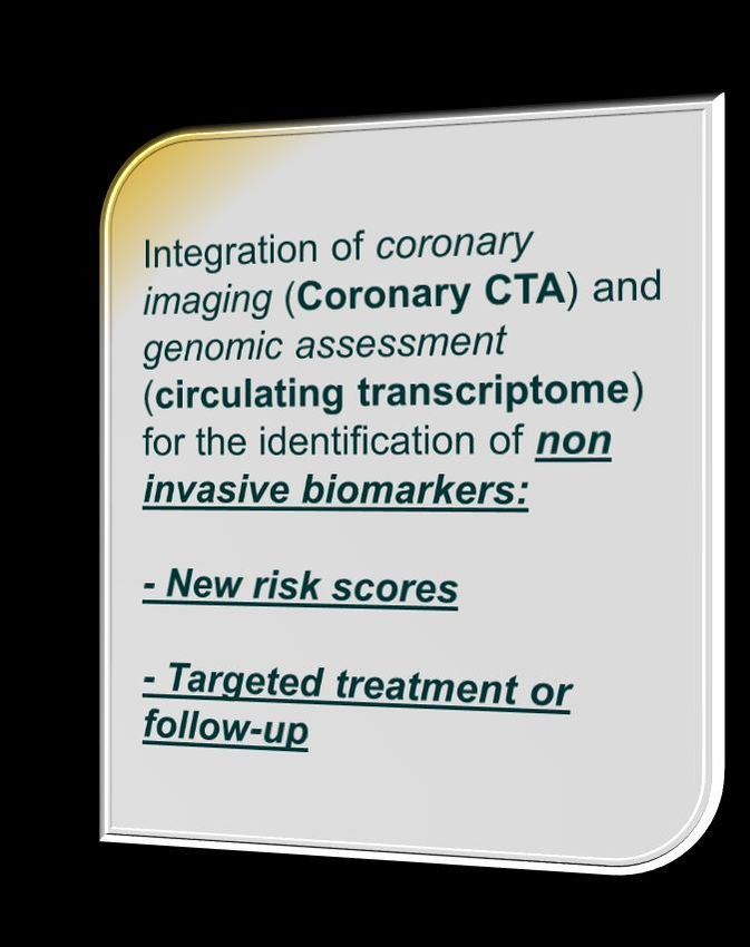
8
2950 patients, mean clinical follow-up of 5 years
1.00

                                                                                        1.00
                                           MACE/LR-free survival
0.90

                                                                                        0.90
0.80

                                                                                        0.80
0.70

                                                                                        0.70

       0          1      2           3                                4           5            0       1      2           3        4           5
                             Years                                                                                Years

              No CAD   NonObs CAD/Low LS                           NonObs CAD/High LS              No CAD   NonObs CAD/Low LS   NonObs CAD/High LS

                                                                                      D. Andreini et al, Int J Cardiol 2017
The most of AMI from
Frequency vs. grade of stenosis
   and rate of complication

                    Naghavi M., Circulation 2003
Background
?
12First Evidence …
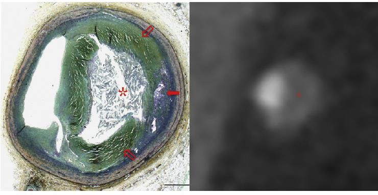
We studied histologic sections of the left main coronary artery in 136 hearts obtained at
autopsy to determine whether atherosclerotic human coronary arteries enlarge in relation to
plaque (lesion) area and to assess whether such enlargement preserves the cross-sectional
area of the lumen.
We conclude that human coronary arteries enlarge in relation to plaque area and that
functionally important lumen stenosis may be delayed until the lesion occupies 40 percent of
the internal elastic lamina area. The preservation of a nearly normal lumen cross-sectional
area despite the presence of a large plaque should be taken into account in evaluating
atherosclerotic disease with use of coronary angiography
N Engl J Med 1987; 316:1371-1375 May 28, 1987 The most common cause of coronary syndrome is plaque rupture with subsequent
thrombosis….
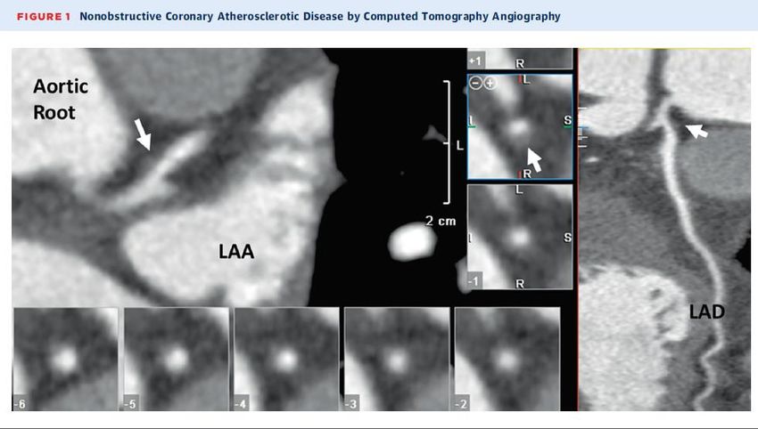
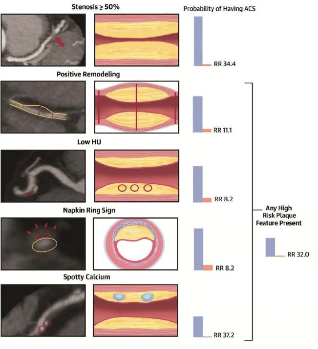
• The non-thrombosed lesion that most
resembles the acute plaque rupture is the
thin cap fibroatheroma (TCFA).
• Vessels demonstrating TCFA do not
usually show severe luminal narrowing
but show positive remodeling.
• Cross-sectional area narrowing in over
75% of TCFA cases is lower than 75%.
J Am Coll Cardiol 2006;47:C13–8Pundziute et al JACC Interv 2008
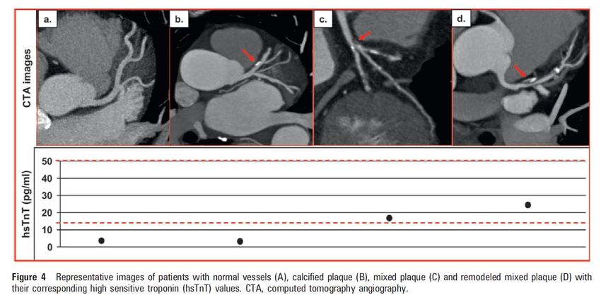
Low attenuation plaques (LAP) /
     Positive Remodelling

                   LAP (plaque with 30 – 50 HU) or intermediate-
                   attenuation plaques (plaque between 50 and
                   150 HU)

                   ratio of the vessel diameter/area at the
                   plaque site to a reference diameter /area
                   proximal to the lesion in a normal-appearing
                   vessel segment.
                   RI>1.1

              JACC:CARDIOVASCULARIMAGING,VOL.6,NO.4,2013
Spotty Calcifications (SCPs) small calcified nodules with length
Napkin-ring sign

                   H. Seifarth et al. / Atherosclerosis 224 (2012) 90e96
Introduz studio placca tramite immagine inj j c
editor
IJC 2017In 1,059 unselected patients who underwent CCTA,
atherosclerotic lesions were analyzed for the
presence of 2 features: PR and LAP. Mean follow-
up of 27 months were valuated. ACS was
independently predicted by PR>1.1 and/or LAP
(hazard ratio: 22.8, 95% confidence interval: 6.9 to
75.2, p 0.001).
Motoyama et al. J Am Coll Cardiol 2009;54:49–57Cardiac CT
Puchner et al. J Am Coll Cardiol. 2014 Aug 19; 64(7): 684–692EHJ im 2016
• Presence of ischemia in not
significant stenosis
• No ischemia in presence of
significant stenosis
Features of CTA-verified high-risk plaques (HRPs), including positive remodeling (PR) and
low attenuation plaque (LAP), were examined for their relationship to the invasive FFR-
verified lesion-specific ischemia. The presence of PR and LAP was highly predictive of
ischemia independent of the severity of stenosis.
Most myocardial infarctions are caused by large volume HRP with large necrotic core and
PR, regardless of the extent of luminal stenosis. Here, we propose that large volume HRP
can be ischemic, independent of the degree of luminal stenosis. Could the strong
predictive value of ischemia testing be a result of indirectly screening for HRP?
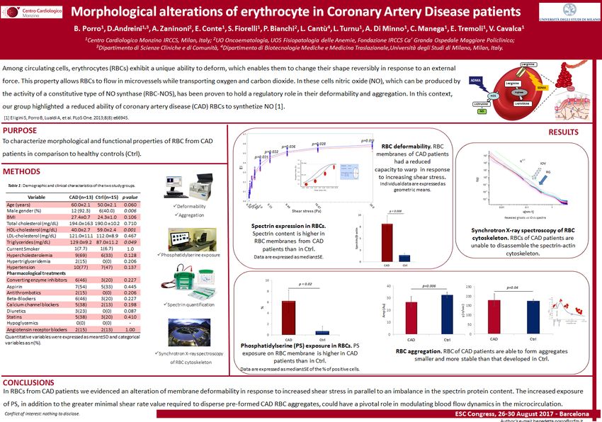
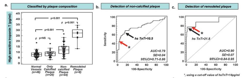
Ahmadi et al. j a c c : c a r d i o v a s c u l a r ima g i n g , v o l . 8 , no. 1 , 2 0 1 5Translational Research: MERCAD project
B. Porro et al, ESC 2017Heart 2017
Am J Cardiol 2016
CAPIRE: Plaque features, Biomarkers and Prognosis
  High-risk features (PR; NRS; LAP)

                                                      High-risk features (PR; NRS; LAP)

                                                                                                            High-risk features (PR; NRS; LAP)
                                          IL-6                                            hs CRP                                                Pentraxin3

P value (Pearson): 0.29                                                                    0.94                                                   0.01

                                                                                                   Andreini D et al. Unpublished data
New perspectives: the EMERALD study
JACC img 2018
31"EPIFANIA" study
(È Prevedibile In quale Forma Avverrà la prima manifestazione clinica Nell’Individuo con Aterosclerosi coronarica non
ostruttiva?)
Coronary CTA
Genomic profile
• 2000 patients:
case-controls
• CTA and
Molecular profile
• At baseline and 2
years f-up
• Clinical follow-up
of 5 yearsMultivariable analysis:
PR was independently related
to impaired hyperemic MBF as
well as an unfavorable FFR
p=0.001 P35
CAPIRE: Plaque features, Biomarkers and Prognosis

                            Andreini D et al. Unpublished data
Chow et al, Arterioscler Thromb Vasc Biol 2015
Jacc 2016
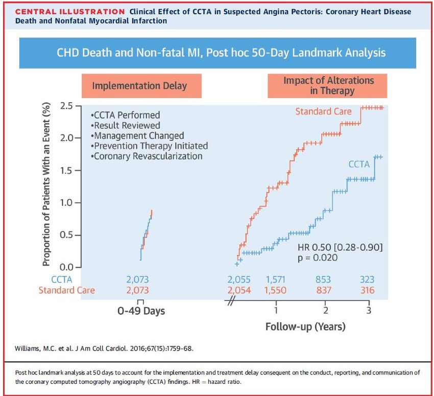
Antiplatelet
                                             Fatal and nonfatal MI

                                          17 vs. 34; hazard ratio: 0.50 [95% c.i.
CCTA leads             to   more   appropriate    use p
Newby et al. NEJM 2018
Am H J 2017
Lee at al, JACC img 2018
Lee at al, JACC img 2018
45
CONCLUSIONS Before the “CCTA era”, patients with non-obstructive CAD, in the absence of inducible ischemia, were included in the same group of those without CAD. Patients with non-obstructive CAD may be divided into two groups, those with low-risk plaque morphology and those in whom plaque characteristics (i.e.higher PRI and/or LAP) are associated with an increased risk of future hard events and could benefit from more aggressive medical therapy. Biomarkers as hsTr (silent rupture) and Pentraxin3 (vascular inflammation) seems promising in the prediction of events together with CCTA. These results support the use of CCTA as a first-line test in patients without known CAD.
CONCLUSIONS
We are quite far from early identification of vulnerable
plaque and vulnerable patient.
Acute coronary events results from a complex interplay in
which atherosclerotic plaque is only one actor toghether
with pro-thrombotic factors.
It is time to go outside the lumen looking for
atherosclerosis.
Circulation. 2012;125:1147-1156This “new” paradigm is not actually new, but simply encourages diagnostic CAD evaluation by a forward rather than backward- looking stance, reflecting the natural history of atherosclerosis progression; namely, that disease severity should be gauged hierarchically: -plaque versus no plaque; -high-risk plaque features versus non-high-risk plaque features; -high-grade stenosis versus non–high-grade stenosis; -ischemia versus no ischemia JACC Vol 67, No. 15, 2016
You can also read