Clinical Protocol for Radiotherapy for Plantar Fasciitis and Achilles Tendonitis
←
→
Page content transcription
If your browser does not render page correctly, please read the page content below
RT-PRO-296
Document Owner: Head of              First Issued: March 2019
Radiotherapy
Document Authoriser: Radiation       Date Next Review: July 2021
Oncology Committee
Version Number: 3.0                  Date Last Review: July 2020

Clinical Protocol for Radiotherapy for
Plantar Fasciitis and Achilles
Tendonitis
UK
1. Introduction and Purpose
To provide clear instructions on the clinical assessment, referral and radiotherapy
treatment of patients with Plantar Fasciitis and Achilles Tendonitis.

2. Terms and Definitions
PF – Plantar Fasciitis
AT – Achilles Tendonitis
MLC – Multi-leaf collimator
OMS – Oncology management system
ClinOnc – Clinical Oncologist

2.1.          Plantar fasciitis (PF) and Achilles Tendonitis (AT)

Plantar Fasciopathy, also known as PF, is a benign inflammatory and degenerative
condition of the plantar fascia that causes heel pain. Achilles tendonitis (AT) refers
to the tendon connecting the heel to the calf.

The main cause is mechanical overload of the plantar fascia or the achilles tendon,
which may be caused by obesity, prolonged standing and walking, intense
exercise, and biomechanical disturbance of the foot.

With conservative treatment, including resting, icing, stretching and better
footwear, 80% of patients have complete resolution of pain within 6 - 12 months.
However, those whose pain does not resolve may need further treatment.

3. Policy – Treatment Considerations
In those who have “resistant” PF/AT i.e. those who have failed conservative
treatment for at least 6 months, two randomised trials have shown a significant
benefit of radiotherapy over either sham (very low dose) radiotherapy or steroid
injection.

Document uncontrolled when printed                                            Page 1 of 7
RT-PRO-296
Document Owner: Head of First Issued: March 2019
Radiotherapy
Document Authoriser: Radiation Date Next Review: July 2021
Oncology Committee
Version Number: 3.0 Date Last Review: July 2020
Treatments for resistant PF/AT include:
• Extracorporeal Shockwave Therapy – This is widely used and funded, and can
be very helpful for some patients, although the evidence is equivocal, and it
may be painful.
• Steroid injections – May provide short-term relief from pain but risks plantar
fascial rupture.
• Surgery – Case series but no randomised evidence and may be associated
with complications.
4. Patient Selection
4.1. Inclusion criteria
a. Pre-consultation
The patient is sent a pre-consultation questionnaire, asking for demographics,
insurance, previous treatment etc. prior imaging and report when available should
be brought to the consultation.
b. Initial consultation
The patient is seen in clinic by the oncologist. A full history is taken, including the
presenting complaint and prior treatment for plantar fasciitis or Achilles tendonitis,
past medical history, whether they have had prior radiotherapy, or have
pacemaker
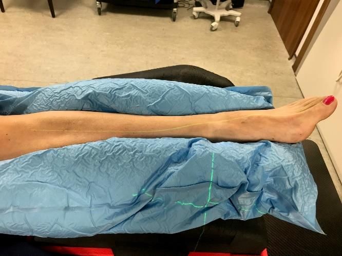
The patient is examined to see if their pain is compatible with plantar fasciitis or
Achilles tendonitis. If there is a site of particular tenderness then this is drawn,
photographed, and documented.
c. Diagnostic imaging
a. If they have not already had diagnostic imaging, then most patients
will undergo a diagnostic ultrasound by the Clinical Oncologist
b. Some may need further radiological investigation e.g. X-ray, MRI
c. If a diagnosis of plantar fasciitis is confirmed, then treatment options
are discussed. Treatments options will include radiotherapy
d. Repeating radiotherapy
For patients who have tolerated the first course of radiotherapy a
repeat course may be considered
4.2. Radiotherapy Dataset
Essential:
1. Radiotherapy Referral detailing site and laterality.
2. Either - Pre-consultation questionnaire returned from patient stating site and
laterality, or – clinic annotation from consultant stating site and laterality.
Document uncontrolled when printed Page 2 of 7
RT-PRO-296
        Document Owner: Head of                 First Issued: March 2019
        Radiotherapy
        Document Authoriser: Radiation          Date Next Review: July 2021
        Oncology Committee
        Version Number: 3.0                     Date Last Review: July 2020

        Desirable:
           1. Any previous imaging reports relevant to diagnosis, this will usually be
              ultrasound.
           2. Referral letter from referring specialist to Clinical Oncology where available.

        4.3.          Consent

        As per GenesisCare Consent form for Dupuytrens disease, Ledderhose disease,
        Plantar Fasciitis (RT-TEM-207).

        4.4.          Scheduling of Patients

        A next-day planning/ treatment pathway is available at GenesisCare UK.

        Cases may start any weekday – preferably not Friday.

        5. Patient Positioning and Localisation
Site               Immobilisation Device     Set-up                CT planning scan   Localisation   Reference mark location

Plantar            Vacbag to be              Pt supine      Scan limits:        As marked            Landmark
Fasciitis/         made prior at             Feet towards Superiorly up to by        Clinical        reference line as
Achilles           CT                        gantry         mid-calf.           Oncology             appropriate
Tendonitis         Affected limb                            Inferiorly to clear                      e.g.          mid
                   to be stabilised          Pt indexed & the entire foot by                         separation heel-
                   in vacbag on              off -set using 3cm.                                     junction of 1st
                   top of indexed            localisation                                            toe-> 2nd toe
                   kneefix & 2               bar            Wire area of                             Define
                   risers                    Unaffected     pain at CT                               proximal/distal
                                             limb to bent                                            and/or edge
                                             at knee to Protocol:
                                             avoid          ‘AbdoPelvis’
                                             concomitant with          manual
                                             dose.          adjustment       of
                                             *Clearance     sup/inf border
                                             to          be as above.
                                             checked*

        Document uncontrolled when printed                                                              Page 3 of 7
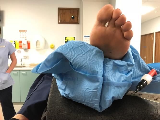
RT-PRO-296
Document Owner: Head of              First Issued: March 2019
Radiotherapy
Document Authoriser: Radiation       Date Next Review: July 2021
Oncology Committee
Version Number: 3.0                  Date Last Review: July 2020

6. Definition of Target
6.1.          Target Volumes PF & AT

PF
Fields cover site of pain plus 2cm all around entire heel and, calcaneus and cuboid
and partial coverage of most distal metatarsal and soft tissue on underside of foot,
MLC to shield other metatarsals and navicular and talus.

AT
Fields cover site pain plus 2cm margin include insertion and lower portion of
Achilles tendon
Modality; 6MV photons with parallel opposed lateral fields

Document uncontrolled when printed                                          Page 4 of 7

RT-PRO-296
Document Owner: Head of                  First Issued: March 2019
Radiotherapy
Document Authoriser: Radiation           Date Next Review: July 2021
Oncology Committee
Version Number: 3.0                      Date Last Review: July 2020

              PF                                                           AT

                                                                       ,

6.2.          Organs at Risk

                          •     not applicable

6.3.          Prescription Dose

                          •     Standard dose = 6Gy in 6 fractions over 3 weeks i.e. 2 fractions
                                per week
                          •     Dose may be given over 2 weeks i.e. 3 fractions per week
                          •     Dose may be lowered to 3Gy in 6 fractions if pain of short
                                duration

6.4.          Plan approval

                          •     Prescription and V-sim sign off as plan approval

7. Pre-treatment Quality Assurance
                          •     Not applicable

8. Pre-treatment Verification/ Checks
Treatment verification is to be undertaken day 1 prior to treatment using 2D
imaging.

10. IGRT
                          •     2D MV imaging daily, prior to treatment delivery, using pre-port.

Document uncontrolled when printed                                                         Page 5 of 7
RT-PRO-296
Document Owner: Head of                  First Issued: March 2019
Radiotherapy
Document Authoriser: Radiation           Date Next Review: July 2021
Oncology Committee
Version Number: 3.0                      Date Last Review: July 2020

11.          Image Review
                          •     Follow process in iView Image Verification Work Instruction (RT-
                                WI-415)

12. Treatment Delivery
                          •     Radiotherapy Treatment Policy (RT-POL-014)
                          •     Weekly patient review documented in OMS

13. Other Considerations
Follow up appointments:

                          •     At 3 months after the end of treatment
                          •     Follow up then yearly for 4 years

14. References

      1. Niewald M, et al. Randomized, multicenter trial on the effect of radiation
         therapy on plantar fasciitis (painful heel spur) comparing a standard dose
         with a very low dose: Mature results after 12 months' follow-up. Int J Radiat
         Oncol Biol Phys 2012; 84: e455-62.
      2. Canyilmaz et al. Prospective Randomized Comparison of the Effectiveness
         of Radiation Therapy and Local Steroid Injection for the Treatment of Plantar
         Fasciitis. Int J Radiation Oncol Biol Phys, Vol. 92, No. 3, pp. 659e666, 2015.
      3. Heyd et al. Radiation Therapy for Painful Heel Spurs. Results of a Prospective
         Randomized Study. Strahlenther Onkol 2007;183:3–9.
      4. A review of the use of radiotherapy in the UK for the treatment of benign
         clinical conditions and benign tumours. London: The Royal College of
         Radiologists, 2015
      5. Dupuytrens radiotherapy consent form (RT-TEM-207)
      6. Radiotherapy Treatment Policy (RT-POL-014)

Document uncontrolled when printed                                                        Page 6 of 7
RT-PRO-296
Document Owner: Head of First Issued: March 2019
Radiotherapy
Document Authoriser: Radiation Date Next Review: July 2021
Oncology Committee
Version Number: 3.0 Date Last Review: July 2020
Revision History
Date
Version Created By Description of change
Created
1.0 March 2019 Portfolio Lead New Protocol
Radiotherapy
2.0 May 2019 Portfolio Lead Document reviewed and
Radiotherapy updated
3.0 July 2020 Rory Walford – Skin & Document update
Benign Specialist
Radiographer
Document uncontrolled when printed Page 7 of 7You can also read