The Convoluted Tubules of the Nephron Must Be Considered Elliptical, and Not Circular, in Stereological Studies of the Kidney
←
→
Page content transcription
If your browser does not render page correctly, please read the page content below
Research Article

                                                             Kidney Blood Press Res 2021;46:229–235                                                   Received: August 4, 2020
                                                                                                                                                      Accepted: February 4, 2021
                                                             DOI: 10.1159/000515051                                                                   Published online: March 31, 2021

The Convoluted Tubules of the Nephron
Must Be Considered Elliptical, and Not Circular,
in Stereological Studies of the Kidney
Marta Ortega-Martinez a Vanessa Gutierrez-Davila a
Esthefania Gutierrez-Arenas b Alberto Niderhauser-Garcia a
Ricardo M. Cerda-Flores c Gilberto Jaramillo-Rangel a
aDepartment   of Pathology, School of Medicine, Autonomous University of Nuevo Leon, Monterrey, Mexico; bSchool
of Biology, Autonomous University of Sinaloa, Culiacan, Mexico; cSchool of Nursing, Autonomous University of
Nuevo Leon, Monterrey, Mexico

Keywords                                                                                         significant differences in the tubular areas in both PCT (F =
Distal convoluted tubule · Kidney · Mouse · Proximal                                             34.843, Sig = 0.000) and DCT (F = 22.390, Sig = 0.000); circular
convoluted tubule                                                                                areas were different from elliptical areas (SCA and LCA vs. EA
                                                                                                 and PA). Conclusion: The convoluted tubules of the nephron
                                                                                                 must not be considered circular, but rather elliptical; care
Abstract                                                                                         should be taken every time the tubules are analyzed in ste-
Introduction: The diameter and area of the proximal convo-                                       reological studies of the kidney, especially when evaluating
luted tubule (PCT) and the distal convoluted tubule (DCT) are                                    their diameters and areas.              © 2021 The Author(s)
of the main parameters analyzed in stereological studies of                                                                                            Published by S. Karger AG, Basel

the kidney. However, there is no consensus about if the PCT
and DCT should be considered circular or elliptical in shape.
Objective: To analyze if there are significant differences in                                        Introduction
the diameter and area of the PCT and DCT, depending on
whether they are considered circular or elliptical. Methods:                                        Morphometry is the term applied to the measurement
Paraffin-embedded sections of kidneys from CD1 mice were                                         of shape or structure. Stereology, sometimes used syn-
stained with hematoxylin and eosin and examined using a                                          onymously, refers to mathematical methods based on
light microscope. Images were captured using a camera                                            geometric probability that allow to measure three-di-
linked to image analysis software. A short diameter (d) and a                                    mensional characteristics from two-dimensional images
long diameter (D) were measured in both PCT and DCT. A                                           [1].
small circular area (SCA), a large circular area (LCA), and an                                      Stereological methods have been used to examine how
elliptical area (EA) were calculated with mathematical for-                                      the kidney reacts to trauma, disease, or experimental con-
mulas that incorporate d and D values, while a program area                                      ditions. Exact knowledge of the number, size, and distri-
(PA) was provided by the software. Results: There was a sig-                                     bution of the kidney components in those situations pro-
nificant difference between d and D in both PCT (F = 1.354,                                      vides important information about its organization and
Sig = 0.000) and DCT (F = 4.989, Sig = 0.000). Also, there were                                  function [2].

karger@karger.com      © 2021 The Author(s)                                                      Correspondence to:
www.karger.com/kbr     Published by S. Karger AG, Basel                                          Gilberto Jaramillo-Rangel, gilberto.jaramillorn @ uanl.edu.mx
                       This is an Open Access article licensed under the Creative Commons
                       Attribution-NonCommercial-4.0 International License (CC BY-NC)
                       (http://www.karger.com/Services/OpenAccessLicense), applicable to
                       the online version of the article only. Usage and distribution for com-
                       mercial purposes requires written permission.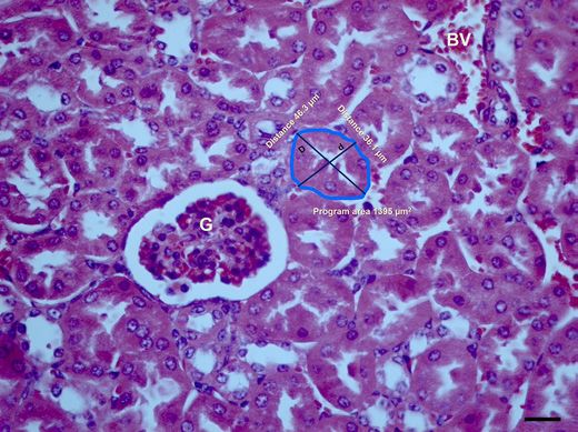
Fig. 1. Schematic representation of a PCT
(A) and a DCT (B) showing the morpho-
metric parameters analyzed. d, the short di-
ameter, is defined as the length of the short-
est straight line that passes through the
center of the tubule and connects 2 oppo-
site extreme points of the BM; D, the large
diameter, is defined as the length of the
largest straight line that passes through the
center of the tubule and connects 2 oppo-
site extreme points of the BM. The tubular
area, defined as the space bounded by the
BM (blue line), was measured in 4 ways.
The SCA was calculated by using the for-
mula for the area of a circle, A = π·r2, where
r is one-half of d. The LCA was calculated
by using the same formula, but replacing
the d value by D. The EA was obtained by
using the formula for the area of an ellipse,
A = π·d′·D′, where d′ and D′ are one-half of
d and D. The PA was provided by the image
analysis software used. PCT, proximal con-
voluted tubule; DCT, distal convoluted tu-
bule; BM, basement membrane; SCA, small
circular area; LCA, large circular area; EA,
elliptical area; PA, program area.

   In addition to the glomerulus, the proximal convolut-         [27], tubules could have another shape. The purpose of
ed tubule (PCT) and the distal convoluted tubule (DCT)           this work was to analyze if there are significant differ-
are the most widely analyzed structures in stereological         ences in the diameter and area of PCT and DCT, depend-
kidney research. In turn, the diameter and less frequently       ing on whether they are considered circular or elliptical
the area are the main measurements evaluated in the PCT          in the stereological analysis.
and DCT.
   However, there is no consensus in the literature on
how tubular diameter (or diameters) should be evaluated.            Materials and Methods
Several authors measure only one diameter, as if the tu-
bules were circular [3–10]. On the contrary, other authors          Animals and Experimental Design
consider that the tubules have an elliptical shape and re-          Animals used were described in previous papers, in
port the short diameter (minimum, proximal), some-               which stereological methods were employed to examine
times along with the large diameter (maximum, distal)            the lung [28, 29]. Male CD1 mice were maintained in stan-
[11–20]. Even more, other authors measure in the same            dard conditions in stainless steel cages (temperature: 18–
sample some tubules as circles and others as ellipses [21,       21°C; relative humidity: 55–60%; circadian rhythm: 12 h
22]. Besides, the calculation of the tubular area may de-        of artificial light per day, 12 h of darkness per day). The
pend on the way the diameter is evaluated [10, 15].              standard chow and water were available ad libitum. Three
   Other approaches to the study of the nephron tubules          animals were sacrificed at the age of 2 months by cervical
also do not consider whether they are circular or elliptical.    dislocation. Only the right-side kidneys were processed
Since the in vivo analysis of the function of the tubules is     and analyzed in order to have a more consistent control in
significantly difficult, mathematical models have been           the selection of samples. Kidneys were divided into 2 equal
created, but they only consider tubules as circular cylin-       halves longitudinally, fixed by immersion in 10% neutral
ders [23, 24]. Interestingly, fractal analysis of nephron tu-    buffered formalin, and embedded in paraffin with the
bules in paraffin-embedded sections stained with routine         samples oriented with the flat surface parallel to the sur-
histological methods has revealed that they are, in fact,        face of the paraffin block to be sectioned. Of note, al-
fractals [25, 26]. Since a circle is a nonfractal structure      though some authors who analyzed the renal tubules mor-

230                     Kidney Blood Press Res 2021;46:229–235                       Ortega-Martinez et al.
                        DOI: 10.1159/000515051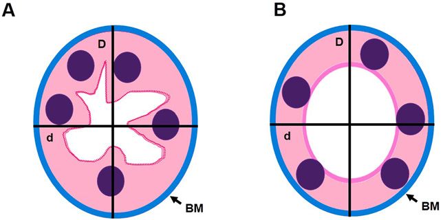
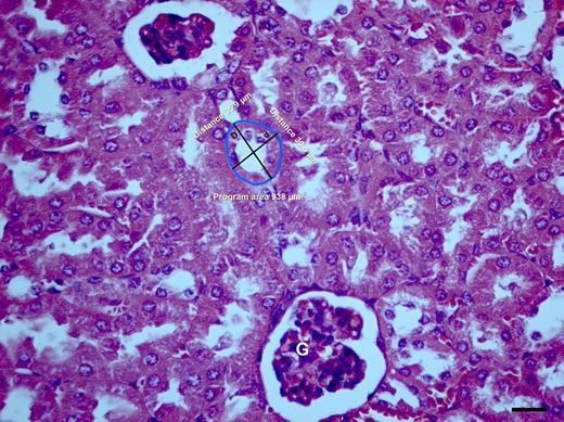
Fig. 2. Morphometric analysis of a PCT. Representative kidney tis-     Fig. 3. Morphometric analysis of a DCT. Representative kidney
sue section from a CD1 mouse stained with hematoxylin and eo-          tissue section from a CD1 mouse stained with hematoxylin and
sin. The short (d) and large (D) diameters are exemplified. The area   eosin. The short (d) and large (D) diameters are exemplified. The
was the space delimited by the blue line. The PA is exemplified.       area was the space delimited by the blue line. The PA is exempli-
The other 3 areas, SCA, LCA, and EA, were obtained with math-          fied. The other 3 areas, SCA, LCA, and EA, were obtained with
ematical formulas using the values of d and D. Bar = 20 μm. G,         mathematical formulas using the values of d and D. Bar = 20 μm.
glomerulus; BV, blood vessel; PCT, proximal convoluted tubule;         G, glomerulus; DCT, distal convoluted tubule; SCA, small circular
SCA, small circular area; LCA, large circular area; EA, elliptical     area; LCA, large circular area; EA, elliptical area; PA, program area.
area; PA, program area.

phometrically fixed the kidneys by perfusion before inclu-             GmbH). All the sections were examined on coded slides
sion in paraffin [4, 11, 22], most used the immersion                  by a single observer.
method for this purpose [6, 8–10, 12–21]. Then, 5-μm                       The parameters analyzed in both PCT and DCT are il-
thick sections were cut, deparaffinized in xylene, hydrated            lustrated in Figures 1–3. The short diameter (d) was de-
in a graded series of alcohol, and stained with hematoxylin            fined as the length of the shortest straight line that passes
and eosin (H&E) according to standard techniques.                      through the center of the tubule and connects 2 opposite
   Analysis was conducted using 3 tissue sections per an-              extreme points of the BM. The large diameter (D) was
imal. In each slide, 20 PCTs and 20 DCTs were examined                 defined as the length of the largest straight line that pass-
in randomly selected microscopic fields at ×400 magnifi-               es through the center of the tubule and connects 2 oppo-
cation. Thus, a total of 120 tubular structures were ana-              site extreme points of the BM. To measure both diame-
lyzed per animal. It is noteworthy that in the literature,             ters, lines d and D were manually drawn on the captured
some authors do not specify the total number of tubules                images described above, and the length of the lines was
examined per subject [5, 6, 8, 9, 11, 20]. When specified,             provided by the software.
most authors report measurements in 100 tubules or few-                    The tubular area was defined as the space bounded by
er [3, 4, 10, 12–18, 21, 22]. Only transverse tubules with             the BM. The small circular area (SCA) was calculated by
complete basement membrane (BM) were evaluated; lon-                   using the formula for the area of a circle, A = π·r2, where
gitudinal tubules were not considered.                                 r is one-half of d. The large circular area (LCA) was cal-
                                                                       culated by using the same formula, but replacing the d
    Morphometry                                                        value by D.
    Sections were examined with a light microscope (Pri-                   The elliptical area (EA) was obtained by using the for-
mo Star; Carl Zeiss Microscopy GmbH, Oberkochen,                       mula for the area of an ellipse, A = π·d′·D′, where d′ and D′
Germany), and high-resolution color images were digi-                  are one-half of d and D. Finally, the program area (PA) was
talized onto a computer screen using a AxioCam ICc1                    obtained using the software mentioned above. To measure
camera (Carl Zeiss Microscopy GmbH) linked to image                    it, a line was drawn manually through the entire tubular
analysis software (Zen lite 2011; Carl Zeiss Microscopy                perimeter, and the area was provided by the software.

Shape of Convoluted Tubules of Nephron                                 Kidney Blood Press Res 2021;46:229–235                            231
                                                                       DOI: 10.1159/000515051
Table 1. Diameters (µm) of the
convoluted tubules Short diameter Large diameter p values
Proximal convoluted tubules 37.4±0.5 44.0±0.7 0.000
Distal convoluted tubules 32.2±0.6 40.2±0.9 0.000
Values are reported as mean±1 standard error. Data were analyzed by Student’s t test.
The results were considered statistically significant at p < 0.05.
Table 2. Areas (µm2) of the convoluted
tubules SCA LCA EA PA p values
Proximal convoluted tubules 1,112±31 1,543±47 1,300±33 1,097±25 0.000a, b
Distal convoluted tubules 829±29 1,313±69 1,027±35 976±20 0.000c
0.001d
0.016e
Values are reported as mean±1 standard error. Data were analyzed by a one-way ANO-
VA followed by a least significant difference test. SCA, small circular area; LCA, large cir-
cular area; EA, elliptical area; PA, program area. The results were considered statistically
significant at p < 0.05. a LCA versus SCA, EA, and PA. b EA versus SCA and PA. c LCA
versus SCA, EA, and PA. d EA versus SCA. e PA versus SCA.
Statistical Analysis 31, 1,300 ± 33, and 1,097 ± 25 μm2, respectively; all p val-
The results are presented as means ±1 standard error. ues of 0.000). Also, EA was significantly greater when
Student’s t test was used to determine statistical signifi- compared to SCA and PA (p values of 0.000).
cance between the tubular diameters (d and D). Compar- The LSD test revealed that in the DCT (Fig. 5), LCA
isons between the tubular areas (SCA, LCA, EA, and PA) (1,313 ± 69 μm2) was significantly greater than SCA, EA,
were made by a one-way ANOVA, followed by post hoc and PA (829 ± 29, 1,027 ± 35, and 976 ± 20 μm2, respec-
analysis using the least significant difference (LSD) test. tively; all p values of 0.000). EA was significantly greater
The significance level for all tests was set at p < 0.05. The when compared to SCA (p value of 0.001). Finally, PA was
data were analyzed using the SPSS for Windows software significantly greater than SCA (p value of 0.016).
(SPSS, Inc., Chicago, IL, USA), release 21.0.
Discussion
Results
The diameter and the area of PCT and DCT are some
The results are summarized in Tables 1 and 2. Stu- of the main parameters analyzed in stereological studies
dent’s t test showed a significant difference between the of the kidney. The results obtained in this work indicate
diameters analyzed in both PCT (F = 1.354, Sig = 0.000) that the accuracy and reproducibility of those analyses
and DCT (F = 4.989, Sig = 0.000). In the PCT (Fig. 4), D may depend on how the shape of the tubules is consid-
(44.0 ± 0.7 μm) was significantly greater in comparison ered.
with d (37.4 ± 0.5 μm). Similarly, in the DCT (Fig. 4), D Several authors considered that the tubules have a cir-
(40.2 ± 0.9 μm) was significantly greater when compared cular shape and therefore measured only one diameter
to d (32.2 ± 0.6 μm). [3–10]. However, our results showed that the convoluted
The ANOVA test showed significant differences in the tubules of the kidney are elliptical in shape; we measured
tubular areas analyzed in both PCT (F = 34.843, Sig = a short diameter (the short axis of the ellipse) and a long
0.000) and DCT (F = 22.390, Sig = 0.000). The LSD test diameter (the long axis of the ellipse) which differed sig-
revealed that in the PCT (Fig. 5), LCA (1,543 ± 47 μm2) nificantly from each other (Table 1; Fig. 4). Therefore,
was significantly greater than SCA, EA, and PA (1,112 ± when analyzing the tubules in stereological studies of the
232 Kidney Blood Press Res 2021;46:229–235 Ortega-Martinez et al.
DOI: 10.1159/000515051Fig. 4. Diameter measurements of the PCT and the DCT. In both PCT (A) and DCT (B), the large diameter was
significantly greater when compared to the short diameter (*p values of 0.000). Values are expressed as means ±
1 standard error. Data were analyzed by Student’s t test. The results were considered statistically significant at
p < 0.05. PCT, proximal convoluted tubule; DCT, distal convoluted tubule.
Fig. 5. Comparison between the areas analyzed in the PCT and in 0.001). Finally, the PA was significantly greater than the SCA (‡p
the DCT. In the PCT (A), the LCA was significantly greater than value of 0.016). Values are expressed as means ± 1 standard error.
the SCA, the EA, and the PA (*all p values of 0.000). The EA was Data were analyzed by a one-way ANOVA followed by a least sig-
significantly greater when compared to the SCA and the PA (†p nificant difference test. The results were considered statistically
values of 0.000). In the DCT (B), the LCA was significantly greater significant at p < 0.05. PCT, proximal convoluted tubule; DCT,
than the SCA, the EA, and the PA (*all p values of 0.000). The EA distal convoluted tubule; SCA, small circular area; LCA, large cir-
was significantly greater when compared to the SCA (†p value of cular area; EA, elliptical area; PA, program area.
kidney, it should be considered that if only one diameter a study when the shape of the tubules is considered circu-
of the tubules is measured and a significant difference is lar because any axis of the ellipse, or both in a random
observed, it may be due to the fact that both axes of the way, could be measured in the replication study.
ellipse were actually measured, either randomly or alter- These problems may not arise if the tubules are con-
natively, and not to the treatment or phenomenon ana- sidered elliptical and the same axis (short or long) is eval-
lyzed per se. Furthermore, it can be difficult to reproduce uated in all tubules. Most authors report the value of the
Shape of Convoluted Tubules of Nephron Kidney Blood Press Res 2021;46:229–235 233
DOI: 10.1159/000515051short axis (short diameter) of the ellipse [12–14, 16–19], but rather elliptical, and care should be taken every time
while others report the value of both axes (short and long the tubules are analyzed in stereological studies of the
diameters) of the ellipse [11, 15, 20]. Furthermore, care kidney, especially when evaluating their diameters and
should be taken to always measure tubules as ellipses and areas.
not some tubules as ellipses and other tubules as circles in
the same sample [21, 22].
The tubules certainly have an elliptical shape. We were Statement of Ethics
able to calculate an EA with a formula that incorporates
the values of a short axis and a long axis, and the line used The present study was performed in accordance with
to measure the area with the software (PA) clearly delim- the principles and procedures defined in the National Re-
ited an ellipse (Fig. 2, 3). search Council Guide for the Care and Use of Laboratory
The SCA and the LCA differed significantly from each Animals (8th edition) and in the Mexican Guidelines
other in both PCT and DCT (Table 2; Fig. 5). SCA and ZOO-062. Experimental protocols were approved by the
LCA were calculated using the value of the short diameter Institutional Review Board and Ethics Committee of the
and the value of the large diameter of the circle, respec- School of Medicine of the Autonomous University of
tively, and this fact would explain the difference encoun- Nuevo Leon (No. PA19-00001).
tered. Therefore, observations regarding accuracy and re-
producibility described above for the circular diameters
could also be applied to the circular area analysis. Conflict of Interest Statement
Interestingly, significant differences were also ob-
served between other areas evaluated. In the PCT, LCA The authors declare that they have no conflicts of in-
was also significantly different from EA and PA, while EA terest.
was significantly different from SCA and PA (Table 2;
Fig. 5). In the DCT, LCA was also significantly different
from EA and PA, while EA and PA were significantly dif- Funding Sources
ferent from SCA (Table 2; Fig. 5). It is likely that the area
of a tubule may vary not only depending on whether it is This research received no external funding.
considered a circle or an ellipse (SCA and LCA vs. EA and
PA) but also if the area is evaluated directly with a piece
of software or with another strategy (PA vs. EA). While Author Contributions
the use of image analysis software to assess morphometric
data might be more exact in comparison with other strat- M.O.-M. and V.G.-D. performed experiments. G.J.-R.
egies, it should be remembered that it is the researcher planned experiments. M.O.-M. and E.G.-A. provided the
who ultimately provides the necessary information to the image analysis. G.J.-R., M.O.-M., and E.G.-A. wrote the
software for the correct execution of the measurements manuscript. R.M.C.-F. provided the statistical analysis of
[30]. In this case, the software would be of little use if the the data. G.J.-R. conceived and designed the study. A.N.-
shape of the tubules were considered circular instead of G. performed the analysis and interpretation of data. All
elliptical. authors read and approved the manuscript.
Finally, although recommendations have been made
for the morphometric analysis of the kidney [2], there is 1 Hamilton PW, Allen DC. Morphometry in
References
no consensus in the literature on how to perform it re- histopathology. J Pathol. 1995 Apr; 175(4):
garding several aspects, such as which method for the fix- 369–79.
2 Nyengaard JR. Stereologic methods and their
ation of the kidney is the correct one, the correct orienta- application in kidney research. J Am Soc
tion of the samples for histological analysis, the number Nephrol. 1999 May;10(5):1100–23.
of structures that should be evaluated, the optimal sam- 3 Hayslett JP, Kashgarian M, Epstein FH.
Changes in proximal and distal tubular reab-
pling strategy, and if the nephron tubules should be con- sorption produced by rapid expansion of ex-
sidered circular or elliptical. With this work, we try to tracellular fluid. J Clin Invest. 1967 Jul; 46(7):
clarify the last-mentioned aspect. In conclusion, the re- 1254–63.
4 Davis RG, Madsen KM, Fregly MJ, Tisher CC.
sults obtained in this work indicate that the convoluted Kidney structure in hypothyroidism. Am J
tubules of the nephron must not be considered circular, Pathol. 1983 Oct;113(1):41–9.
234 Kidney Blood Press Res 2021;46:229–235 Ortega-Martinez et al.
DOI: 10.1159/0005150515 Lebrecht D, Venhoff AC, Kirschner J, Wiech 14 Lane PH. Long-term furosemide treatment in 22 Kang J, Dai XS, Yu TB, Wen B, Yang ZW. Gly-
T, Venhoff N, Walker UA. Mitochondrial tu- the normal rat: dissociation of glomerular hy- cogen accumulation in renal tubules, a key
bulopathy in tenofovir disoproxil fumarate- pertrophy and glomerulosclerosis. Am J Kid- morphological change in the diabetic rat kid-
treated rats. J Acquir Immune Defic Syndr. ney Dis. 1999 Jun;33(6):1058–63. ney. Acta Diabetol. 2005 Jun;42(2):110–6.
2009 Jul;51(3):258–63. 15 Czekaj P, Pałasz A, Lebda-Wyborny T, 23 Praljak N, Ryan SD, Resnick A. Pulsatile flow
6 Saraga M, Vukojević K, Krželj V, Puretić Z, Nowaczyk-Dura G, Karczewska W, Florek E, through idealized renal tubules: fluid-struc-
Bočina I, Durdov MG, et al. Mechanism of et al. Morphological changes in lungs, placen- ture interaction and dynamic pathologies.
cystogenesis in nephrotic kidneys: a histo- ta, liver and kidneys of pregnant rats exposed Math Biosci Eng. 2019 Dec;17(2):1787–807.
pathological study. BMC Nephrol. 2014 Jan; to cigarette smoke. Int Arch Occup Environ 24 Weinstein AM. A mathematical model of the
15:3. Health. 2002 Oct;75 Suppl:S27–35. rat proximal tubule. Am J Physiol. 1986 May;
7 Hemmi S, Matsumoto N, Jike T, Obana Y, 16 Sagen JV, Bostad L, Njølstad PR, Søvik O. En- 250(5 Pt 2):F860–73.
Nakanishi Y, Soma M, et al. Proximal tubule larged nephrons and severe nondiabetic ne- 25 Nigro M, Viggiano D, Ragone V, Trabace T,
morphology in rats with renal congestion: a phropathy in hepatocyte nuclear factor-1β di Palma A, Rossini M, et al. A cross-section-
study involving the in vivo cryotechnique. (HNF-1β) mutation carriers. Kidney Int. al study on the relationship between hemato-
Med Mol Morphol. 2015 Jun;48(2):92–103. 2003 Sep;64(3):793–800. logical data and quantitative morphological
8 Amjad Z, Yasmin T, Ashraf I, Perveen K, Mir- 17 Okada K, Matsumoto K. Effect of dietary salt indices from kidney biopsies in different glo-
za T, Shoro AA. Lead-induced morphometric restriction on tubular hypertrophy in rats merular diseases. BMC Nephrol. 2018 Mar;
changes in the kidneys of albino rats amelio- with early-stage chronic renal failure. Scand J 19(1):62.
rated by ginkgo biloba extract (EGb 761). J Urol Nephrol. 2004;38(4):326–31. 26 Gil J, Gimeno M, Laborda J, Nuviala J, Be-
Pak Med Assoc. 2017 Jan;67(1):58–65. 18 Okada K, Matsumoto K, Takahashi S. Oral lanche I. Tangential algorithm for calculation
9 Momeni HR, Eskandari N. Effect of curcumin adsorbent prevents reduction of anionic sites of the fractal dimension of kidney tubuli sec-
on kidney histopathological changes, lipid of the glomerular basement membrane in dia- tions. Int. J. Morphol.. 2006;24(1):31–4.
peroxidation and total antioxidant capacity of betic nephropathy. Nephron Exp Nephrol. 27 Karolj A, Wu HT, Radisic M. A healthy dose
serum in sodium arsenite-treated mice. Exp 2005;99(2):e56–62. of chaos: using fractal frameworks for engi-
Toxicol Pathol. 2017 Feb;69(2):93–7. 19 Leh S, Hultström M, Rosenberger C, Iversen neering higher-fidelity biomedical systems.
10 Truter D, Chellan N, Strijdom H, Webster I, BM. Afferent arteriolopathy and glomerular Biomaterials. 2019 Oct;219:119363.
Rawstorne J, Kotzé SH. Histomorphological collapse but not segmental sclerosis induce 28 Ortega-Martínez M, Rodríguez-Flores LE,
changes in the pancreas and kidney and his- tubular atrophy in old spontaneously hyper- Ancer-Arellano A, Cerda-Flores RM, de-la-
topathological changes in the liver in male tensive rats. Virchows Arch. 2011 Jul;459(1): Garza-González C, Ancer-Rodríguez J, et al.
Wistar rats on antiretroviral therapy and mel- 99–108. Analysis of cell turnover in the bronchiolar
atonin treatment. Acta Histochem. 2018 May; 20 Alkharfy KM, Ahmed M, Yakout SM, Al- epithelium through the normal aging process.
120(4):347–55. Daghri NM. Effects of calcitriol on structural Lung. 2016 Aug;194(4):581–7.
11 Welling LW, Welling DJ. Shape of epithelial changes of kidney in C57BL/6J mouse model. 29 Ortega-Martínez M, Gutiérrez-Dávila V, Ni-
cells and intercellular channels in the rabbit Int J Clin Exp Med. 2015 Aug;8(8):12390–6. derhauser-García A, Cerda-Flores RM, Gar-
proximal nephron. Kidney Int. 1976 May; 21 Rangan GK, Wang Y, Tay YC, Harris DC. In- cía-Juárez J, de-la-Garza-González C, et al.
9(5):385–94. hibition of nuclear factor-kappaB activation Morphometric analysis of the non-epithelial
12 Okada K, Takahashi S. Measurement of prox- reduces cortical tubulointerstitial injury in areas of mouse bronchioles through the nor-
imal tubular diameter for evaluation of tubu- proteinuric rats. Kidney Int. 1999 Jul; 56(1): mal aging process. Am J Transl Res. 2019 Jun;
lar hypertrophy. Nephron. 1995;70(1):122. 118–34. 11(6):3637–44.
13 Fujimaki M, Nagase M, Uchida S. Long-term 30 Mandarim-de-Lacerda CA, Del-Sol M. Tips
effect of manidipine on renal function and for studies with quantitative morphology
structure in uninephrectomized spontane- (morphometry and stereology). Int. J. Mor-
ously hypertensive rats. Clin Exp Pharmacol phol.. 2017;35(4):1482–94.
Physiol. 1997 Jul;24(7):506–12.
Shape of Convoluted Tubules of Nephron Kidney Blood Press Res 2021;46:229–235 235
DOI: 10.1159/000515051You can also read