Successful delivery of subretinal aflibercept (new surgical technique) for the treatment of submacular hemorrhage in idiopathic polypoidal ...
←
→
Page content transcription
If your browser does not render page correctly, please read the page content below
Journal of Surgical Case Reports, 2021;8, 1–4

                                                                             https://doi.org/10.1093/jscr/rjab358
                                                                             Surgical Technique

SURGICAL TECHNIQUE

Successful delivery of subretinal aflibercept (new

                                                                                                                                                         Downloaded from https://academic.oup.com/jscr/article/2021/8/rjab358/6353045 by guest on 07 November 2021
surgical technique) for the treatment of submacular
hemorrhage in idiopathic polypoidal choroidal
vasculopathy
K.V. Chalam* and Suzie Gasparian
Department of Ophthalmology, Loma Linda University Medical School, Loma Linda, CA, USA
*Correspondence address. Department of Ophthalmology, Loma Linda University School of Medicine, 11370 Anderson Street, ste 2025, Loma Linda, CA
92354, USA. Tel: +(904) 244-9361; Fax: +(904) 244-9391; E-mail: kchalam@llu.edu

Abstract
Submacular hemorrhage (SMH) is often a result of trauma, wet age-related macular degeneration or IPCV and frequently
leads to blindness secondary to extreme toxicity of hemoglobin products on photoreceptors. We describe a new technique
of subretinal af libercept injection during pars plana vitrectomy for the treatment of SMH in idiopathic polypoidal choroidal
vasculopathy (IPCV). A 55-old male presented with sudden loss of vision (HM) secondary to massive subretinal hemorrhage
associated with IPCV. Subretinal injection of af libercept with a 25 g/42 g cannula coupled to the viscous f luid control unit of a
standard vitrectomy system was performed during parsplana vitrectomy. Controlled injection of af libercept intra-operatively
has resulted in a resolution of SMH (confirmed with OCT and ICG). Visual acuity improved from HM to 20/20. This combined
approach delivered anti-VEGF agent to target tissue in controlled fashion with the assistance of VFC system (similar to gene
therapy) and restored full vision with resolution of SMH.

INTRODUCTION
                                                                             into subretinal space with the aid of a 25 g/42 g cannula coupled
Submacular hemorrhage (SMH) is a major vision-threatening                    to the viscous fluid control (VFC) unit of a standard vitrectomy
complication of a variety of retinal diseases, including wet age-            system. We believe that the delivery of biologic agent (afliber-
related macular degeneration (AMD) and Idiopathic polypoidal                 cept) into subretinal space resolved the underlying pathology
choroidal vasculopathy (IPCV) [1–3]. Subretinal blood induces                and limited the toxic effect of hemoglobin products on photore-
irreversible functional and anatomical damage to photorecep-                 ceptors through dilution of hemorrhage.
tors (from iron toxicity) and often leads to blindness [4].
    Treatment of SMH remains a difficult challenge [5]. Currently,
combination of vitrectomy with intra-vitreal or subretinal t-PA
                                                                             CASE REPORT
with or without gas and intravitreal anti-VEGF therapy is utilized           A 55-year-old Caucasian male presented with sudden-onset of
[1–3, 5, 6]. Alternatively, less invasive treatment options include          loss of vision in his left eye. Best-corrected visual acuity (BCVA)
intravitreal injections of gas, t-PA and/or anti-VEGF agents [2, 7].         was 20/30 in the right eye (OD) and hand movements (HM) in the
    In this report, we describe a new surgical technique for effi-           left eye (OS). Slit-lamp examination revealed nuclear sclerotic
cient and controlled delivery of an anti-VEGF agent (aflibercept)            cataracts in both eyes.

Received: June 18, 2021. Accepted: July 27, 2021
Published by Oxford University Press and JSCR Publishing Ltd. © The Author(s) 2021.
This is an Open Access article distributed under the terms of the Creative Commons Attribution License (https://creativecommons.org/licenses/by/4.0/),
which permits unrestricted reuse, distribution, and reproduction in any medium, provided the original work is properly cited.
                                                                                                                                                1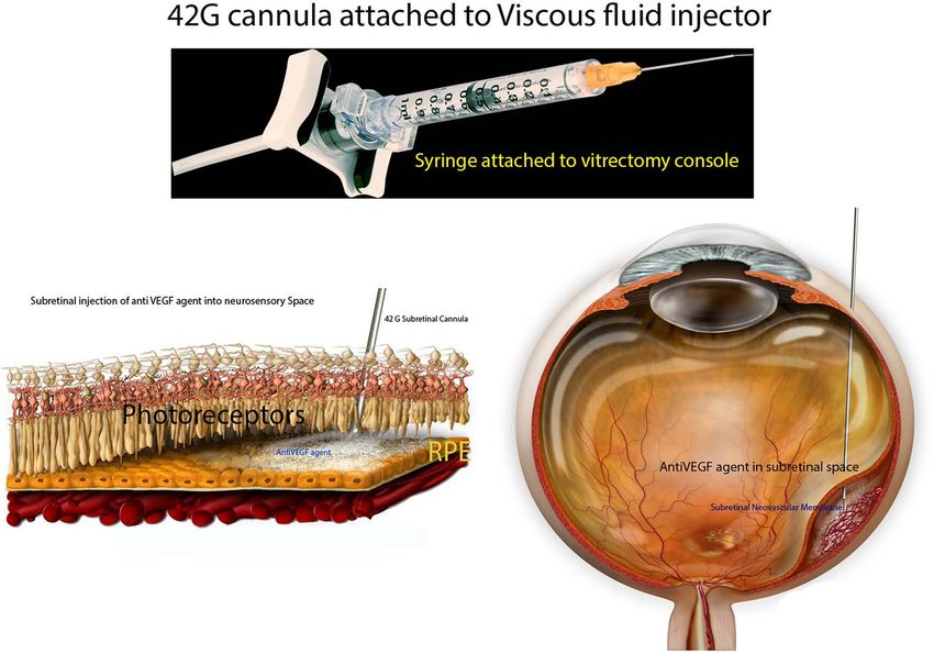
2      K.V. Chalam and S. Gasparian

                                                                                                                                                                         Downloaded from https://academic.oup.com/jscr/article/2021/8/rjab358/6353045 by guest on 07 November 2021
Figure 1: Preparation of the subretinal agent delivery system. (A)Prepared 1-mL syringe coupled with a 42 g/25 g cannula connected to the VFC unit of the vitrectomy
device. (B) Histologic schematic of delivery of anti VEGF agent into subretinal space. (C) Schematic of delivery of anti-VEGF agent during vitreoretinal surgery.

    Fundus examination of the left eye revealed a large dense
subretinal hemorrhage, ∼12 disc-diameters in size, covering the
entire posterior pole with evidence of fresh subretinal hem-
orrhage along with multiple polypoidal lesions (Fig. 2A). Fun-
doscopy of the right eye was normal. Optical Coherence Tomog-
raphy (OCT; Heidelberg Engineering Inc., Heidelberg, Germany)
of the right eye was normal. It could not be performed in left eye
due to large subretinal hematoma.

Surgical technique
About 0.05 cc (2 mg; Eylea; Bayer, Basel, Switzerland) of filterd
aflibercept was placed in a custom 1 cc syringe (Medone; Tampa,
FL) prior to connection to the VFC unit of a standard vitrectomy
(Constellation; Alcon Laboratories, Inc., Fort Worth, TX) system;
25 g/42 g cannula was attached to the 1-mL syringe of the VFC
unit (Fig. 1).
    Standard 3-port, 23-gauge pars plana vitrectomy was then
performed. After completion of core vitrectomy and induction                         Figure 2:   Demonstration of successful resolution of SMH in IPCV by Optos
of posterior vitreous detachment, the superotemporal aspect of                       ultra-wide field fundus photography (Optos, Marlborough, MA) and Heidelberg
                                                                                     Spectralis OCT (Heidelberg Engineering Inc., Heidelberg, Germany). (A) Fundus
the macula (about 1 disc-diameter superior to the superior edge
                                                                                     photograph at initial visit demonstrating a massive dense subretinal hemorrhage
of the subretinal hemorrhage) was penetrated with the 25 g/42 g                      covering entire posterior pole of right eye. (B) Fundus photograph 3 months
cannula (Fig. 1C). Cannula insertion is facilitated by engaging                      after pars plana vitrectomy with delivery of subretinal aflibercept demonstrating
the retina at a beveled 45–60◦ angle. VFC was activated with                         complete resolution of SMH. (C) Swept source OCT of the macula 1 month post-
the foot pedal (10 PSI) and aflibercept was delivered into the                       operatively demonstrating return of near-normal macular contour with polyps
subretinal space (with the aid of intraoperative OCT); dilution of                   associated with idiopathic polypoidal choroidopathy.

subretinal hemorrhage was noted. Complete gas-fluid exchange
was performed and sclerotomies were closed.

                                                                                     persistent polypoidal lesions in the macula during the early
Post-operative course
                                                                                     phases of the angiogram (Fig. 3C). Complete resolution of the
Complete resolution of subretinal hemorrhage along with                              hemorrhagic manifestations attributable to the polypoidal
anatomical improvement was confirmed by ultra-wide field                             lesions and scarring of few polyps was noted (Fig. 2B). OCT
fundus photography and wide field OCT imaging (Fig. 2C and D).                       angiography (OCT-A) demonstrated active and inactive polyps
BCVA was 20/200 on first post-operative day and improved                             with scarring along with branching vascular network (BVN) on
to 20/20 at 3 month follow-up visit. FA and ICGA revealed                            corresponding B-scan (Fig. 4).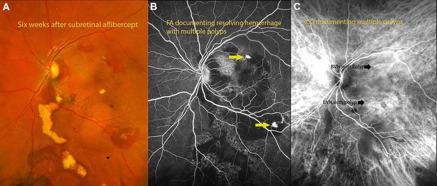
Successful delivery of subretinal af libercept (new surgical technique) 3
Downloaded from https://academic.oup.com/jscr/article/2021/8/rjab358/6353045 by guest on 07 November 2021
Figure 3: (A) fundus photograph 6 weeks after surgery documenting resolving hemorrhage with residual exudates from polyps. (B) Flourescein angiography documenting
polyps with residual hemorrhage. (C) Indocyanin angiography demonstrating areas of polyps with BVNs in the areas of polyps.
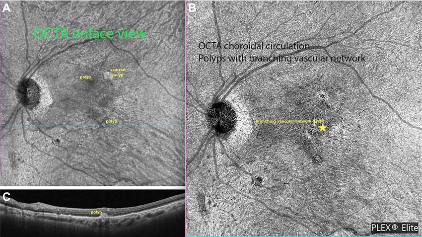
Figure 4: Ziess Plexelite OCT angiography images of IPCV before and after intervention. (A) OCT angiography enface image of a polyp with subretinal hemorrhage before
surgery. (B) OCT angiography enface image of resolved hemorrhage with regressed polyp.
DISCUSSION We modified previously reported surgical procedures and
combined pars plana vitrectomy with delivery of aflibercept into
SMH is a relatively common and challenging complication of
the submacular space (site of pathology) in efficient and con-
IPCV with poor prognosis [3, 8]. We report the first case of suc-
trolled manner with the aid of VFC system to control neovascular
cessful displacement and resolution of SMH using a combined
process as well as dilute subretinal blood. Unlike with intra-
vitrectomy and subretinal anti-VEGF delivery technique in IPCV.
vitreal administration (where drug has to penetrate retina to
Subretinal hemorrhage typically arises from the choroidal reach subretinal space), high concentration of anti VEGF agent is
or retinal circulation and is an accumulation of blood between delivered to target tissue with this modified approach. VFC unit
the neurosensory retina and RPE. Multiple techniques for SMH of the vitrectomy system provided a controlled, efficacious anti-
displacement have been proposed, but data pertaining to man- VEGF delivery into the submacular space, similar to techniques
agement of subretinal hemorrhage in IPCV remains limited. employed in gene therapy protocols. Aflibercept has proven
Intra-vitreal or subretinal t-PA is employed in the management successful in IPCV for management of recurrence of polyps. Once
of subretinal hematoma in conjunction with intravitreal anti- bleeding occurs in IPCV, re-bleeds are more likely to occur within
VEGF agents (many in the setting of neovascular AMD) in var- a short period [10]. Our patient had no recurrence of hemorrhage
ious studies [1–3, 5, 7–10]. One study reported successful SMH during follow-up.
displacement with subretinal application of t-PA followed by In view of extreme iron toxicity to photoreceptors, timing
intra-vitreal injection in neovascular AMD in 35 of 41 eyes [6]. is a crucial factor in adequate displacement of SMH. Our
Kitahashi et al. compared pneumatic displacement (PD) with patients’ visual acuity considerably improved with prompt
intravitreal bevacizumab to PD alone for the treatment of SMH surgical intervention, which further highlights the association
in IPCV and found complete displacement from under the fovea between duration of SMH displacement and visual outcomes.
in 86.4% in PD with intra-vitreal bevacizumab versus 50% in the Independent factors affecting BCVA 6 months after treatment
PD only group [9]. included pre-treatment BCVA, pre-treatment central ellipsoid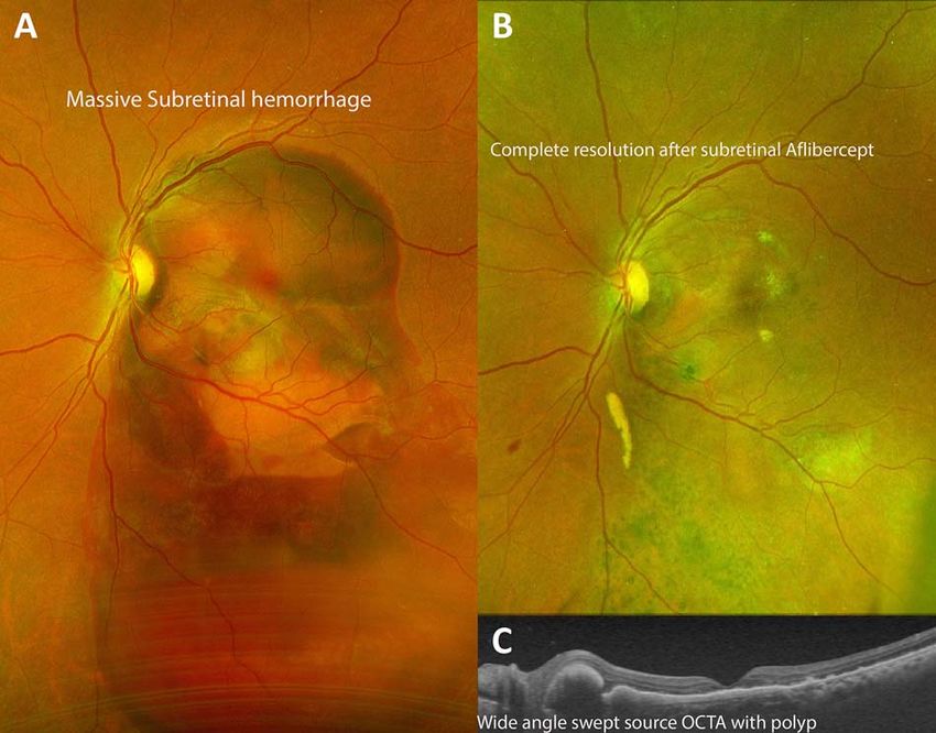
4     K.V. Chalam and S. Gasparian

zone, pre-treatment central PED thickness and post-treatment           3. Kimura S, Morizane Y, Hosokawa MM, Shiode Y, Doi
central PED thickness [9], which further substantiates our                S, Hosogi M, et al. Outcomes of vitrectomy combined
hypothesis.                                                               with subretinal tissue plasminogen activator injection
    Our result suggest that vitrectomy with subretinal injection          for submacular hemorrhage associated with polypoidal
of aflibercept is a safe and effective treatment option for resolu-       choroidal vasculopathy. Jpn J Ophthalmol 2019;63:
tion of SMH associated with IPCV.                                         382–8.
    In conclusion, we report a new, safe, effective and affordable     4. Glatt H, Machemer R. Experimental subretinal hemorrhage
method of subretinal delivery of aflibercept with a 25 g/42 g             in rabbits. Am J Ophthalmol 1982;94:762–73.
cannula coupled to the VFC unit of a standard vitrectomy sys-          5. Lu AQ, Prensky JG, Baker PS, Scott IU, Mahmoud TH,
tem in the management of SMH in IPCV. This novel surgical                 Todorich B. Update on medical and surgical management
approach facilitated successful delivery of therapeutic agent into        of submacular hemorrhage. Expert Rev Ophthalmol 2020;15:
subretinal space with complete resolution of the SMH and good             43–57.
recovery of visual acuity. We believe that this method may also        6. Treumer F, Roider J, Hillenkamp J. Long-term outcome of

                                                                                                                                          Downloaded from https://academic.oup.com/jscr/article/2021/8/rjab358/6353045 by guest on 07 November 2021
be used in instances of SMH in monocular patients with wet                subretinal coapplication of rtPA and bevacizumab followed
macular degeneration.                                                     by repeated intravitreal anti-VEGF injections for neovas-
                                                                          cular AMD with submacular haemorrhage. Br J Ophthalmol
                                                                          2012;96:708–13.
CONFLICT OF INTEREST STATEMENT                                         7. Hattenbach LO, Klais C, Koch FH, Gümbel HO. Intravitreous
None declared.                                                            injection of tissue plasminogen activator and gas in the
                                                                          treatment of submacular hemorrhage under various condi-
                                                                          tions. Ophthalmology 2001;108:1485–92.
FUNDING                                                                8. Sho K, Takahashi K, Yamada H, Wada M, Nagai Y, Otsuji T,
None.                                                                     et al. Polypoidal choroidal vasculopathy: incidence, demo-
                                                                          graphic features, and clinical characteristics. Arch Ophthal-
                                                                          mol 2003;121:1392–6.
REFERENCES                                                             9. Kitahashi M, Sakurai M, Yokouchi H, Kubota-Taniai M, Mita-
 1. Juncal VR, Hanout M, Altomare F, Chow DR, Giavedoni LR,               mura Y, Yamamoto S, et al. Pneumatic displacement with
    Muni RH, et al. Surgical management of submacular hem-                intravitreal bevacizumab for massive submacular hemor-
    orrhage: experience at an academic Canadian centre. Can J             rhage due to polypoidal choroidal vasculopathy. Clin Ophthal-
    Ophthalmol 2018;53:408–14.                                            mol 2014;8:485–92.
 2. Chen CY, Hooper C, Chiu D, Chamberlain M, Karia N, Heriot         10. Sakamoto T, Sheu SJ, Arimura N, Sameshima S, Shimura
    WJ. Management of submacular hemorrhage with intravit-                M, Uemura A, et al. Vitrectomy for exudative age-related
    real injection of tissue plasminogen activator and expansile          macular degeneration with vitreous hemorrhage. Retina
    gas. Retina 2007;27:321–8.                                            2010;30:856–64.
You can also read


