Strong and widespread cycloheximide resistance in Stichococcus like eukaryotic algal taxa - Nature
←
→
Page content transcription
If your browser does not render page correctly, please read the page content below
www.nature.com/scientificreports
OPEN Strong and widespread
cycloheximide resistance
in Stichococcus‑like eukaryotic algal
taxa
Nur Hidayu Syuhada1, Faradina Merican1,2*, Syazana Zaki1, Paul A. Broady3,
Peter Convey4,5 & Narongrit Muangmai6,7
This study was initiated following the serendipitous discovery of a unialgal culture of a Stichococcus-
like green alga (Chlorophyta) newly isolated from soil collected on Signy Island (maritime Antarctica)
in growth medium supplemented with 100 µg/mL cycloheximide (CHX, a widely used antibiotic active
against most eukaryotes). In order to test the generality of CHX resistance in taxa originally identified
as members of Stichococcus (the detailed taxonomic relationships within this group of algae have
been updated since our study took place), six strains were studied: two strains isolated from recent
substrate collections from Signy Island (maritime Antarctica) (“Antarctica” 1 and “Antarctica” 2), one
isolated from this island about 50 years ago (“Antarctica” 3) and single Arctic (“Arctic”), temperate
(“Temperate”) and tropical (“Tropical”) strains. The sensitivity of each strain towards CHX was
compared by determining the minimum inhibitory concentration (MIC), and growth rate and lag time
when exposed to different CHX concentrations. All strains except “Temperate” were highly resistant
to CHX (MIC > 1000 µg/mL), while “Temperate” was resistant to 62.5 µg/mL (a concentration still
considerably greater than any previously reported for algae). All highly resistant strains showed no
significant differences in growth rate between control and treatment (1000 µg/mL CHX) conditions.
Morphological examination suggested that four strains were consistent with the description of the
species Stichococcus bacillaris while the remaining two conformed to S. mirabilis. However, based
on sequence analyses and the recently available phylogeny, only one strain, “Temperate”, was
confirmed to be S. bacillaris, while “Tropical” represents the newly erected genus Tetratostichococcus,
“Antarctica 1” Tritostichococcus, and “Antarctica 2”, “Antarctica 3” and “Arctic” Deuterostichococcus.
Both phylogenetic and CHX sensitivity analyses suggest that CHX resistance is potentially widespread
within this group of algae.
Resistance to antibiotic agents is an ancient and widespread phenomenon in the natural e nvironment1. Its
evolution is stimulated by the selection pressure of sharing an ecological niche with an antibiotic-producing
organism2. An organism is considered naturally resistant to an antibiotic agent when it has developed a mecha-
nism to mitigate the toxic effects of that agent and continues to function in its p resence3. Organisms that were
originally susceptible to an agent may also later acquire resistance through mechanisms such as chromosomal
mutation or by acquisition from external genetic elements obtained from naturally resistant organisms present
in the e nvironment3.
Cycloheximide (CHX) is an antibiotic originally discovered in studies of the bacterium Streptomyces griseus4,
where it was found to be effective in killing fungal pathogens at a concentration as low as 0.2 µg/mL but pos-
sessed little or no antibiotic activity against bacteria4. CHX inhibits the growth of most eukaryotes by interfer-
ing with 80S ribosomes during protein s ynthesis5. Studies using cultures have confirmed the antibiotic activity
of CHX against a range of eukaryotic algae and fungi and that it has little or no effect on prokaryotes6–9. CHX
1
School of Biological Sciences, Universiti Sains Malaysia, Gelugor, Penang, Malaysia. 2National Antarctic Research
Centre, University of Malaya, Kuala Lumpur, Malaysia. 3School of Biological Sciences, University of Canterbury,
Christchurch, New Zealand. 4British Antarctic Survey, Cambridge, UK. 5Department of Zoology, University of
Johannesburg, Johannesburg, South Africa. 6Department of Fishery Biology, Faculty of Fisheries, Kasetsart
University, Bangkok, Thailand. 7Graduate School of Integrated Sciences for Life, Hiroshima University, Hiroshima,
Japan. *email: faradina@usm.my
Scientific Reports | (2022) 12:1080 | https://doi.org/10.1038/s41598-022-05116-y 1
Vol.:(0123456789)www.nature.com/scientificreports/
Strains CHX concentration (µg/mL)
Stichococcus-like
“Antarctica 1” > 1000
“Antarctica 2” > 1000
“Antarctica 3” > 1000
“Arctic” > 1000
“Tropical” > 1000
“Temperate” 62.5
Controls
Chlorella 31.3
Coccomyxa 15.6
Table 1. Minimum inhibitory concentrations (MIC) of cycloheximide for each of six strains of Stichococcus-
like algae and two control green algal strains assessed by visual observation on Day 14 of growth in culture.
completely inhibited the growth of four of 10 species of Chlorophyta at concentrations as low as 1 µg/mL and
none were tolerant to more than 50 µg/mL8. At concentrations of 20 µg/mL or less, CHX inhibited the growth of
yellow-green algae and diatoms (Ochrophyta (= Heterokontophyta))8. Complete lysis of cells of Euglena gracilis
occurred within seven days in broth containing 100 µg/mL CHX9. These investigations have led to the standard
and widespread use of CHX at 20–200 µg/mL in bacterial and cyanobacterial cultures to eliminate eukaryotic
algae and fungi10–14 against which it is regarded as one of the most effective antibiotics9.
Resistance to CHX has now been reported in some groups of yeasts, where it has also been used as taxonomic
marker15. Cloning of the CHX resistance gene from a naturally resistant strain of yeast into a sensitive strain
has provided a convenient dominant vector marker for recombinant DNA t echnology16,17. Naturally occurring
resistance to CHX in yeasts is known in Saccharomyces (200 µg/mL) and Kluyveromyces (500 µg/mL)18. In con-
trast, CHX resistance has been recorded in only one wild-type alga, the unicellular rhodophyte Cyanidioschyzon
merolae, which was isolated from an acidic hot s pring19. This was resistant to only very low CHX concentration
(0.5 µg/mL) after an extended lag time in culture of up to 10 days.
Polar microalgae in terrestrial habitats can be exposed to harsh environmental conditions such as freezing
temperatures, low water availability, and continuous daylight during summer and darkness during w inter20.
Antarctic green algae have evolved both avoidance and protection/resistance strategies, as well as mechanisms for
repair of damage, which enable them to tolerate these extremes21,22. However, to our knowledge, research has yet
to address the adaptive strategies developed by polar microalgae in the presence of natural antibiotic compounds,
despite the presence in Antarctic soils of compounds that can inhibit growth of e ukaryotes23.
In an initial isolation of cyanobacteria from samples obtained from Signy Island, South Orkney Islands,
Antarctica, we observed growth of a small number of discrete colonies of green algae on multiple culture plates
of agarised BBM supplemented with 100 µg/mL CHX. Based on light microscopy examination of morphologi-
cal features, these were identified as a Stichococcus-like alga. This study set out to confirm the identity of these
algae and the presence of resistance to CHX, and to extend the number of strains studied in order to assess how
widespread resistance is within other representatives assigned at the time to this genus. The fitness of algae in
the presence of CHX was measured by assessing the Minimum Inhibitory Concentration (MIC), one of the
most commonly used methods of quantifying microbial fitness24. Growth rate and lag period of cultures in
different CHX concentrations were also measured and compared. As morphological variability in Stichococcus
is very low25–27, morphological assessment was combined with molecular phylogenetic analyses to confirm the
generic identity of the strains examined here. We note that, subsequent to this study being carried out, a new
molecular phylogenetic analysis has become available that has erected several new genera within the original
genus ‘Stichococcus’28.
Results
The minimum inhibitory concentration (MIC) of cycloheximide. Visible green growth was observed
at up to the maximum tested CHX concentration (1000 µg/mL) for all strains except “Temperate” (Table 1). The
control Chlorella and Coccomyxa strains were the least resistant. Visual observations were supported by growth
assessment as indicated by both chlorophyll fluorescence and expressed as cell density using regression analysis.
The scatterplots (Fig. S1) showed a strong positive linear relationship between chlorophyll fluorescence and cell
density in all tested strains, which was confirmed by Pearson’s correlation analysis (Table S1).
The MIC values of the susceptible strains (“Temperate”, Chlorella and Coccomyxa) were determined by the null
value of chlorophyll fluorescence. Figure 1 shows the mean cell density achieved by each studied strain exposed
to different CHX concentrations after incubation for 2 weeks. One-way ANOVA with post hoc Tukey’s pairwise
comparisons indicated no significant differences in the cell densities achieved between all CHX concentrations
including the control for strains “Antarctica 3” and “Antarctica 1”, indicating that exposure to CHX had no
negative effect on growth for these strains. The cell densities were significantly different between the controls
and the treatments for strains “Antarctica 2” and “Arctic” but no differences were detected between the different
concentrations of CHX. A similar trend was also seen in strain “Tropical”, except that 1000 µg/mL CHX had a
significantly greater negative effect on growth. No growth was recorded at or above 62.5 µg/mL CHX, 31.3 µg/
Scientific Reports | (2022) 12:1080 | https://doi.org/10.1038/s41598-022-05116-y 2
Vol:.(1234567890)www.nature.com/scientificreports/
Figure 1. Mean (± standard error) of cell density achieved by each of six strains of Stichococcus-like algae
and single strains of Chlorella and Coccomyxa in BBM liquid medium supplemented with different CHX
concentrations after two weeks’ incubation. Means sharing the same letter (a, b or c) are not significantly
different (Tukey’s HSD, p < 0.05).
Scientific Reports | (2022) 12:1080 | https://doi.org/10.1038/s41598-022-05116-y 3
Vol.:(0123456789)www.nature.com/scientificreports/
Figure 2. Mean (± standard error) of the growth rate of each of five strains of Stichococcus-like algae in control
(BBM) versus BBM + 1000 µg/mL CHX. Means sharing the same letter (a, b) are not significantly different
(Tukey’s HSD).
Figure 3. Mean (± standard error) of the lag time for each of five strains of Stichococcus-like algae to reach
constant growth rate in control BBM, BBM + 0.5 µg/mL CHX and BBM + 1000 µg/mL CHX. Means with
different letters (a, b, c) are significantly different (Tukey’s HSD, p < 0.05).
mL CHX and 15.6 µg/mL CHX in “Temperate”, Chlorella and Coccomyxa, respectively, consistent with the visual
MIC result.
Growth rate assessment. The growth rates achieved by each strain in BBM (control) and BBM + 1000 µg/
mL (treatment) CHX are presented in Fig. 2. One-way Welch’s ANOVA indicated there was a significant dif-
ference in the growth rates achieved between the strains. Based on Games-Howell post-hoc test, there was a
significantly greater growth rate in “Antarctica 1” compared with the “Tropical” strain control and treatment
growth rates (Table S2).
The times required to reach a constant growth rate by these strains were compared between BBM,
BBM + 0.5 µg/mL CHX and BBM + 1000 µg/mL CHX (Fig. 3). Both low and high CHX concentrations had
significant effects on the lag time of strain “Tropical”, but similar effects were not apparent in strains “Antarctica
1”, “Antarctica 2” and “Arctic”. Although post hoc testing identifies marginally non-significant differences in lag
time between control and treatments in “Antarctica 3” (p = 0.06), possibly suggesting greater initial resistance
to exposure to CHX, the overall shape of the response was visually very similar to that of strains “Antarctica 1”
and “Antarctica 2”.
Confirmation of generic assignment of Stichococcus‑like algal strains. Morphological evaluations
conducted on all six strains conform closely to the previous descriptions of Stichococcus Nageli 1849 (Fig. S2).
Scientific Reports | (2022) 12:1080 | https://doi.org/10.1038/s41598-022-05116-y 4
Vol:.(1234567890)www.nature.com/scientificreports/
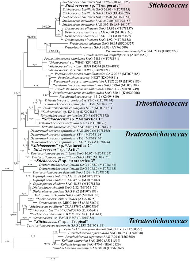
Figure 4. Maximum-likelihood tree based on concatenated 18S rDNA and ITS 2 sequences obtained in
this study (in boldface). Numbers next to branches indicate statistical support value (maximum likelihood
bootstrap/Bayesian posterior probabilities). Chlorella-like algal strains including Pseudochlorella, Koliella and
Edaphachlorella were used as the outgroup. Scale bar represents 0.2 changes per site.
Scientific Reports | (2022) 12:1080 | https://doi.org/10.1038/s41598-022-05116-y 5
Vol.:(0123456789)www.nature.com/scientificreports/
The sequence dataset consisted of concatenated 18S rDNA and ITS2 sequences (12,917 bp including gaps).
Both ML and BI analyses yielded identical topologies hence only the ML tree is presented here (Fig. 4). “Temper-
ate” was the only strain that can be confidently identified as S. bacillaris, located within a strongly supported clade
(99/0.99 for ML and BI, respectively) containing confirmed strains of S. bacillaris29. The other Stichococcus-like
strains were located within the newly erected c lades28 with “Antarctica 1” being within the Tritostichococcus
clade, “Antarctica 2”, “Antarctica 3” and “Arctic” clustered within the Deuterostichococcus clade and “Tropical”
being within the Tetratostichococcus clade.
Discussion
The data presented here conclusively demonstrate that multiple Stichococcus-like algal strains obtained across a
global range of locations are able to grow in the presence of the widely used eukaryotic growth inhibitor cyclohex-
imide. This is the first demonstration of chlorophytes being able to grow in the presence of this antibiotic. In
susceptibility testing, it is common practice to use the performance standards provided by the Clinical Laboratory
Standards Institute (CLSI)30 or European Committee on Antimicrobial Susceptibility Testing (EUCAST)31 as a
guideline for MIC breakpoints in order to determine whether a strain is resistant, intermediate or susceptible to
an antibiotic compound. However, the existing standards are limited to bacteria and fungi. From the standard
MIC list for unicellular fungi (yeasts), most strains have MIC less than 10 µg/mL. This value is far below the
MIC recorded in most microalgal strains in the present study (> 1000 µg/mL), and even considerably lower than
the less resistant “Temperate” strain (62.5 µg/mL). Previously8, eukaryotic algae have been recorded as being
completely inhibited at all concentrations of CHX.
In the present study strains “Antarctica 1”, “Antarctica 2”, “Antarctica 3”, “Arctic” and “Tropical” were highly
resistant to CHX and exhibited an MIC > 1000 µg/mL. Most of these strains showed statistically indistinguishable
growth rates, the only exception being between “Antarctica 1” treatment and “Tropical” control and treatment
conditions, with even then this difference being small. The presence of CHX resulted in an extended lag period
in each strain relative to their control, suggesting the cells were initially stressed by the treatment32. However,
based on the growth rate of each strain, normal growth was then resumed. This temporary inhibition of cell divi-
sion may be a form of physiological adaptation to the toxicants or due to death of susceptible c ells33. The growth
rates measured in the present study indicated that cells of all strains were able to grow normally once they had
adapted to CHX. The lack of significant differences in the lag times of cultures exposed to the lowest and highest
CHX concentrations in strains “Antarctica 1”, “Antarctica 2” and “Antarctica 3” suggested that growth was only
affected by presence of CHX, not the concentration. Only the “Temperate” strain had a lower MIC of 62.5 µg/mL.
Resistance to high CHX concentration was also observed in the “Tropical” strain, isolated from tropical rain-
forest. Although this strain required nine days to adapt to 1000 µg/mL CHX (Fig. 3), the subsequent growth rate
at this concentration was not significantly different from that of the control (Fig. 2). This response is analogous
to previous studies in which growth of Stichococcus cells was recorded only after 4–5 days’ exposure to multiple
herbicides32 or aluminum33.
Morphological combined with molecular genetic assessment of the six Stichococcus-like strains was able
to place only one of the studied strains, “Temperate”, definitively within a known species. Its characteristics
conformed to S. bacillaris Nägeli 1849. The species identity of the other five Stichococcus-like strains lie within
independent lineages that have now been separated in a newly-available molecular phylogenetics analysis from
the redefined Stichococcus clade28. The morphologically simple features that characterize Stichococcus-like strains
restrict reliable identification of species within the genus.
Soils are a reservoir for many natural antibiotic compounds34–37. In Antarctica there is evidence that soil
communities have yet to be exposed to the extensive microbial and chemical contamination that is now wide-
spread on other c ontinents38,39. There has not been extensive release of human manufactured antibiotics in the
region and, hence, the presence of antibiotics is unlikely to be a human artefact. Polar soils are characteristically
nutrient-limited40 and competition between microorganisms is likely to be an important stressor. It is therefore
plausible that there has been evolution of antagonistic activity against potential c ompetitors41–43. Accurate deter-
mination of antibiotic diversity and concentration in soil is difficult, especially when the compounds are made up
of complex mixtures of small molecules with different properties44. Exposure to natural antibiotics is considered
to be the major driver in the evolutionary selection for antibiotic resistance in the soil microbial community45–47.
Although no such instances have been reported in microalgae, an analogous response is known in microalgae
naturally exposed to heavy metals, which are another important environmental stressor33,48. The data presented
here provide strong evidence of the potential for natural evolution of antibiotic resistance in eukaryotic algae.
Antibiotic resistance genes (ARGs) function to protect an organism from the inhibitory or harmful effects
of an antibiotic produced by another organism. ARGs may evolve in nature in response to antibiotics produced
by neighboring cells or be acquired through horizontal gene transfer49. Antibiotic resistance genes found in
environments lacking human impact are potentially ancient genes that are transferred vertically from parent
to offspring, with limited or no horizontal transfer between s pecies37. This is supported by the present study
in which all four strains from the polar regions and the tropical strain showed the highest resistance to CHX.
This investigation has confirmed and extended our preliminary observation of CHX resistance in eukaryotic
Antarctic microalgae isolated from CHX-supplemented growth medium. Within the genera tested here, this
novel antibiotic resistance was restricted to strains of Stichococcus-like algae from globally widespread locations.
Methods
Sample collection. Six Stichococcus-like algal strains were isolated from samples originating from Antarc-
tica, the Arctic, temperate and tropical regions. Two control algae with susceptibility to low concentrations of
CHX, Chlorella and Coccomyxa, were isolated from Antarctica. Details of each strain are presented in Table 2.
Scientific Reports | (2022) 12:1080 | https://doi.org/10.1038/s41598-022-05116-y 6
Vol:.(1234567890)www.nature.com/scientificreports/
GenBank accession
number
Strain 18S ITS-2 Location Habitat Year of isolation Source
Signy Island, Antarctica 60°43″S,
“Antarctica 1” MN968057 MN968730 Soil 2016 Field material
45°37″W
Signy Island, Antarctica 60°43″S,
“Antarctica 2” MN968346 MN968731 Soil 2016 Field material
45°37″W
Signy Island, Antarctica 60°43″S, Culture Collection of Algae and
“Antarctica 3” MN968320 MN968556 Soil ~ 1972
45°37″W Protozoa (CCAP)
Prudhoe Bay, Alaska 70°19′32″N, Culture Collection of Algae at Uni-
“Arctic” MN968500 MN968719 – –
148°42′41″W versity of Texas (UTEX)
Bernburg, Germany 51°47′40″N, Culture Collection of Algae at Göt-
“Temperate” MN968499 MN968718 Melt water before 1936
11°44′24″E tingen University (SAG)
Penang Hill, Malaysia 5°25′28″N,
“Tropical” MN968502 MN968732 Corticolous on tree Fragea fragans 2017 Field material
100°16′08″E
Signy Island, Antarctica 60°43″S,
Chlorella – – Soil 2016 Field material
45°37″W
Signy Island, Antarctica 60°43″S,
Coccomyxa – – Soil 2016 Field material
45°37″W
Table 2. Original collection localities of the six Stichococcus-like algal strains and the two control algae used in
this study.
Cultures were established on 1% agarised full strength Bold’s Basal Medium (BBM)50,51. These were incubated
under a cool white fluorescent lamp (12:12 h light:dark cycle at 27 µmol/m2/s) at 18 °C for polar strains and 24 °C
for temperate and tropical strains.
General experimental procedures. Seven days prior to the growth inhibition assay, cells of each strain
were introduced to fresh BBM liquid medium to ensure that cultures were in exponential phase. CHX powder
was added directly to BBM. The mixture was filter sterilized using an 0.22 µm microfilter (HmbG, cat no. P0376).
Cultures of all strains were incubated at 24 °C under continuous light provided by white fluorescent lamps with
27 µmol/m2/s light intensity in a controlled temperature culture room.
Microalgal growth inhibition assay. Serial dilutions of CHX were prepared by two-fold dilutions follow-
icrodilution52, resulting in final concentrations of 1000, 500, 250, 125, 62.5, 31.3, 15.6,
ing the protocol of broth m
8, 4, 2, 1 and 0.5 µg/mL. Three replicates were prepared at each concentration. The assay was carried out for 15 d
using sterile, transparent, 96-well polystyrene microtitre plates (TPP, cat. no. 92096).
A week-old culture was adjusted to 1 × 10³ cells/mL and 100 µL was injected into three replicate wells at
each CHX concentration and three replicate wells of a positive control of BBM without CHX. Plates were sealed
using Parafilm® M in order to prevent evaporation and the plates were illuminated from above and below dur-
ing incubation.
The growth of cells in the control and at each CHX concentration was assessed using three different meth-
ods: visual assessment, chlorophyll fluorescence and cell counts. Chlorophyll fluorescence was measured daily.
Cultures were transferred from transparent to black 96-well microplates53. The cultures were mixed thoroughly
by repeatedly loading in the pipette tips before transferring. Chlorophyll fluorescence was measured using a
microplate reader (Tecan infinite M1000 PRO; Tecan Austria GmbH) with excitation wavelength of 485 nm and
emission wavelength of 680 nm53. The fluorescence data were expressed as relative fluorescence units (RFU). Cell
density was assessed only on the positive control using a Neubauer haemocytometer. Cells were counted from
the four corner squares of the chamber and density calculated using the given formula54. Growth in the wells
was visually assessed by the unaided eye on day 15.
The strength of the relationship between chlorophyll fluorescence and cell number was assessed using Pear-
son’s correlation. A regression analysis was conducted to determine cell density estimates derived from chloro-
phyll fluorescence of the treatments followed by one-way ANOVA. Differences were accepted as significant at
p < 0.05. Post hoc analyses were performed using Tukey’s test for multiple comparisons of means. All statistical
analyses were performed using SPSS.
Determinations were made of the minimum inhibitory concentration (MIC), population growth rate and lag
time. The visual assessment at day 15 was used to determine MIC. Population growth rates were evaluated for
strains that were resistant to up to 1000 µg/mL CHX. The daily relative fluorescence unit (RFU) measurements
were used to calculate the population growth rate, α, of each strain in all three replicates of both the control and
BBM + 1000 µg/mL CHX using the GrowthRates program55. This also estimated the duration of the lag phase by
extrapolating the slope of the exponential phase back to the initial RFU. The generated growth rates and lag time
of the control and treatments were compared using ANOVA. Differences were accepted as significant at p < 0.05.
Morphological examination using light microscopy. Morphological assessment was made of all
strains using an Olympus BX53 light microscope at 80–2000× magnification. Observations and measurements
were made of cell shape, cell dimensions, size of chloroplast, presence of pyrenoid, formation and fragmentation
Scientific Reports | (2022) 12:1080 | https://doi.org/10.1038/s41598-022-05116-y 7
Vol.:(0123456789)www.nature.com/scientificreports/
of filaments and cell division. Size measurements were made on 30 randomly chosen cells. Photomicrographs
were taken. Identification of morphospecies was carried out based on the relevant taxonomic literature56–58.
Molecular analyses. DNA was extracted using the Tiangen DNAsecure Plant Kit (Beijing) following the
manufacturer’s instructions. The extracted DNA was stored frozen at − 20 °C. The quality and purity of the
extracted DNA were determined using a Nanodrop Quawell UV Spectrophotometer Q3000. The 18S rDNA gene
and the internal transcribed spacer 2 (ITS-2) region were amplified using polymerase chain reaction (PCR) and
the combination of primers 20F (5′-GTA GTC ATA TGC TTG TCT C-3′) and 18L (5′-CAC CTA CGG AAA
CCT TGT TAC GAC TT-3′) for the 18S rRNA gene59 and primers ITS_f (5′-AGG AGA AGT CGT AAC AAG
GT-3′) and ITS_r (5′-TCCTCCGCTTATTGATATGC-3′) for the ITS-2 region60. This resulted in products of
approximately 1700 bp for the 18S rDNA and 300 bp for the ITS-2 region. The reaction mix comprised 2 μL of
extracted DNA used in 50 μL reactions containing 1 μL of each forward and reverse primer, 21 μL of ultrapure
water and 25 μL of MyTaq™ Red Mix, which is a pre-prepared mixture of buffer, dNTPs and Taq polymerase
(Bioline, United Kingdom).
PCR was carried out using a Bio-Rad Thermal Cycler with standard parameters. Thermal cycling conditions
to amplify the 18S region were set at 95 °C for 5 min for pre-denaturation, followed by 35 cycles of denaturation
at 94 °C for 1 min, 56 °C for 1 min, 72 °C for 3 min with a final extension at 72 °C for 10 min. For the ITS-2
region, the conditions used were 96 °C for 5 min for pre-denaturation, followed by 40 cycles of denaturation at
96 °C for 1 min, 56 °C for 1 min, 72 °C for 1 min with a final extension at 72 °C for 5 min. Once the reaction was
completed, the integrity of the PCR product was verified using a 2% agarose gel. Amplified DNA was purified
using the MEGAquick-spinTM Total Fragment DNA Purification Kit (iNtRON Biotechnology, Korea).
Phylogenetic tree analyses. All sequences were edited and assembled using the Geneious 11.0 software
package (Biomatters, http://www.geneious.com). Sequence alignments were prepared using the MUSCLE algo-
rithm in Geneious 11.0 and then manually checked by eye. The closest related sequences were identified from
GenBank using the Basic Local Alignment Search Tool (BLAST) a lgorithm61.
The alignment, which included the sequences newly obtained in this study together with additional sequences
of closely related species from GenBank, contained 73 sequences for the 18S rDNA analysis and 46 sequences for
the ITS-2 analysis. All new sequences generated in this study have been deposited in GenBank under accession
numbers listed in Table 2.
Phylogenetic analyses were conducted based on the concatenated 18S rDNA and ITS-2 dataset, using two
different methods: maximum likelihood (ML) and Bayesian inference (BI). Before carrying out these analyses,
the best-fit model of DNA substitution was determined using the program K akusan462. ML analyses were per-
formed with RaxML v763 in Geneious 11.0 using the general time-reversible invariant-sites (GTRI) nucleotide
substitution model with the default parameters. The bootstrap probability of each branch was calculated using
1000 replications. BI analyses were performed using the program MrBayes v3.1.264. Two independent analyses,
each consisting of four Markov chains, were run simultaneously for 3,000,000 generations, sampling every 100
generations. A burn-in of 25% of saved trees was removed, and the remaining trees were used to calculate the
Bayesian posterior probability values. ML and BI trees were edited with the program FigTree v1.3.165. Chlorella-
like strains were used as outgroup to root the tree.
Received: 5 April 2021; Accepted: 4 January 2022
References
1. D’Costa, V. M. et al. Antibiotic resistance is ancient. Nature 477, 457–461 (2011).
2. Kaur, P. & Peterson, E. Antibiotic resistance mechanisms in bacteria: Relationships between resistance determinants of antibiotic
producers, environmental bacteria, and clinical pathogens. Front. Microbiol. 9, 2928 (2018).
3. Munita, J. M. & Arias, C. A. Mechanisms of antibiotic resistance. Microbiol. Spectr. 4, 1–24 (2016).
4. Leach, B. E., Ford, J. H. & Whiffen, A. J. Actidione, an antibiotic from Streptomyces griseus. J. Am. Chem. Soc. 69, 474 (1947).
5. Schneider-Poetsch, T. et al. Inhibition of eukaryotic translation elongation by cycloheximide and lactimidomycin. Nat. Chem.
Biol. 6, 209–217 (2010).
6. Whiffen, A. J. The production, assay, and antibiotic activity of actidione, an antibiotic from Streptomyces griseus. J. Bacteriol. 56,
283 (1948).
7. Palmer, C. M. & Maloney, T. E. Preliminary screening for potential algicides. Ohio J. Sci. 55, 1–8 (1955).
8. Zehnder, A. & Hughes, E. O. The antialgal activity of actidione. Can. J. Microbiol. 4, 399–408 (1958).
9. Hunter, E. O. Jr. & McVeigh, I. The effects of selected antibiotics on pure cultures of algae. Am. J. Bot. 48, 179–185 (1961).
10. Vaara, T., Vaara, M. & Niemelä, S. Two improved methods for obtaining axenic cultures of cyanobacteria. Appl. Environ. Microbiol.
38, 1011–1014 (1979).
11. Castenholz, R. W. Culturing methods for cyanobacteria. Methods Enzymol. 167, 68–93 (1988).
12. Bolch, C. J. & Blackburn, S. I. Isolation and purification of Australian isolates of the toxic cyanobacterium Microcystis aeruginosa
Kützing. J. Appl. Phychol. 8, 5–13 (1996).
13. Choi, G. G., Bae, M. S., Ahn, C. Y. & Oh, H. M. Induction of axenic culture of Arthrospira (Spirulina) platensis based on antibiotic
sensitivity of contaminating bacteria. Biotechnol. Lett. 30, 87–92 (2008).
14. Katoh, H., Furukawa, J., Tomita-Yokotani, K. & Nishi, Y. Isolation and purification of an axenic diazotrophic drought-tolerant
cyanobacterium, Nostoc commune, from natural cyanobacterial crusts and its utilization for field research on soils polluted with
radioisotopes. Biochim. Biophys. Acta. 1817, 1499–1505 (2008).
15. Mutoh, E., Ohta, A. & Takagi, M. Studies on cycloheximide-sensitive and cycloheximide-resistant ribosomes in the yeast Candida
maltosa. Gene 224, 9–15 (1998).
16. Takagi, M., Kawai, S., Shibuya, I., Miyazaki, M. & Yano, K. Cloning in Saccharomyces cerevisiae of a cycloheximide resistance gene
from the Candida maltosa genome which modifies ribosomes. J. Bacteriol. 68, 417–419 (1986).
Scientific Reports | (2022) 12:1080 | https://doi.org/10.1038/s41598-022-05116-y 8
Vol:.(1234567890)www.nature.com/scientificreports/
17. Dehoux, P., Davies, J. & Cannon, M. Natural cycloheximide resistance in yeast: The role of ribosomal protein L41. Eur. J. Biochem.
213, 841–848 (1993).
18. Adoutte-Panvier, A. & Davies, J. E. Studies of ribosomes of yeast species: Susceptibility to inhibitors of protein synthesis in vivo
and in vitro. Mol. Gen. Genet. 194, 310–317 (1984).
19. Yagisawa, F. et al. Isolation of cycloheximide-resistant mutants of Cyanidioschyzon merolae. Cytologia 69, 97–100 (2004).
20. Thomas, D. N. et al. The Biology of Polar Regions 2nd edn. (Oxford University Press, 2008).
21. Hughes, K. A. Solar UV-B radiation, associated with ozone depletion, inhibits the Antarctic terrestrial microalga, Stichococcus
bacillaris. Polar Biol. 29, 327–336 (2006).
22. Karsten, U. & Holzinger, A. Green algae in alpine biological soil crust communities: Acclimation strategies against ultraviolet
radiation and dehydration. Biodivers. Conserv. 23, 1845–1858 (2014).
23. Shekh, R. M., Singh, P., Singh, S. M. & Roy, U. Antifungal activity of Arctic and Antarctic bacteria isolates. Polar Biol. 34, 139–143
(2011).
24. Wiser, M. J. & Lenski, R. E. A comparison of methods to measure fitness in Escherichia coli. PLoS ONE 10, 10 (2015).
25. Fritsch, F. E. The Structure and Reproduction of the Algae Vol. II (Cambridge University Press, 1959).
26. Fukushima, H. Studies on cryophytes in Japan. J. Yokohama Munic Univ. Ser. C 43, 1–146 (1963).
27. Hoham, R. W. Pleiomorphism in the snow alga, Raphidonema nivale Lagerh (Chlorophyta), and a revision of the genus Raphi-
donema Lagerh. Syesis 6, 255–263 (1973).
28. Pröschold, T. & Darienko, T. The green puzzle Stichococcus (Trebouxiophyceae, Chlorophyta): New generic and species concept
among this widely distributed genus. Phytotaxa 2, 113–142 (2020).
29. Hodač, L. et al. Widespread green algae Chlorella and Stichococcus exhibit polar-temperate and tropical-temperate biogeography.
FEMS Microbiol. Ecol. 92, 1–16 (2016).
30. Clinical and Laboratory Standards Institute. Performance Standards for Antimicrobial Susceptibility Testing: 27th Informational
Supplement (Clinical and Laboratory Standards Institute, 2017).
31. European Committee for Antimicrobial Susceptibility Testing (EUCAST) of the European Society of Clinical Microbiology and
Infectious Diseases (ESCMID). EUCAST discussion document E.Dis. 5.1. Determination of minimum inhibitory concentrations
(MIC’s) of antibacterial agents by broth dilution. Clin. Microbiol. Infect. 9, 1–7 (2003).
32. Bertrand, R. L. Lag phase is a dynamic, organized, adaptive, and evolvable period that prepares bacteria for cell division. J. Bacteriol.
201, e00697-e718 (2019).
33. Claesson, A. & Törnqvist, L. The toxicity of aluminium to two acido-tolerant green algae. Water Res. 22, 977–983 (1988).
34. Knapp, C. W. et al. Antibiotic resistance gene abundances correlate with metal and geochemical conditions in archived Scottish
soils. PLoS ONE 6, e27300 (2011).
35. Su, J. Q., Wei, B., Xu, C. Y., Qiao, M. & Zhu, Y. G. Functional metagenomic characterization of antibiotic resistance genes in agri-
cultural soils from China. Environ. Int. 65, 9–15 (2014).
36. Tomova, I., Stoilova-Disheva, M., Lazarkevich, I. & Vasileva-Tonkova, E. Antibiotic activity and resistance to heavy metals and
antibiotics of heterotrophic bacteria isolated from sediment and soil samples collected from two Antarctic islands. Front. Life Sci.
8, 348–357 (2015).
37. Van Goethem, M. W. et al. A reservoir of ‘historical’ antibiotic resistance genes in remote pristine Antarctic soils. Microbiome 6,
40 (2018).
38. Cowan, D. A. et al. Non-indigenous microorganisms in the Antarctic: Assessing the risks. Trends Microbiol. 19, 540–548 (2011).
39. Convey, P. et al. The spatial structure of Antarctic biodiversity. Ecol. Monogr. 84, 203–244 (2014).
40. Cowan, D. A., Makhalanyane, T. P., Dennis, P. G. & Hopkins, D. W. Microbial ecology and biogeochemistry of continental Antarctic
soils. Front. Microbiol. 5, 154 (2014).
41. Lo Giudice, A., Bruni, V. & Michaud, L. Characterization of Antarctic psychrotrophic bacteria with antibacterial activities against
terrestrial microorganisms. J. Basic Microbiol. 47, 496–505 (2007).
42. Bell, T., Callender, K., Whyte, L. & Greer, C. Microbial competition in polar soils: A review of an understudied but potentially
important control on productivity. Biology 2, 533–554 (2013).
43. Núñez-Montero, K. & Barrientos, L. Advances in Antarctic research for antibiotic discovery: A comprehensive narrative review of
bacteria from Antarctic environments as potential sources of novel antibiotic compounds against human pathogens and micro-
organisms of industrial importance. Antibiotics 7, 90 (2018).
44. Davies, J. Are antibiotics naturally antibiotics?. J. Ind. Microbiol. Biotechnol. 33, 496–499 (2006).
45. Levy, S. B. Antibiotic resistance: Consequences of inaction. Clin. Infect. Dis. 33, 124–129 (2001).
46. Marshall, B. M. & Levy, S. B. Food animals and antimicrobials: Impacts on human health. Clin. Microbiol. Rev. 24, 718–733 (2011).
47. Andersson, D. I. & Hughes, D. Evolution of antibiotic resistance at non-lethal drug concentrations. Drug Resist. Updates 15, 162–172
(2012).
48. Kuwabara, J. S. & Leland, H. V. Adaptation of Selenastrum capricornutum (Chlorophyceae) to copper. Environ. Toxicol. Chem. 5,
197–203 (1986).
49. Martínez, J. L., Coque, T. M. & Baquero, F. What is a resistance gene? Ranking risk in resistomes. Nat. Rev. Microbiol. 13, 116
(2015).
50. Bold, H. C. The morphology of Chlamydomonas chlamydogama sp. nov. Bull. Torrey Bot. Club 76, 101–108 (1949).
51. Bischoff, H. & Bold, H. C. Phycological studies IV. Some soil algae from enchanted rock and related algal species. Univ. Texas Publ.
6318, 95 (1963).
52. Balouiri, M., Sadiki, M. & Ibnsouda, S. K. Methods for in vitro evaluating antibiotic activity: A review. J. Pharm. Anal. 6, 71–79
(2016).
53. Zhao, Q. et al. Microalgal microscale model for microalgal growth inhibition evaluation of marine natural products. Sci. Rep. 8,
10541 (2018).
54. LeGresley, M. & McDermott, G. Counting chamber methods for quantitative phytoplankton analysis—Haemocytometer, Palmer-
Maloney cell and Sedgewick-Rafter cell. In Microscopic and Molecular Methods for Quantitative Phytoplankton Analysis (eds
Karlson, B. et al.) 25–30 (IOC Manuals and Guides. Intergovernmental Oceanographic Commission of UNESCO, 2010).
55. Hall, B. G., Acar, H., Nandipati, A. & Barlow, M. Growth rates made easy. Mol. Biol. Evol. 31, 232–238 (2010).
56. Mattox, K. R. & Bold, H. C. Phycological studies III. The taxonomy of certain ulotrichacean algae. Univ. Texas Publ. 6222, 1–67
(1962).
57. Prescott, G. W. Algae of the Western Great Lakes area (C Bron Company Publishers, 2010).
58. John, D. M., Whitton, B. A. & Brook, A. J. The Freshwater Algal Flora of the British Isles 2nd edn. (Cambridge University Press,
2011).
59. Hallmann, C. et al. Molecular diversity of phototrophic biofilms on building stone. FEMS Microbiol. Ecol. 84, 355–372 (2013).
60. Liu, J., Gerken, H. & Li, Y. Single-tube colony PCR for DNA amplification and transformant screening of oleaginous microalgae.
J. Appl. Phycol. 26, 1719–1726 (2014).
61. Altschul, S. F. et al. Gapped BLAST and PSI-BLAST: A new generation of protein database search programs. Nucleic Acids Res. 25,
3389–3402 (1997).
62. Kanehisa, M., Goto, S., Sato, Y., Furumichi, M. & Tanabe, M. KEGG for integration and interpretation of large-scale molecular
data sets. Nucleic Acids Res. 40, D109–D114 (2011).
Scientific Reports | (2022) 12:1080 | https://doi.org/10.1038/s41598-022-05116-y 9
Vol.:(0123456789)www.nature.com/scientificreports/
63. Stamatakis, A. RAxML-VI-HPC: Maximum likelihood-based phylogenetic analyses with thousands of taxa and mixed models.
Bioinformatics 22, 2688–2690 (2006).
64. Ronquist, F. & Huelsenbeck, J. P. MrBayes 3: Bayesian phylogenetic inference under mixed models. Bioinformatics 19, 1572–1574
(2003).
65. Rambaut, A. FigTree. Version 1.3.1 [software]. Available from: http://www.treebioedacuk/software/figtree (2009).
Acknowledgements
We thank Dr Japareng Lalung for collection of soil samples from Signy Island and the staff at the British Ant-
arctic Survey (BAS), Signy Island research station for logistical and other practical support. This study received
funding support from YPASM Berth Support, YPASM Fellowship 304/PBIOLOGI/650963 and RUI grant 1001/
PBIOLOGI/811305. Faradina Merican is supported by The Habitat Foundation research grant. Peter Convey
is supported by NERC core funding to the BAS ‘Biodiversity, Evolution and Adaptation’ Team. We gratefully
acknowledge the support of Professor Wan Maznah Wan Omar (Universiti Sains Malaysia) in obtaining funding
for the Antarctic expedition. We also thank The Habitat, Penang Hill for funding and facilitating the tropical
sample collection during the BioBlitz event. This study contributes to the international SCAR ‘State of the Ant-
arctic Ecosystem’ (AntEco) research programme.
Author contributions
N.H.S.: conducted the laboratory work and data analyses, and wrote the first draft of the manuscript; F.M.: project
supervision, data analyses and manuscript editing; S.Z.: data analyses and manuscript editing; P.A.B.: data inter-
pretation, manuscript editing and proofreading; P.C.: project supervision, data interpretation, and manuscript
editing and proofreading; N.M.: data analyses and manuscript editing.
Competing interests
The authors declare no competing interests.
Additional information
Supplementary Information The online version contains supplementary material available at https://doi.org/
10.1038/s41598-022-05116-y.
Correspondence and requests for materials should be addressed to F.M.
Reprints and permissions information is available at www.nature.com/reprints.
Publisher’s note Springer Nature remains neutral with regard to jurisdictional claims in published maps and
institutional affiliations.
Open Access This article is licensed under a Creative Commons Attribution 4.0 International
License, which permits use, sharing, adaptation, distribution and reproduction in any medium or
format, as long as you give appropriate credit to the original author(s) and the source, provide a link to the
Creative Commons licence, and indicate if changes were made. The images or other third party material in this
article are included in the article’s Creative Commons licence, unless indicated otherwise in a credit line to the
material. If material is not included in the article’s Creative Commons licence and your intended use is not
permitted by statutory regulation or exceeds the permitted use, you will need to obtain permission directly from
the copyright holder. To view a copy of this licence, visit http://creativecommons.org/licenses/by/4.0/.
© The Author(s) 2022
Scientific Reports | (2022) 12:1080 | https://doi.org/10.1038/s41598-022-05116-y 10
Vol:.(1234567890)You can also read