Stereotactic surgery for neurocysticercosis of the 4th ventricle: illustrative cases
←
→
Page content transcription
If your browser does not render page correctly, please read the page content below
J Neurosurg Case Lessons 2(3):CASE21279, 2021
DOI: 10.3171/CASE21279

Stereotactic surgery for neurocysticercosis of the 4th ventricle: illustrative cases
Luis J. Saavedra, MD,1 Carlos M. Vasquez, MD,1 Hector H. Garcıa, MD, PhD,2 Luis A. Antonio, MD,1 Yelimer Caucha, MD,1
Jesus Felix, MD,1 Jorge E. Medina, MD,1 and William W. Lines, MD1

Departamento de Investigacion, Docencia y Atencion Especializada en Neurocirugıa and 2Cysticercosis Unit, Instituto Nacional de Ciencias Neurologicas, Lima, Peru
1

    BACKGROUND Neurocysticercosis, caused by the larval stage of Taenia solium, affects the cerebral ventricles in 20–30% of cases and may lead to
    hydrocephalus and other neurological morbidity. Conventional treatment for cysts in the 4th ventricle includes open surgery (suboccipital approach) and
    neuroendoscopy, with the latter being the option of choice. Stereotactic surgery, minimally invasive, offers a good alternative for this type of deep lesion.
    OBSERVATIONS The authors report the cases of two women, 30 and 45 years old, who presented with headache, dizziness, and ataxia and
    were diagnosed with 4th ventricle cysticercosis. Magnetic resonance imaging (MRI) revealed dilated 4th ventricles (approximately 2.5 cm in
    both cases, with cystic images inside the ventricular cavity). Both patients were treated with stereotactic surgery via a suboccipital
    transcerebellar approach. Cyst material was extracted, and the diagnosis was confirmed by pathological examination. The surgeries had no
    complications and resulted in clinical improvement. Control MRI scans showed reduction of the volume of the ventricle without residual cysts.
    LESSONS Minimally invasive stereotactic surgery provided a safe alternative for 4th ventricle neurocysticercosis cysts, with more benefits than risks in
    comparison with conventional techniques.

    https://thejns.org/doi/abs/10.3171/CASE21279
    KEYWORDS neurocysticercosis; stereotactic surgery; 4th ventricle

     Neurocysticercosis (NCC), a zoonosis caused by the invasion of the                    Illustrative Cases
human central nervous system by the cystic larval stage of the pork tape-                  Case 1
worm (Taenia solium), is the most frequent neurological helminth infection                     A 30-year-old woman with a 1-year history of hydrocephalus treated
worldwide and one of the most frequent causes of epilepsy and hydro-                       with peritoneal ventricular shunt consulted for headache, nausea, vomiting,
cephalus in adult patients.1–4 NCC can be parenchymal, ventricular, or                     dizziness, and progressive walking instability of 2 months’ evolution. Neu-
subarachnoid. Ventricular NCC can be life threatening due to the risk of
                                                                                           rological examination revealed global disorientation and cerebellar ataxia
obstructive hydrocephalus, either because of the cyst or by induced epen-
                                                                                           without other neurological signs. Magnetic resonance imaging (MRI) of the
dymitis, hence the importance of prompt diagnosis and treatment.5,6 Ven-
tricular NCC occurs in 20–30% of all NCC patients. Cysts may be                            brain (Fig. 1) in fast imaging employing steady-state acquisition (FIESTA),
adhered to the ependyma or free inside the ventricular cavity.7,8 Neuroen-                 fluid-attenuated inversion recovery (FLAIR), and T1-weighted sequences
doscopic exeresis is the treatment option recommended by current man-                      before and after gadolinium injection revealed multiple cystic lesions in the
agement guidelines, with subsequent medical treatment with albendazole                     4th ventricle, associated with an increase in size (2.6 cm in anteroposterior
if there is reason to believe that viable cysts remain after surgery.2,4,8,9 To            diameter). Immunodiagnosis was performed using enzyme-linked immu-
the best of our knowledge, there are no published reports of stereotactic                  noelectrotransfer blot (EITB) analysis (Western blot; sensitivity of 98% and
surgery for 4th ventricular NCC. We report two patients with 4th ventricular               specificity of 100% in patients with two or more viable brain lesions), which
NCC who were successfully treated with stereotactic surgery.                               showed reactivity to all seven diagnostic antibody bands (Table 1). Blood

ABBREVIATIONS CSF = cerebrospinal fluid; EITB = enzyme-linked immunoelectrotransfer blot; FIESTA = fast imaging employing steady-state acquisition;
FLAIR = fluid-attenuated inversion recovery; MRI = magnetic resonance imaging; NCC = neurocysticercosis.
INCLUDE WHEN CITING Published July 19, 2021; DOI: 10.3171/CASE21279.
SUBMITTED May 11, 2021. ACCEPTED May 28, 2021.
© 2021 The authors, CC BY-NC-ND 4.0 (http://creativecommons.org/licenses/by-nc-nd/4.0/).

                                                                                                                     J Neurosurg Case Lessons | Vol 2 | Issue 3 | July 19, 2021 | 1

                                                                                                                          Unauthenticated | Downloaded 12/19/21 06:54 PM UTC
FIG. 1. MRI studies of case 1. A–C: Preoperative imaging. Dilated 4th ventricle with images suggestive of
cystic lesions in its interior. D–F: Control MRI at 3 months after surgery. The diameter of the 4th ventricle has
decreased, and there are no remnant cysts (A and D, axial FIESTA; B and E, axial FLAIR; C and F, sagittal
postcontrast T1).
counts and biochemistry values were within normal parameters. The cystic lesion inside the 4th ventricle with an apparent scolex, associated
patient’s treatment was planned for cyst exeresis by stereotactic surgery. with increased ventricular size (2.5 cm in anteroposterior diameter). The
(Details are described below under Surgery.) result of serum EITB for neurocysticercosis was strongly positive (seven
There were no complications, and she was discharged on day 4 bands). The patient’s blood counts and biochemistry values were within
after surgery without any neurological deficit. Pathology confirmed the normal parameters. She was programmed for cyst excision by stereotac-
diagnosis (Fig. 2). Two months after surgery, she received antiparasitic tic surgery. (Details are described below under Surgery.)
treatment (albendazole 400 mg/day for 30 days, with concomitant dexa- There were no complications, and she was discharged on day 3 after
methasone at 8 mg/day for 2 weeks and then gradually withdrawn for 6 surgery without any neurological deficit. Pathology confirmed the diagnosis
weeks). At 3 and 24 months, control brain MRI was performed, which (Fig. 2). Two months after surgery, she received antiparasitic treatment as
showed that the 4th ventricle had recovered its size and that there were part of a trial comparing albendazole alone with combined albendazole
no remnant cystic lesions inside. At her 2-year follow-up visit, she was plus praziquantel with concomitant dexamethasone. At 4 and 12 months,
asymptomatic (modified Rankin scale score 0). control brain MRI was performed, which showed that the 4th ventricle had
recovered its size and that there were no remnant cystic lesions inside. In
Case 2 her follow-up visits at 4, 9, and 12 months, she reported mild intermittent
A 45-year-old woman with no past medical history consulted for a 15- headache only (modified Rankin scale score 1).
month history of progressive headache and dizziness. Her neurological
examination revealed disorientation in time and cerebellar ataxia without Surgery
other neurological findings. Brain MRI (Fig. 3) in FIESTA, FLAIR, and Individuals with ventricles with a diameter >2 cm were consid-
T1-weighted sequences before and after gadolinium injection revealed a ered eligible for this procedure. Local anesthesia plus sedation was
TABLE 1. Epidemiological, clinical, and surgical characteristics of the patients
Surgical Modified Rankin
Ventricular Time Scale score
Case Sex, Age (yrs) Symptoms Neurological Findings Diameter (mm) Anesthesia (mins) at 12 Mos
1 F, 30 Headache, nausea, vomiting, Global disorientation, 26.6 24.1 Local 95 0
dizziness, walking instability cerebellar ataxia
2 F, 45 Headache, dizziness Disorientation in time, 21.9 25.6 Local 86 1
cerebellar ataxia
2 | J Neurosurg Case Lessons | Vol 2 | Issue 3 | July 19, 2021
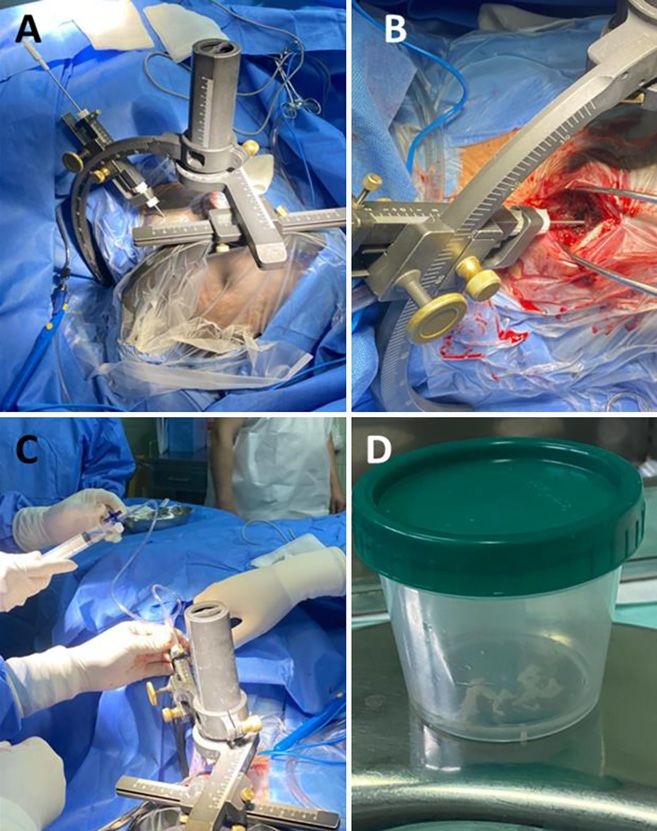
Unauthenticated | Downloaded 12/19/21 06:54 PM UTCused in both patients. The Micromar stereotactic frame was used; it
has a 2-mm diameter biopsy cannula composed of two hollow can-
nulas, one inside the other. Both cannulas have a 1-cm rectangular
side window located 1 mm from the lower end. The stereotactic
frame was placed at the level of the superior temporal line after
infiltration with lidocaine without epinephrine at the points where the
pins were fixed. Once the frame was set, contrast-enhanced com-
puted tomography was performed with a standard protocol for ster-
eotaxis, and the target (midpoint of the 4th ventricle) coordinates
were calculated (Fig. 4).
We used a transcerebellar suboccipital approach with the patient
in the lateral decubitus position. The incision site was marked 3 cm
lateral to the midline and 3 cm below the projection of the trans-
verse sinus. Once in the bone plane (occipital scale), a trepan, an
“X” durotomy, and finally a small corticotomy were performed. The
stereotactic frame arch and guide were placed with the previously
calculated coordinates, and the closed cannula was introduced
(connected to a triple-line system with extension coupled to a 20-ml
syringe with saline solution) across the cerebellar hemisphere direct
to the 4th ventricle. Once the cannula was set in position, the posi-
tion was verified by initial aspiration, obtaining cerebrospinal fluid
(CSF) in both cases, and then very gentle aspiration was applied
FIG. 2. Cysticercal membranes extracted by stereotaxis. A and B: until cyst membranes were extracted. If any resistance is noted, the
Case 1. C and D: Case 2. Typical double-layer eosinophilic mem- membrane is fragmented with the cutter cannula mechanism, and
branes are shown. Hematoxylin and eosin stain; Original magnifications
10 (A and C) and 40 (B and D). the process continues until only CSF comes out. No other traction
instrumentation (such as forceps) was used. This procedure was
repeated at 0°, 90°, 180°, and 270° until only CSF was obtained
without greater resistance. The cyst membranes were extracted
without difficulty in both cases, and there were no complications.
FIG. 3. MRI scans of case 2. A–C: Preoperative imaging. Dilated 4th ventricle with images suggestive of cys-
tic lesions in its interior. D–F: Control MRI at 4 months after surgery. The diameter of the 4th ventricle has
decreased, and there are no remnant cysts (A and D, axial FIESTA; B and E, axial FLAIR; C and F, sagittal
postcontrast T1).
J Neurosurg Case Lessons | Vol 2 | Issue 3 | July 19, 2021 | 3
Unauthenticated | Downloaded 12/19/21 06:54 PM UTCthe brainstem reported a rate of complications of 8.9%.27 Another
report of 78 patients foundof stereotaxis to safely treat these two patients with NCC cysts in the 4th 18. Hanak BW, Walcott BP, Codd PJ, et al. Fourth ventricular neurocys-
ventricle. tercercosis presenting with acute hydrocephalus. J Clin Neurosci.
2011;18(6):867–869.
References 19. Franko LR, Pandian B, Gupta A, et al. Posterior fossa craniotomy
1. Mahale RR, Mehta A, Rangasetty S. Extraparenchymal (racemose) for adherent fourth ventricle neurocysticercosis. Oper Neurosurg
neurocysticercosis and its multitude manifestations: a comprehen- (Hagerstown). 2019;16(5):E154–E158.
sive review. J Clin Neurol. 2015;11(3):203–211. 20. Apuzzo ML, Dobkin WR, Zee CS, et al. Surgical considerations in
2. Garcia HH, Nash TE, Del Brutto OH. Clinical symptoms, diagnosis, treatment of intraventricular cysticercosis. An analysis of 45 cases.
and treatment of neurocysticercosis. Lancet Neurol. 2014;13(12): J Neurosurg. 1984;60(2):400–407.
1202–1215. 21. Bergsneider M. Endoscopic removal of cysticercal cysts within the
3. Gripper LB, Welburn SC. Neurocysticercosis infection and dis- fourth ventricle. Technical note. J Neurosurg. 1999;91(2):340–345.
ease—a review. Acta Trop. 2017;166:218–224. 22. Coyle CM. Neurocysticercosis: an update. Curr Infect Dis Rep.
4. Jensen TO, Post JJ. Intraventricular neurocysticercosis: presenta- 2014;16(11):437.
tion, diagnosis and management. Asian Pac J Trop Med. 23. Rajshekhar V. Surgical management of neurocysticercosis. Int J
2016;9(8):815–818. Surg. 2010;8(2):100–104.
5. Gupta P, Agrawal M, Sinha VD, Gupta A. Intraventricular racemose 24. Nash TE, Ware JM, Mahanty S. Intraventricular neurocysticercosis:
type neurocysticercosis with anterior interhemispheric fissure experience and long-term outcome from a tertiary referral center in
cyst: a rare case report. J Neurosci Rural Pract. 2015;6(2): the United States. Am J Trop Med Hyg. 2018;98(6):1755–1762.
234–237. 25. Psarros TG, Zouros A, Coimbra C. Neurocysticercosis: a neurosur-
6. Citow JS, Johnson JP, McBride DQ, Ammirati M. Imaging features gical perspective. South Med J. 2003;96(10):1019–1022.
and surgery-related outcomes in intraventricular neurocysticercosis. 26. Fengqiang L, Jiadong Q, Yi L. Computer-assisted stereotactic neu-
Neurosurg Focus. 2002;12(6):e6. rosurgery with framework neurosurgery navigation. Clin Neurol
7. Peralta D, Banegas J, Miller E. Neurocisticercosisintraventricular tra- Neurosurg. 2008;110(7):696–700.
tada eficazmente con agentes anticestodos. Rev Med Hondur. 27. Samadani U, Stein S, Moonis G, et al. Stereotactic biopsy of brain
2012;80(3):108–110. stem masses: Decision analysis and literature review. Surg Neurol.
8. Del Brutto OH. Neurocysticercosis: up-dating in diagnosis and treat- 2006;66(5):484–491.
Ordo~nez Rubiano EG,
28. Zorro O, Camacho JE, et al. Procedimientos
ment. Article in Spanish. Neurologia. 2005;20(8):412–418.
9. Sandoval-Balanzario MA, Rincon-Navarro RA, Granados-Lopez R, neuroquirurgicos cerebrales guiados por estereotaxia realizados en
Santos-Franco JA. Endoscopic third ventriculostomy for chronic el Hospital Universitario de San Ignacio (HUSI): evolucion a corto y
communicating hydrocephalus in adults. Article in Spanish. Rev mediano plazo. Univ Medica. 2012;54(1):39–52.
Med Inst Mex Seguro Soc. 2015;53(3):280–285. 29. White AC Jr, Coyle CM, Rajshekhar V, et al. Diagnosis and Treat-
10. Campbell BR, Reynoso D, White AC. Intraventricular neurocysticer- ment of Neurocysticercosis: 2017 Clinical Practice Guidelines by
cosis and Bruns’ syndrome: a review. J Rare Dis Res Treat. the Infectious Diseases Society of America (IDSA) and the
2017;2(2):1–5. American Society of Tropical Medicine and Hygiene (ASTMH). Clin
11. Rana S, Prasad A, Brar R, et al. Caught in the act: migrating intra- Infect Dis. 2018;66(8):e49–e75.
ventricular neurocysticercosis causing intermittent unilateral hydro-
cephalus due to foramen of Monro obstruction. Acta Neurol Belg. Disclosures
2018;118(3):509–511. The authors report no conflict of interest concerning the materials or
12. Araujo AL, Rodrigues RS, Marchiori E, et al. Migrating intraventricu- methods used in this study or the findings specified in this paper.
lar cysticercosis: magnetic resonance imaging findings. Arq Neuro-
psiquiatr. 2008;66(1):111–113. Author Contributions
13. Thomas B, Krishnamoorthy T. Migrating intraventricular cysticercus Conception and design: Lines, Saavedra, Vasquez. Acquisition of data:
during MRI. Neurology. 2005;65(8):1321. Lines, Caucha, Felix. Analysis and interpretation of data: Lines, Garcıa,
14. Garcıa HH, Evans CAW, Nash TE, et al. Current consensus guide- Medina. Drafting the article: Lines, Garcıa. Critically revising the article:
lines for treatment of neurocysticercosis. Clin Microbiol Rev. Lines, Garcıa. Reviewed submitted version of manuscript: Lines,
2002;15(4):747–756. Garcıa, Antonio. Approved the final version of the manuscript on behalf
15. Kaif M, Husain M, Ojha BK. Endoscopic management of of all authors: Lines. Administrative/technical/material support: Lines.
intraventricular neurocysticercosis. Turk Neurosurg. 2019;29(1): Study supervision: Lines. Operating surgeon: Saavedra.
59–65.
16. Goel RK, Ahmad FU, Vellimana AK, et al. Endoscopic management Correspondence
of intraventricular neurocysticercosis. J Clin Neurosci. 2008;15(10): William W. Lines: Instituto Nacional De Ciencias Neurologicas, Lima,
1096–1101. Peru. williamincn@hotmail.com.
17. Loyo M, Kleriga E, Esta~nol B. Fourth ventricular cysticercosis.
Neurosurgery. 1980;7(5):456–458.
J Neurosurg Case Lessons | Vol 2 | Issue 3 | July 19, 2021 | 5
Unauthenticated | Downloaded 12/19/21 06:54 PM UTCYou can also read