MIS revisional surgery for gastro-esophageal reflux disease: how I do it
←
→
Page content transcription
If your browser does not render page correctly, please read the page content below
Review Article
                                                                                                                                    Page 1 of 6

MIS revisional surgery for gastro-esophageal reflux disease:
how I do it
Sarah K. Thompson, Rippan N. Shukla

Flinders University Discipline of Surgery, College of Medicine & Public Health, Flinders Medical Centre, Bedford Park, Australia
Contributions: (I) Conception and design: SK Thompson; (II) Administrative support: Both authors; (III) Provision of study materials or patients:
SK Thompson; (IV) Collection and assembly of data: RN Shukla; (V) Data analysis and interpretation: RN Shukla; (VI) Manuscript writing: Both
authors; (VII) Final approval of manuscript: Both authors.
Correspondence to: Sarah Thompson, MD, PhD, FRACS. College of Medicine & Public Health, Rm 5E221.3, Flinders Medical Centre, Bedford Park,
Australia. Email: sarah.thompson@flinders.edu.au.

                Abstract: Minimally invasive surgery to control gastro-esophageal reflux disease (GERD) was introduced
                in 1991, and quickly became mainstream as an excellent option for patients with breakthrough symptoms
                on maximal medical therapy. However, a small subset of patients will be unhappy with the result of
                laparoscopic fundoplication, and will present for consideration of a laparoscopic revisional procedure. A
                failed fundoplication may result in one of three situations: (I) true recurrent reflux symptoms, often due to an
                anatomical cause; (II) residual “reflux” symptoms post laparoscopic fundoplication, often due to symptoms
                mistakenly attributed to reflux; and (III) new symptoms post laparoscopic fundoplication, such as bloating,
                increased flatulence and dysphagia. With an increased morbidity and mortality rate for laparoscopic revisional
                fundoplication, it is critical to select the right patient for a redo procedure. Mandatory investigations when
                dealing with a presentation of recurrent reflux symptoms, include: endoscopy (ideally performed by the
                responsible surgeon), 24-hour pH study (if no evidence of reflux on endoscopy), esophageal manometry, and
                barium swallow. In this paper, we discuss our definition of a failed fundoplication, we outline our operative
                approach to a minimally invasive revisional fundoplication, and we discuss our postoperative management.
                With these steps, 86% of patients undergoing a laparoscopic revisional fundoplication in our institution are
                satisfied with the result.

                Keywords: Recurrent reflux; failed fundoplication; laparoscopic redo fundoplication; laparoscopic revisional
                fundoplication

                Received: 28 March 2021. Accepted: 01 May 2021.
                doi: 10.21037/aoe-21-26
                View this article at: http://dx.doi.org/10.21037/aoe-21-26

Introduction                                                                 carry a higher morbidity rate (23%), dysphagia rate (25%),
                                                                             and mortality rate (1%) (2).
Minimally invasive surgery to control gastro-esophageal
                                                                                In this paper, we discuss our definition of a failed
reflux disease (GERD) was introduced in 1991, and quickly                    fundoplication, and we outline our operative approach
became mainstream as an excellent option for patients                        to a minimally invasive redo fundoplication. From open
with breakthrough symptoms on maximal medical therapy.                       surgery (either via a thoracotomy or a laparotomy) to
However, a small subset of patients will be unhappy with                     now laparoscopic surgery (sometimes with 3-dimensional
the result of laparoscopic fundoplication (3–6%), and                        cameras or robotic-assisted surgery), the approach to
will present for consideration of a laparoscopic revisional                  revisional fundoplication has evolved rapidly over the past
procedure (1). Careful patient selection for laparoscopic                    two decades. With these advances, tips and tricks have been
revisional surgery is critical because revisional procedures                 picked up and will be highlighted here. We also discuss our

© Annals of Esophagus. All rights reserved.                                            Ann Esophagus 2021 | http://dx.doi.org/10.21037/aoe-21-26
Page 2 of 6 Annals of Esophagus, 2021
approach to postoperative management. operative symptoms continue to persist following surgery.
These patients are often those with atypical reflux symptoms
(i.e., cough, hoarseness, etc), or those with negative
What is a failed fundoplication?
objective testing for GERD prior to surgical intervention.
One of the biggest challenges in the management of GERD It is this group of patients whom the surgeon should
patients post fundoplication is that there is no accepted try to avoid operating on in the first place, as this group
standard definition of what constitutes a failure of anti- often lack the correct indications for primary laparoscopic
reflux surgery. Failure can encompass all of the following: fundoplication, resulting in persistent symptoms and patient
recurrent or residual reflux symptoms, use of anti-reflux dissatisfaction.
medication, or de novo symptoms (including different reflux
symptoms or side-effect symptoms from the procedure).
Laparoscopic revisional surgery: technique
Failure can be defined subjectively, through the patient’s
description of their symptoms or objectively, through Patient selection
endoscopy, acidic pH or total reflux studies, esophageal
manometry, and barium studies. A failed procedure may The most important first step for the surgeon is to take a
not necessarily have an identifiable anatomical cause. The thorough history. In particular, what was the indication for
International Society of the Diseases of the Esophagus primary anti-reflux surgery? Further, was there objective
(ISDE) is currently working on a document to further evidence of reflux pre-operatively (either ulcerative
define the failed fundoplication—an essential step towards esophagitis on endoscopy or a positive pH study)? The
accurate reporting of failure from fundoplication, and the surgeon must not re-operate on a patient whose “recurrent
management of the failed fundoplication. reflux” is not true reflux (see “Choosing the right patient for
laparoscopic fundoplication: a review of preoperative predictors” in
this special edition). If the recurrent symptoms are different
Our definition of a failed fundoplication to those experienced by the patient pre-operatively, further
A laparoscopic fundoplication can often result in symptoms investigations are warranted to exclude other causes
of bloating, increased flatulence, and dysphagia. These such as biliary colic and peptic ulcer disease. Mandatory
are side-effects of the procedure itself as circumferential investigations when dealing with a presentation of recurrent
swelling of the esophago-gastric junction (secondary to reflux symptoms, include: endoscopy (ideally performed by
manipulation of the esophagus as well as the intra-operative the responsible surgeon), 24-hour pH study (if no evidence
use of an esophageal sling) can impede the passage of food of reflux on endoscopy), esophageal manometry, and barium
into the stomach, and may result in a fundoplication which swallow (3).
is air-tight as well as “acid-tight”. That said, these symptoms
should subside once the postoperative swelling has subsided, Preoperative preparation
usually within the first 3 months, and certainly by the
6-month mark. However, persistence of these symptoms, in If the decision has been made to perform laparoscopic
particular dysphagia and bloating, for greater than 6 months revisional surgery, optimize all conditions. All patients
is considered by many as a failed fundoplication. should commence a very low-calorie diet (VLCD) to
The second group of patients with a failed fundoplication shrink the liver, reduce visceral fat, and improve access
are those with recurrent reflux symptoms, often due to an to the hiatus (4). Most patients will require at least
anatomical problem in the peri-operative period. Common 2 weeks on a VLCD, whilst those with a BMI over 35 may
causes include a slipped wrap, a recurrent hiatus hernia, benefit from at least 4 weeks. Enlist the assistance of an
or perhaps the creation of a partial fundoplication where experienced second operator, and book adequate time for
a total fundoplication should have been performed. This the procedure.
group of patients is relatively straightforward to manage as
a laparoscopic revisional fundoplication should resolve the
Positioning and trocar placement
patient’s reflux symptoms.
The final group of patients with a failed fundoplication The patient is positioned in the supine lithotomy (French)
are those with residual reflux symptoms, i.e., their pre- position to allow the surgeon to operate from between
© Annals of Esophagus. All rights reserved. Ann Esophagus 2021 | http://dx.doi.org/10.21037/aoe-21-26Annals of Esophagus, 2021 Page 3 of 6
used instead of a bougie, depending on surgeon preference.
Operative technique—key steps
Our usual approach is to first restore normal anatomy.
We start at the right pars flaccida and identify the right
crus. We then carry the dissection across the anterior
esophagus to the left crus. Be alert and prepared for braided
sutures placed at the hiatus during primary laparoscopic
fundoplication (e.g., Ethibond®), or mesh in situ. There will
Figure 1 The right and left crus of the diaphragm must be cleaned be far more scarring and fibrosis for braided sutures than
off entirely down to the decussation. A 5-French feeding tube is for monofilament sutures used at primary operation. These
used as an esophageal sling. patients should be made aware of the higher conversion
rate to laparotomy for their revisional procedure. Consider
the use cautery and ultrasonic shears judiciously around the
esophagus. If the short gastric vessels were not divided at
the first operation, this provides a nice uninterrupted plane
to locate the left pillar of the crural diaphragm.
Once the normal anatomy has been restored, a sling
(we prefer a pediatric 5-French feeding tube) is placed
around the esophagus and care is taken to identify and
preserve the posterior vagus. The right and left pillars
of the diaphragm need to be identified clearly down to
the crural decussation (Figure 1). The crural diaphragm
is closed posteriorly with non-absorbable monofilament
Figure 2 The diaphragmatic defect is closed with permanent, sutures (e.g., 2-0 NovafilTM), taking care not to angulate
monofilament sutures. the esophagus (Figure 2). If this is a concern (i.e., the
anesthetist cannot advance the bougie in a straight line), a
combined posterior and anterior closure should be done.
the legs. Steep reverse Trendelenburg positioning is then We use a 52-French or 54-French bougie to calibrate the
achieved with the patient on a gel mat. Consider port diaphragmatic closure. With the bougie advanced into the
placement carefully. Do not reuse the old port sites if they stomach, there should be just enough space between the
are in the wrong place—it is best to optimize the field of esophagus and the pillars to allow the easy passage of the
view for the revisional procedure! We prefer a closed Veress tip of a laparoscopic grasper.
technique in the left upper quadrant with insufflation to A fundoplication is then re-created, ensuring there is
10–14 mmHg. Port placement is as follows: 11 mm camera adequate intra-abdominal esophageal length. We believe
port 15 cm from the xiphoid, to the left of midline; 12 mm that with adequate peri-esophageal dissection 5–6 cm up
working port in the left upper quadrant 11 cm from the into the chest, cases of true shortened esophagus are rare.
xiphoid along the costal margin; 5 mm working port in the With many revisional cases, the cause of a slipped wrap is
right upper quadrant 11 cm from the xiphoid along the due to misidentification of the fundus of the stomach at
costal margin; and a 5 mm assistant port in the left lateral primary surgery (Figure 3). Always ensure the top-most
abdomen. A Nathanson liver retractor is placed at the level aspect of the stomach is used to create the fundoplication,
of the xiphoid. whether for a partial or total fundoplication. For a Nissen
It is a good idea to make sure the anesthetist has a 52- fundoplication, avoid excess stomach above the wrap
or 54-French bougie in the mid-esophagus to help with (Figure 4). The key is a shoe-shine manoeuvre (Figure 5)
laparoscopic localisation of the esophagus. Primary anti- which avoids inadvertent excess stomach posterior to
reflux surgery often displaces the esophagus to a more the esophagus (Figure 6). We prefer to calibrate a total
anterior location. An intra-operative endoscope may also be fundoplication over a 54-French bougie. It is important
© Annals of Esophagus. All rights reserved. Ann Esophagus 2021 | http://dx.doi.org/10.21037/aoe-21-26Page 4 of 6 Annals of Esophagus, 2021
Figure 3 A malpositioned 180-degree wrap where the body of the Figure 6 The correct appearance of a 360-degree fundoplication
stomach was used to create the wrap. The fundus of the stomach once the bougie has been retracted into the mid-esophagus. Note
had re-herniated into the mediastinum as a para-esophageal hernia. the wrap faces the caudate lobe of the liver when the short gastric
vessels are left intact.
Figure 4 Excess stomach above a 360-degree fundoplication.
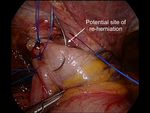
Figure 7 A common site of re-herniation after a partial 180-degree
fundoplication.
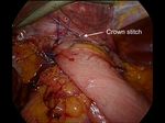
crown stitch (Figure 8).
Postoperative care
Post-operative care should focus on early mobilization
and avoidance of nausea. We prefer to use Ondansetron or
Figure 5 A shoe-shine maneuver to avoid leaving excess stomach Tropisetron routinely for the first 24–48 hours post-surgery.
above the wrap. After 12–24 hours, no pain relief should be necessary aside
from paracetamol. Our institutional protocol is to perform
a contrast swallow the following morning. This provides
to recognize that if the short gastric vessels have not been confirmation of correct positioning of the fundoplication
divided, the Nissen fundoplication will face towards the below the diaphragm, and confirms the absence of a leak.
patient’s caudate lobe. For a partial anterior 180-degree We have recently shown that routine postoperative contrast
fundoplication, the most common site of re-herniation of swallows, primarily in patients with a hiatus hernia, reduces
the gastric fundus into the mediastinum occurs at the 1 or the morbidity related to early and late reoperations (5).
2 o’clock position (Figure 7). Consider the insertion of a Once the swallow has been performed and reviewed, the
© Annals of Esophagus. All rights reserved. Ann Esophagus 2021 | http://dx.doi.org/10.21037/aoe-21-26Annals of Esophagus, 2021 Page 5 of 6
recurrent symptoms.
Thorough investigation with endoscopy, barium swallow,
pH testing, and esophageal manometry is advisable prior
to laparoscopic revisional surgery. Our key steps are
highlighted above, and our postoperative management
including routine anti-emetics, postoperative contrast
swallow, and careful dietary counselling. With these steps,
86% of patients undergoing a laparoscopic revisional
fundoplication in our institution are satisfied or highly
satisfied with the result (6).
Acknowledgments
Figure 8 A ‘crown stitch’ to close off the potential site of re-
herniation. Funding: None.
Footnote
patients start a fluid diet and are discharged home on
puréed/vitamised food for 1 to 2 weeks. They then continue Provenance and Peer Review: This article was commissioned
onto a soft diet for a further 4 weeks. Care is taken to avoid by the Guest Editors (Timothy M. Farrell and Geoffrey
constipation in the early post-operative period and our Kohn) for the series “Minimally Invasive Procedures for
patients are counselled to take a stool softener for the first Gastroesophageal Reflux Disease” published in Annals of
couple of weeks if needed. Esophagus. The article has undergone external peer review.
Conflicts of Interest: Both authors have completed the
Conclusions
ICMJE uniform disclosure form (available at http://dx.doi.
As highlighted above, the ISDE (International Society org/10.21037/aoe-21-26). The series “Minimally Invasive
of the Diseases of the Esophagus) is currently working Procedures for Gastroesophageal Reflux Disease” was
on a document to define the failed fundoplication— commissioned by the editorial office without any funding
an essential step towards accurate reporting of failure or sponsorship. SKT serves as an unpaid editorial board
from fundoplication, and the management of the failed member of Annals of Esophagus from Sep. 2019 to Aug.
fundoplication. We believe there are three categories of 2021. The authors have no other conflicts of interest to
patients with a “failed fundoplication”: (I) patients with declare.
troublesome and persistent side-effects of a laparoscopic
fundoplication. Most commonly, this includes bloating, Ethical Statement: The authors are accountable for all
increased flatulence, and dysphagia; (II) patients with aspects of the work in ensuring that questions related
recurrent reflux symptoms, often due to an anatomical to the accuracy or integrity of any part of the work are
problem such as a slipped wrap, a recurrent hiatus hernia, or appropriately investigated and resolved.
a mal-positioned fundoplication; (III) patients with residual
reflux symptoms, often those with atypical symptoms that Open Access Statement: This is an Open Access article
may not have been due to reflux in the first place (e.g., distributed in accordance with the Creative Commons
cough). Attribution-NonCommercial-NoDerivs 4.0 International
Laparoscopic revisional fundoplication is indicated for License (CC BY-NC-ND 4.0), which permits the non-
patients with true recurrent reflux, as well as those with commercial replication and distribution of the article with
distressing complications of surgery including dysphagia the strict proviso that no changes or edits are made and
and bloating. Of utmost importance is patient selection, the original work is properly cited (including links to both
both at the primary and revisional procedure. It is wise not the formal publication through the relevant DOI and the
to assume the correct indication was present at the initial license). See: https://creativecommons.org/licenses/by-nc-
procedure, and prudent to fully investigate any patient with nd/4.0/.
© Annals of Esophagus. All rights reserved. Ann Esophagus 2021 | http://dx.doi.org/10.21037/aoe-21-26Page 6 of 6 Annals of Esophagus, 2021
References 4. Lewis MC, Phillips ML, Slavotinek JP, et al. Change in
liver size and fat content after treatment with Optifast very
1. Carlson MA, Frantzides CT. Complications and results of
low calorie diet. Obes Surg 2006;16:697-701.
primary minimally invasive antireflux procedures: a review
5. Liu DS, Wee MY, Grantham J, et al. Routine
of 10,735 reported cases. J Am Coll Surg 2001;193:428-39. esophagograms after hiatus hernia repair minimizes
2. Yadlapati R, Hungness ES, Pandolfino JE. reoperative morbidity: a multicenter comparative
Complications of Antireflux Surgery. Am J Gastroenterol cohort study. Ann Surg 2021. doi: 10.1097/
2018;113:1137-47. SLA.0000000000004812. [Epub ahead of print].
3. Allaix ME, Rebecchi F, Schlottmann F, et al. Secrets for 6. Lamb PJ, Myers JC, Jamieson GG, et al. Long-term
successful laparoscopic antireflux surgery: adequate follow- outcomes of revisional surgery following laparoscopic
up. Ann Laparosc Endosc Surg 2017;2:57-60. fundoplication. Br J Surg 2009;96:391-7.
doi: 10.21037/aoe-21-26
Cite this article as: Thompson SK, Shukla RN. MIS revisional
surgery for gastro-esophageal reflux disease: how I do it. Ann
Esophagus 2021.
© Annals of Esophagus. All rights reserved. Ann Esophagus 2021 | http://dx.doi.org/10.21037/aoe-21-26You can also read


