Combined spinal and video-assisted thoracoscopic (VATS) approach to thoracic neurogenic dumbbell tumour in the prone position: a case report
←
→
Page content transcription
If your browser does not render page correctly, please read the page content below
Case Report
                                                                                                                                      Page 1 of 5

Combined spinal and video-assisted thoracoscopic (VATS)
approach to thoracic neurogenic dumbbell tumour in the prone
position: a case report
Thirugnanam Agasthian, James Chee Min Khoo

Mt Elizabeth Medical Centre, Singapore
Correspondence to: Thirugnanam Agasthian, MBBS, MMed (Surgery), FRCS (Ed), FRCS (G). Mt Elizabeth Medical Center, Singapore.
Email: t.agasthian@gmail.com.

                 Abstract: Neurogenic tumours are the third commonest cause of all mediastinal tumours with 10% of
                 them having intraspinal extension with variable spinal and mediastinal components. This is often referred to
                 as a dumbbell neurogenic tumour. In such cases the surgical team comprises a neurosurgeon and a thoracic
                 surgeon. Though several surgical approaches have been described for dumbbell tumours of the thoracic
                 region, the optimal surgical approach is a single stage posterior laminectomy combined with video-assisted
                 thoracoscopic (VATS). In this approach the posterior spinal approach is done in the prone position by the
                 neurosurgeon. This is then followed by repositioning to a lateral decubitus position for excision by VATS
                 by the thoracic surgeon. We describe a case of a 63-year-old male presenting with mild weakness of the
                 lower limbs and right sciatica. MRI whole spine showed with an 8.4 cm × 4.5 cm × 3.7 cm dumbbell thoracic
                 schwannoma arising from the T10 thoracic root. This was excised first by a single stage combined posterior
                 laminectomy by the neurosurgeon followed by the VATS approach, both being performed in the prone
                 position. Postoperative course was complicated by right femoral nerve neuropraxia from compression nerve
                 injury to the groin. He made a almost complete recovery from the neuropraxia on subsequent follow -up.

                 Keywords: Case report; video-assisted thoracoscopic (VATS); prone; schwannoma

                 Received: 25 June 2020; Accepted: 28 December 2020.
                 doi: 10.21037/jovs-20-141
                 View this article at: http://dx.doi.org/10.21037/jovs-20-141

Introduction                                                                    combined posterior and VATS approach in the prone position.
                                                                                   We present the following article in accordance with
Neurogenic tumours are the third commonest cause of all
                                                                                the CARE reporting checklist (available at http://dx.doi.
mediastinal tumours with 10% having intraspinal extension
                                                                                org/10.21037/jovs-20-141).
with variable mediastinal and spinal components (1,2). The
surgical team usually involves a thoracic and a neurosurgeon
if there is an intraspinal extension. The surgical technique is                 Case presentation
either a single stage posterior only approach or a Single stage
combined posterior approach with video-assisted thoracoscopic                   Patient is a 63-year-old male presenting with mild weakness
(VATS) depending on the tumour size, invasiveness and                           of the lower limbs and right sciatica. He has a past history
extend of intraspinal and mediastinal components (3). In the                    of hypertension, treated syphilis, obesity (BMI 35), smoking
combined approach the posterior spinal part is done first                       and hyperlipidaemia. MRI of the whole spine revealed a
followed by VATS and involves repositioning the patient from                    right 8.4 cm × 4.5 cm × 3.7 cm dumbbell neurogenic tumour
prone to lateral decubitus position which prolongs surgical                     arising from T10-T11 neural foramina with associated
time, re-draping and increases costs (4). We describe a case of a               widening of adjacent neural foramina and extension into the
thoracic neurogenic dumbbell tumour excised by a single stage                   spinal canal with displacement of the thoracic spinal cord

© Journal of Visualized Surgery. All rights reserved.                                      J Vis Surg 2021 | http://dx.doi.org/10.21037/jovs-20-141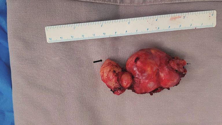
Page 2 of 5 Journal of Visualized Surgery, 2021
Figure 1 Preoperative axial T1 weighted post contrast T10
dumbbell neurogenic tumour.
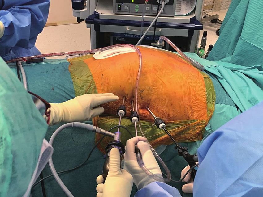
Figure 2 Operative view of right video assisted thoracoscopic
to the left. There were also associated degenerative changes surgery in prone position.
throughout the spine with multilevel disc desiccation
and protrusions facet joint arthrosis and hypertrophy of
noted. The overlying mediastinal pleura was first incised. En
ligamentum flavum, and borderline anterolisthesis with
bloc removal of the tumour was done with division of distal
multilevel mild spinal canal foraminal stenosis causing his
T10 nerve root and other smaller nerve tributary extensions
right sciatic (Figure 1).
from adjacent intercostal and sympathetic nerves. Care was
He underwent resection through a combine single stage
taken not to injure the adjacent intercostal vessels. The
posterior and VATS approach. After general anaesthesia,
tumour was removed via an endo-bag by enlarging one of
a left sided double lumen endotracheal tube was placed.
the 5-mm incision and a F20 chest tube was inserted through
He was positioned in a prone position with extra care and
the same incision. Intraoperative blood loss was minimal and
padding taken to protect vital compression points.
operative time for VATS was 115 minutes (Video 1). Post
Posterior midline laminectomy was first performed by the
operatively he had femoral nerve neuropraxia probably due
neurosurgeon for the extramedullary, intra and extradural
to the prone position exacerbated by his high BMI. Chest
component of the neurilemmoma. Intraoperatively the
tube was removed on 3rd POD. Due to limb physiotherapy
lesion was noted to originate extramedullary from the right
and rehabilitation for his femoral nerve neuropraxia
T10 root. It extended through the dura into the enlarged
he was discharged on 14 th POD. Final histology was a
intervertebral foramen with the major extradural component 9.5 cm × 4.5 cm × 2.5 cm benign schwannoma (Figure 3).
eroding the pedicle and facet joint and extending anteriorly The post-operative MRI scan showed no residual tumour.
to the retro pleural space and the posterior mediastinum. (Figure 4). At follow-up in clinic he had near complete
The firm tumour was excised piecemeal with the dumbbell resolution of his femoral nerve neuropraxia.
origin divided from its origin from the T10 intraspinal All procedures performed in studies involving human
nerve root. The affected bony edges were resected, and the participants were in accordance with the ethical standards of
main component was dissected free from its surrounding the institutional and/or national research committee(s) and
attachments and delivered into the retro pleural space, with the Helsinki Declaration (as revised in 2013). Written
sparing the intercostal vessels. The dura and the wound informed consent was obtained from the patient.
were then closed after meticulous hemostasis.
This was followed by VATS also in the prone position.
Discussion
The right lung was isolated. One 5-mm camera port was
placed directly over the T10 level at midaxillary line. Two Neurogenic tumours are the third commonest cause of
5mm working port incisions were made at T8 and T12 levels mediastinal tumours with about 75% occurring in the
respectively in the posterior axillary line. A 5-mm 30 degrees posterior mediastinum due to the presence of multiple
camera was used (Figure 2). The previously divided intraspinal neural plexuses. About 10% have intraspinal extension
and mediastinal component of the neurogenic tumour was with varying mediastinal and spinal components. Up to
identified and was noted to bilobed with cystic components. 40% of intraspinal dumbbell tumours are asymptomatic
Associated scalloping of the adjacent rib by the tumour was even with radiological displacement of spinal cord.
© Journal of Visualized Surgery. All rights reserved. J Vis Surg 2021 | http://dx.doi.org/10.21037/jovs-20-141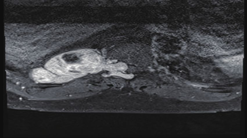
Journal of Visualized Surgery, 2021                                                                                                            Page 3 of 5

                                                Bilobed mediastinal
                                                    Component
                                                                        Intraspinal
                                                                          Divided
              Extrathoracic Distal Nerve Root                         Proximal Nerve
                                                                           Root

Figure 3 Schwannoma.                                                                   Figure 4 Postoperative axial T2 weighted MRI scan showing no
                                                                                       residual disease.

Therefore preoperative MRI of the spine is mandatory in
all paravertebral neurogenic tumours to rule out intraspinal                           disadvantages are need for spinal instrumentation due to
extension and to plan appropriate surgical approach. Spinal                            spinal instability from the extended bony resections and
schwannomas account for 25% of all spinal tumours and                                  possible injuries to intrathoracic structures due to limited
commonly occurs in the thoracic region. It can occur solely                            exposure. It is also generally contraindicated in malignant
in the spinal canal or extend along the nerve root to the                              cases due to possible adequacy of surgical margins (7).
extra vertebral space through the intervertebral foramen                                    Combined posterior laminectomy and VATS is the
and become dumbbell or hour glass shaped tumour. They                                  procedure of choice for most dumbbell tumours with
can be classified into 4 sub types depending on the degree of                          large mediastinal component. It allows for less posterior
spinal and mediastinal extensions. The intraspinal extension                           bone removal and the need for complex expensive spinal
can be intra or extramedullary, intra or extradural or                                 instrumentation to reduce spinal instability (3). Though
foraminal. The mediastinal part is usually well encapsulated                           some advocate doing the thoracic part first followed by spinal
and can be bilobed with cystic components. It can vary from                            in combined cases, a posterior approach should always be
small to large and rarely invasive in malignant cases. Up to                           performed first to decompress the spinal cord and release the
10% of dumbbell tumours are malignant (1,2).                                           tumour from the intraspinal nerve root. This reduces risk of
    Historically several approaches have been described                                spinal cord injury during subsequent mediastinal component
for dumbbell tumours in the thoracic region. The surgical                              removal (3,6). Since the advent of VATS, thoracotomy
approaches are either a single stage posterior only approach,                          is rarely used except in large invasive malignant tumours
single stage anterior approach or a Single stage combined                              where an anterior only approach is preferred. Advantages of
posterior approach with VATS or thoracotomy (3-7).                                     VATS are it causes less pain, pulmonary dysfunction, chest-
Optimal approach will depend on tumour size, invasiveness                              wall trauma and immune derangement. It provides excellent
and extend of intraspinal and mediastinal components (5).                              exposure of the whole chest for safe dissection under direct
In cases where the mediastinal extension is only foraminal                             visualization. Its disadvantages are extra incisions and need
or a small mediastinal component it can be removed in                                  for repositioning (4) This can be avoided as shown in our case
a single stage posterior approach by laminectomy alone                                 by doing both combined procedures in the prone position as
by an experienced neurosurgeon. Most times a combined                                  it saves operative time and cost.
approach involving both a neurosurgical and thoracic team                                   Prone position in thoracic surgery was popular in the early
                                                                                           th
is needed (3-7).                                                                       20 century for airway management in patients undergoing
    Single stage posterior approach is effective for resecting                         lung surgery for bronchiectasis and lung abscess. It helped to
tumours with a predominantly intraspinal component.                                    prevent the spillage of purulent material into healthy parts of
It involves laminectomy. Exposure can be enhanced by                                   the lung during pulmonary resections, but became obsolete
additional facetectomy, pediculectomy costotransversectomy                             with the advent of double-lumen endotracheal tubes. It was
or rib resection depending on the size and invasiveness of                             revived again in the 1990’s for mobilization of the thoracic
the mediastinal component. Advantages of this approach                                 oesophagus by VATS for esophageal cancers. In the prone
are single incision with less muscle damage, blood                                     position the lung naturally falls away from the posterior
loss, operative time and postoperative pain (7). Main                                  mediastinum under gravity giving excellent exposure to

© Journal of Visualized Surgery. All rights reserved.                                               J Vis Surg 2021 | http://dx.doi.org/10.21037/jovs-20-141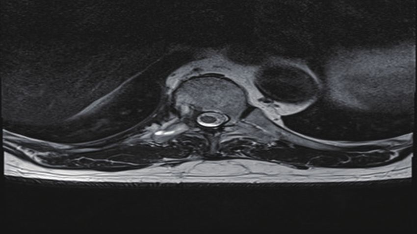
Page 4 of 5                                                                                        Journal of Visualized Surgery, 2021

the posterior mediastinum. It also minimises lung trauma              reporting checklist. Available at http://dx.doi.org/10.21037/
by abolishing the need for excessive lung retraction by an            jovs-20-141
additional surgical assistant. Hemodynamically it is well
tolerated due to less ventilation perfusion mismatch (8,9).           Conflicts of Interest: Both authors have completed the
    Although rare prone position can cause serious                    ICMJE uniform disclosure form (available at http://dx.doi.
complications which can result in long term disability.               org/10.21037/jovs-20-141). TA serves as an unpaid editorial
It includes hypoperfusion hemodynamic changes,                        board member of Journal of Visualized Surgery from June
ophthalmologic injuries, peripheral nerve compression                 2019 to May 2021. The other author has no conflicts of
injuries, compartmental syndrome, pressure ulcers and airway          interest to declare.
swelling (10). Meralgia paraesthesia, femoral artery thrombosis
and avascular necrosis of the hip have also been reported             Ethical Statement: The authors are accountable for all
due to excessive pressure over the groin (10-13). Excessive           aspects of the work in ensuring that questions related
compression pressure over the groin and the anterior superior         to the accuracy or integrity of any part of the work are
iliac spine occurs due to the pelvic padding bolsters especially if   appropriately investigated and resolved. All procedures
placed too closely (10). Injury to the lateral cutaneous nerve of     performed in studies involving human participants were in
the thigh in the prone position causing meralgia paraesthesia         accordance with the ethical standards of the institutional
is seen in up to 24% of patients (10). Post-operatively, patients     and/or national research committee(s) and with the Helsinki
complain of paraesthesia in the thigh with complete resolution        Declaration (as revised in 2013). Written informed consent
of symptoms occurring in most cases within 6 months (10).             was obtained from the patients.
Risk factors for compression pressure groin injury in the
prone position increases with obesity, degenerative spinal            Open Access Statement: This is an Open Access article
disease, inadequate padding and duration of surgery greater           distributed in accordance with the Creative Commons
than 3.5 hours (10). Our patient developed a right femoral            Attribution-NonCommercial-NoDerivs 4.0 International
nerve neuropraxia which is extremely rare and not reported            License (CC BY-NC-ND 4.0), which permits the non-
previously. It was probably caused by excessive groin pressure        commercial replication and distribution of the article with
from his obesity (BMI 35), degenerative spine disease and             the strict proviso that no changes or edits are made and the
duration of surgery. He however made rapid neuromuscular              original work is properly cited (including links to both the
recovery in the first 2 weeks with mild residual weakness in          formal publication through the relevant DOI and the license).
subsequent follow-ups. This complication can be avoided by            See: https://creativecommons.org/licenses/by-nc-nd/4.0/.
careful extra padding of the groin especially in obese patients
as well minimising operative time in the prone position.
                                                                      References

                                                                      1.   Topçu S, Alper A, Gülhan E, et al. Neurogenic tumours
Conclusions
                                                                           of the mediastinum: a report of 60 cases. Can Respir J
Most neurogenic dumbbell tumours can be completely and                     2000;7:261-5.
safely removed by a single stage combined posterior and VATS          2.   Akwari OE, Payne WS, Onofrio BM, et al. Dumbbell
approach in the prone position avoiding the need for excessive             neurogenic tumors of the mediastinum. Diagnosis and
bone removal and complex spinal instrumentation. Meticulous                management. Mayo Clin Proc 1978;53:353-8.
care and extra precautionary measures should be taken to              3.   Payer M, Radovanovic I, Jost G. Resection of thoracic
prevent nerve injuries in the groin during prone position.                 dumbbell neurinomas: single postero-lateral approach or
                                                                           combined posterior and transthoracic approach? J Clin
                                                                           Neurosci 2006;13:690-3.
Acknowledgments
                                                                      4.   Konno S, Yabuki S, Kinoshita T, et al. Combined
Funding: None.                                                             laminectomy and thoracoscopic resection of dumbbell-type
                                                                           thoracic cord tumor. Spine (Phila Pa 1976) 200;26:E130-4.
                                                                      5.   Eden K. The dumb-bell tumours of the spine. BJS
Footnote
                                                                           1941;28:549-70.
Reporting Checklist: The authors have completed the CARE              6.   Nam KH, Ahn HY, Cho JS, et al. One Stage Posterior

© Journal of Visualized Surgery. All rights reserved.                             J Vis Surg 2021 | http://dx.doi.org/10.21037/jovs-20-141
Journal of Visualized Surgery, 2021 Page 5 of 5
Minimal Laminectomy and Video-Assisted Thoracoscopic associated with prone positioning in elective spinal surgery.
Surgery (VATS) for Removal of Thoracic Dumbbell World J Orthop 2015;6:351-9.
Tumor. J Korean Neurosurg Soc 2017;60:257-61. 11. Tseng MD, Cappetto L, Majid K, et al. Bilateral
7. Thorat JD, Rajendra T, Thirugnanam A, et al. Single- femoral artery ischemia detected by multimodality
stage posterior midline approach for dumbbell tumors of neuromonitoring during posterior scoliosis surgery: a case
the thoracic spine, with intraoperative CT guidance. Surg
report. Spine (Phila Pa 1976) 2010;35:E799-E803.
Neurol Int 2011;2:31.
12. Orpen N, Walker G, Fairlie N, et al. Avascular necrosis of
8. Cuschieri A, Shimi S, Banting S. Endoscopic
the femoral head after surgery for lumbar spinal stenosis.
oesophagectomy through a right thoracoscopic approach.
J R Coll Surg Edinb 1992;37:7-11. Spine (Phila Pa 1976) 2003;28:E364-E367.
9. Agasthian T. Revisiting the prone position in video- 13. Cho KT, Lee HJ. Prone position-related meralgia
assisted thoracoscopic surgery. Asian Cardiovasc Thorac paresthetica after lumbar spinal surgery: a case report
Ann 2010 Aug;18:364-7. and review of the literature. J Korean Neurosurg Soc
10. DePasse JM, Palumbo MA, Haque M, et al. Complications 2008;44:392-5.
doi: 10.21037/jovs-20-141
Cite this article as: Agasthian T, Khoo JCM. Combined spinal
and video-assisted thoracoscopic (VATS) approach to thoracic
neurogenic dumbbell tumour in the prone position: a case
report. J Vis Surg 2021.
© Journal of Visualized Surgery. All rights reserved. J Vis Surg 2021 | http://dx.doi.org/10.21037/jovs-20-141You can also read