Seizures Related to Neurocysticercosis and Cocaine Use
←
→
Page content transcription
If your browser does not render page correctly, please read the page content below
Open Access Case
                               Report                                                  DOI: 10.7759/cureus.22488

                                                Seizures Related to Neurocysticercosis and
                                                Cocaine Use
Review began 02/07/2022
                                                Benjamin S. Daines 1 , Katherine G. Holder 1 , Balaji Mohanakrishnan 1 , James W. Walker 1
Review ended 02/13/2022
Published 02/22/2022                            1. Internal Medicine, Texas Tech University Health Sciences Center, Amarillo, USA
© Copyright 2022
Daines et al. This is an open access article    Corresponding author: Benjamin S. Daines, benjamin.daines@ttuhsc.edu
distributed under the terms of the Creative
Commons Attribution License CC-BY 4.0.,
which permits unrestricted use, distribution,
and reproduction in any medium, provided
the original author and source are credited.    Abstract
                                                Neurocysticercosis (NCC) is an infection of the central nervous system with Taenia solium cysts that most
                                                commonly results in seizures. In stable patients without recent symptoms, these seizures may be provoked
                                                by seizure threshold-lowering drugs such as cocaine. This case details a 38-year-old male with a past
                                                medical history of epilepsy presenting with seizures due to comorbid NCC and cocaine use. This case was
                                                complicated by the lack of available information regarding the patient’s past medical history and medication
                                                use. We highlight the importance of obtaining a full work-up, including brain imaging, to provide optimal
                                                treatment for patients with seizures despite a history of drug use.

                                                Categories: Internal Medicine, Infectious Disease, Substance Use and Addiction
                                                Keywords: drug-induced seizure, taenia solium, infectious and parasitic diseases, seizure, cocaine use,
                                                neurocysticercosis

                                                Introduction
                                                Neurocysticercosis (NCC) is the most common helminthic central nervous system infection in humans and is
                                                a common cause of epilepsy throughout South America [1,2]. While NCC is a rare diagnosis in the United
                                                States with an estimated incidence of 0.2 to 0.6 cases per 100,000, it disproportionately impacts Hispanic
                                                populations in the United States with an incidence up to 5.8 cases per 100,000 [3]. With a growing Hispanic
                                                community in the United States, NCC has become an increasingly important clinical consideration due to
                                                significant associated morbidity via seizures, hydrocephalus, and cerebral edema [4]. Early recognition and
                                                treatment of NCC with cysticidal drug therapy can reduce recurrence of seizures and aid in resolution of
                                                parenchymal cysts [5].

                                                Cocaine use can also result in seizures or exacerbate an underlying seizure disorder by lowering the seizure
                                                threshold due to indirect sympathomimetic effects [6]. Patients in the emergency department who had used
                                                cocaine presented due to seizures in nearly 10% of cases [7]. While cases of cocaine-induced seizures have
                                                decreased, cocaine use is still an important consideration in a patient presenting with seizures [8]. Currently,
                                                there are no peer-reviewed cases in the literature describing comorbid NCC and cocaine use. Here, we
                                                present a case of a 38-year-old male with seizures due to NCC and cocaine use.

                                                Case Presentation
                                                A 38-year-old male with a past medical history of epilepsy presented after two episodes of seizures 3 hours
                                                apart on the day of presentation. The seizures were witnessed by family. Each episode lasted approximately
                                                5 minutes and resulted in loss of consciousness with post-ictal confusion. The patient denied nausea,
                                                vomiting, bowel changes, chest pain, shortness of breath, paresthesia, weakness, vision changes, and
                                                headache. He endorsed some fatigue. The patient was asleep prior to the episodes and had no recent
                                                changes in physical activity, eating, drinking, or stressful inciting events. He denied recent sick contacts and
                                                travel. He endorsed drinking alcohol 1-2 times weekly but denied tobacco and recreational drug use. One
                                                year ago, the patient moved to the United States from Mexico where he had lived his entire life. He stopped
                                                taking his seizure medication for epilepsy one year ago. He does not know what seizure medication he was
                                                taking nor any details of his epilepsy diagnosis from childhood. He says he has not had a seizure since he
                                                was a child. He endorsed no family history of epilepsy or other medical conditions.

                                                On physical examination, there were no focal neurological deficits. Cranial nerves II through XII were intact
                                                with normal sensation and strength in all extremities bilaterally. Deep tendon reflexes were 2+ bilaterally.
                                                The patient was alert and oriented to person, place, and time. Vital signs were stable and initial laboratory
                                                values in Table 1 revealed mild leukocytosis without eosinophilia. Urine drug screen was positive for
                                                cocaine. When this result was brought up, the patient admitted to using cocaine a few times in the past, but
                                                not habitually. He said his most recent cocaine use was a few days ago. The previous cocaine use with
                                                normal physical examination findings indicated a likely drug-induced seizure, but additional imaging was
                                                ordered due to the unknown etiology of the patient’s childhood epilepsy diagnosis.

                               How to cite this article
                               Daines B S, Holder K G, Mohanakrishnan B, et al. (February 22, 2022) Seizures Related to Neurocysticercosis and Cocaine Use. Cureus 14(2):
                               e22488. DOI 10.7759/cureus.22488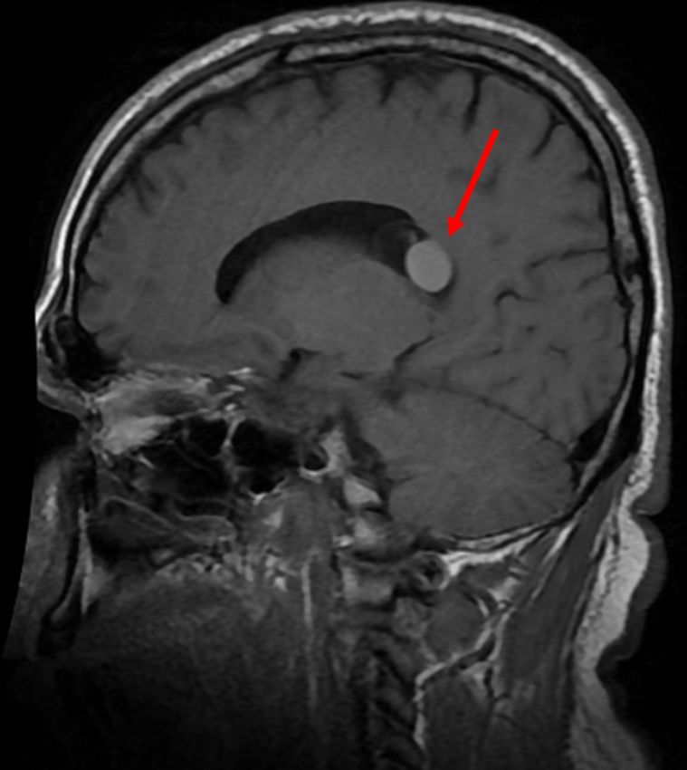
Investigation Value Reference Range
WBC (k/µL) 12.8 4.8-10.8
Hemoglobin (g/dL) 14.8 14.0-18.0
Eosinophils (%) 0.5 0.0-5.0
Platelets (k/µL) 251 150-400
Sodium (mmol/L) 140 135-145
Potassium (mmol/L) 4.0 3.5-5.0
Chloride (mmol/L) 109 98-107
Bicarbonate (mmol/L) 20 21-31
ALP (U/L) 67 25-100
ALT (U/L) 40 10-40
AST (U/L) 31 12-38
TABLE 1: Laboratory Results
WBC: white blood cells; ALP: alkaline phosphatase; ALT: alanine transaminase; AST: aspartate transaminase
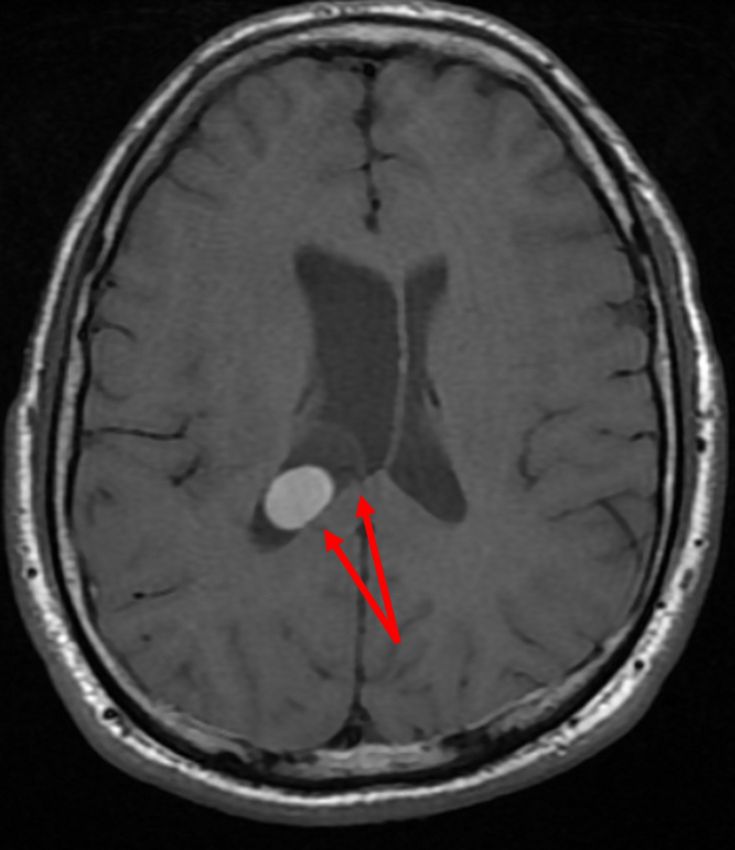
Brain MRI demonstrated a non-enhancing T1 hyperintense 1.7 cm diameter lesion (Figure 1) within a larger
cystic 2.6 cm diameter lesion in the atrium of the right lateral ventricle adherent to the choroid (Figure 2).
There was asymmetric enlargement of the right ventricle. These MRI findings combined with the previous
diagnosis of epilepsy and social history of growing up in Mexico indicated a clinical diagnosis of NCC. The
patient was subsequently started on lorazepam 2 mg IV as needed and levetiracetam 500 mg IV every 12
hours while inpatient for seizure prevention. Neurosurgery determined surgical intervention was
unnecessary at the time and requested a follow-up MRI in six months to determine cyst stability. Infectious
disease recommended a 14-day and 21-day course of albendazole and dexamethasone, respectively, with
neurology follow-up for seizure control. The patient appeared clinically well with no new seizures or
neurological changes at 24 hours, so they were discharged on albendazole 600 mg twice a day for 14 days,
dexamethasone 2 mg three times a day for 21 days, and levetiracetam 500 mg twice a day until neurology
follow-up. The patient was advised to stop recreational drug use to help prevent seizure recurrence. Follow-
up imaging and neurology appointments were scheduled.
2022 Daines et al. Cureus 14(2): e22488. DOI 10.7759/cureus.22488 2 of 6FIGURE 1: Sagittal T1-weighted brain MRI demonstrating non-enhancing
hyperintense lesion.
2022 Daines et al. Cureus 14(2): e22488. DOI 10.7759/cureus.22488 3 of 6FIGURE 2: Axial T1-weighted brain MRI demonstrating hyperintense
lesion within larger cystic lesion and enlargement of the right ventricle.
Discussion
NCC is a common cause of seizures worldwide and occurs due to ingestion of eggs from the cestode Taenia
solium [1]. These eggs are transmitted fecal-orally from a human carrier of the tapeworm [1]. T. solium is
responsible for both cysticercosis via ingestion of eggs and taeniasis, a mild gastrointestinal infection via
ingestion of cysts from undercooked pork [1]. NCC is cysticercosis that develops in the central nervous
system. The variety in number, size, stage, and location of cysts along with differences in host immune
response results in varied pathophysiologic effects [9]. While vestibular cysticerci tend to result in little
inflammation, colloidal and meningeal cysticerci can cause intense inflammatory responses resulting in
gliosis and edema [9]. The chronic nature of NCC due to the persistence of cysts for years along with a varied
disease progression makes diagnosis a significant challenge [9].
Symptomatic NCC most commonly presents with seizures, but many will present with hydrocephalus, focal
neurologic deficits, or headache [10]. Diagnosis may be a challenge due to non-specific presentation coupled
with difficulty in obtaining a biopsy specimen for histopathologic analysis [9]. Initial diagnosis begins with
neuroimaging via CT and MRI [9]. Visualization of several cysts displaying “hole-with-dot” patterns
indicative of the tapeworm scolex can confirm a diagnosis of NCC [9,11]. Locating such an image for
diagnosis is often unattainable due to variability in stage of cyst involution [9]. Cysticerci often appear as
non-enhancing hyperdense lesions [9]. MRI with fluid-attenuated inversion recovery tends to better
visualize intraventricular cysts, such as in this case [11]. Gold standard for diagnosis with 98% sensitivity
and 100% specificity is an enzyme-linked immunoelectrotransfer blot (EITB) for T. solium antibodies in
serum [12].
While EITB was not obtained in this case, a clinical diagnosis could be made with characteristic imaging
2022 Daines et al. Cureus 14(2): e22488. DOI 10.7759/cureus.22488 4 of 6findings alongside the patient’s social history. Epidemiologic factors play a critical role in the diagnosis of
NCC. NCC is endemic to South America, parts of Central America, and Asia [3]. NCC rates in autopsies from
these countries are as high as 3.6% [9]. In Mexico, records from a large neurologic hospital found an NCC
rate of 2.5% [13]. With changes in immigration patterns, 90% of NCC diagnoses in the United States and
Europe are Central American immigrants [3]. In this case, the patient was an immigrant from Mexico with
epilepsy of unknown origin, so NCC was an important consideration. Diagnosis should be supplemented by
history of immigration from endemic areas or contacts with known carriers of T. solium.
No previous literature has linked NCC-induced seizures to cocaine use. While the patient in this case
endorsed a lack of seizures since childhood with and without their anti-seizure medication, the recent
cocaine use likely contributed to the new-onset seizures. Cocaine can lower the seizure threshold via
indirect sympathomimetic effects [6]. While most cocaine-induced seizures appear to occur within hours of
use, cocaine metabolites can persist for days before clearance [14,15]. This persistent adrenergic
hyperactivity may lower the seizure threshold for an extended period. Previous studies investigating the
effects of cocaine use on grey and white matter have shown inconsistent results and cocaine appears to have
no significant impact on overall brain structure [16]. Further investigation is necessary to determine the
impact of cocaine and brain masses like NCC on provocation of seizures.
Typical treatment of NCC typically consists of cysticidal drug therapy, anti-seizure medication, and anti-
inflammatories. Albendazole (15 mg/kg/day) has been identified as the superior cysticidal therapy for
reducing the burden of cystic brain lesions due to NCC [17]. Praziquantel (50 mg/kg/day) is another available
therapy, albeit a less efficacious option for reducing cysts [17]. For seizure prevention, a first-line anti-
epileptic, such as levetiracetam in this case, is adequate [3]. The length of anti-epileptic treatment is a
continuing matter of debate with a previous study finding 50% of patients with successfully treated brain
cysticerci experiencing seizures after withdrawal of anti-epileptics [18]. The patient in this case was
discharged on levetiracetam to be adjusted at the discretion of his neurologist. Anti-inflammatories, such as
steroids, have shown efficacy in reducing seizures due to cyst degeneration via albendazole or praziquantel
[19]. In this case, dexamethasone (0.1 mg/kg/day) was used for the duration of cysticidal therapy to
successfully prevent seizures.
Conclusions
This patient presented with a seizure and evidence of comorbid NCC and cocaine use. While NCC is
exceedingly rare in the United States, it should be an important consideration in any patient from an NCC-
endemic region presenting with seizures of unknown origin. Despite initial urine drug screen findings,
thorough work-up including brain imaging is necessary to appropriately diagnose and treat patients with
seizures. Treatment with cysticidal therapy and drug cessation counseling can prevent subsequent seizures
and significantly improve patient symptoms.
Additional Information
Disclosures
Human subjects: Consent was obtained or waived by all participants in this study. Conflicts of interest: In
compliance with the ICMJE uniform disclosure form, all authors declare the following: Payment/services
info: All authors have declared that no financial support was received from any organization for the
submitted work. Financial relationships: All authors have declared that they have no financial
relationships at present or within the previous three years with any organizations that might have an
interest in the submitted work. Other relationships: All authors have declared that there are no other
relationships or activities that could appear to have influenced the submitted work.
References
1. Del Brutto OH: Neurocysticercosis: a review. ScientificWorldJournal. 2012, 2012:159821.
10.1100/2012/159821
2. Medina MT, Durón RM, Martínez L, et al.: Prevalence, incidence, and etiology of epilepsies in rural
Honduras: the Salamá Study. Epilepsia. 2005, 46:124-31. 10.1111/j.0013-9580.2005.11704.x
3. Serpa JA, White AC Jr: Neurocysticercosis in the United States . Pathog Glob Health. 2012, 106:256-60.
10.1179/2047773212Y.0000000028
4. Wallin MT, Kurtzke JF: Neurocysticercosis in the United States: review of an important emerging infection .
Neurology. 2004, 63:1559-64. 10.1212/01.wnl.0000142979.98182.ff
5. Del Brutto OH, Roos KL, Coffey CS, García HH: Meta-analysis: cysticidal drugs for neurocysticercosis:
albendazole and praziquantel. Ann Intern Med. 2006, 145:43-51. 10.7326/0003-4819-145-1-200607040-
00009
6. Koppel BS, Samkoff L, Daras M: Relation of cocaine use to seizures and epilepsy . Epilepsia. 1996, 37:875-8.
10.1111/j.1528-1157.1996.tb00041.x
7. Derlet RW, Albertson TE: Emergency department presentation of cocaine intoxication . Ann Emerg Med.
1989, 18:182-6. 10.1016/s0196-0644(89)80111-8
8. Thundiyil JG, Kearney TE, Olson KR: Evolving epidemiology of drug-induced seizures reported to a Poison
Control Center System. J Med Toxicol. 2007, 3:15-9. 10.1007/BF03161033
9. Pittella JE: Neurocysticercosis. Brain Pathol. 1997, 7:681-93. 10.1111/j.1750-3639.1997.tb01083.x
2022 Daines et al. Cureus 14(2): e22488. DOI 10.7759/cureus.22488 5 of 610. Carabin H, Ndimubanzi PC, Budke CM, et al.: Clinical manifestations associated with neurocysticercosis: a
systematic review. PLoS Negl Trop Dis. 2011, 5:e1152. 10.1371/journal.pntd.0001152
11. Lerner A, Shiroishi MS, Zee CS, Law M, Go JL: Imaging of neurocysticercosis. Neuroimaging Clin N Am.
2012, 22:659-76. 10.1016/j.nic.2012.05.004
12. Tsang VC, Brand JA, Boyer AE: An enzyme-linked immunoelectrotransfer blot assay and glycoprotein
antigens for diagnosing human cysticercosis (Taenia solium). J Infect Dis. 1989, 159:50-9.
10.1093/infdis/159.1.50
13. Fleury A, Moreno García J, Valdez Aguerrebere P, de Sayve Durán M, Becerril Rodríguez P, Larralde C,
Sciutto E: Neurocysticercosis, a persisting health problem in Mexico . PLoS Negl Trop Dis. 2010, 4:e805.
10.1371/journal.pntd.0000805
14. Pascual-Leone A, Dhuna A, Altafullah I, Anderson DC: Cocaine-induced seizures. Neurology. 1990, 40:404-
7. 10.1212/wnl.40.3_part_1.404
15. Jufer RA, Wstadik A, Walsh SL, Levine BS, Cone EJ: Elimination of cocaine and metabolites in plasma, saliva,
and urine following repeated oral administration to human volunteers. J Anal Toxicol. 2000, 24:467-77.
10.1093/jat/24.7.467
16. Narayana PA, Datta S, Tao G, Steinberg JL, Moeller FG: Effect of cocaine on structural changes in brain: MRI
volumetry using tensor-based morphometry. Drug Alcohol Depend. 2010, 111:191-9.
10.1016/j.drugalcdep.2010.04.012
17. Sotelo J, del Brutto OH, Penagos P, Escobedo F, Torres B, Rodriguez-Carbajal J, Rubio-Donnadieu F:
Comparison of therapeutic regimen of anticysticercal drugs for parenchymal brain cysticercosis . J Neurol.
1990, 237:69-72. 10.1007/BF00314663
18. Del Brutto OH: Prognostic factors for seizure recurrence after withdrawal of antiepileptic drugs in patients
with neurocysticercosis. Neurology. 1994, 44:1706-9. 10.1212/wnl.44.9.1706
19. White AC Jr, Coyle CM, Rajshekhar V, et al.: Diagnosis and treatment of neurocysticercosis: 2017 clinical
practice guidelines by the Infectious Diseases Society of America (IDSA) and the American Society of
Tropical Medicine and Hygiene (ASTMH). Clin Infect Dis. 2018, 66:e49-75. 10.1093/cid/cix1084
2022 Daines et al. Cureus 14(2): e22488. DOI 10.7759/cureus.22488 6 of 6You can also read