Preoperative interventional artery embolization for the treatment of a giant malignant phyllodes tumor: A case report
←
→
Page content transcription
If your browser does not render page correctly, please read the page content below
MOLECULAR AND CLINICAL ONCOLOGY 15: 133, 2021

           Preoperative interventional artery embolization for the
       treatment of a giant malignant phyllodes tumor: A case report
                        YUE TIAN1,2*, LINLIN LIU3*, LIANG CHEN2, SHENGDI ZHAO1,2, RUIJUN SU1,2,
                             WENZHU ZHANG1,2, AIMEI JIANG1,2, WENLIN CHEN4 and FEI GE2

                    1
                 No. 1 School of Clinical Medicine, Kunming Medical University; 2First Affiliated Hospital of
       Kunming Medical University, Kunming, Yunnan 650032; 3School of Forensic Medicine, Kunming Medical University;
             4
              The Third Affiliated Hospital of Kunming Medical University, Kunming, Yunnan 650118, P.R. China

                                        Received August 27, 2020; Accepted April 1, 2021

                                                    DOI: 10.3892/mco.2021.2295

Abstract. Phyllodes tumors (PTs) are rare but complex fibro‑        Introduction
epithelial lesions of the breast. The present report describes an
unusual case of a giant malignant PT with a rich blood supply       The American Cancer Society estimates that ~10% of women
treated with dominant blood supply internal thoracic artery         will have a detectable breast mass in their lifetime (1). A phyl‑
interventional embolization before surgery. A 41‑year‑old           lodes tumor (PT) of the breast is a rare fibroepithelial neoplasm,
woman without underlying systemic disease presented with a          accounting for 0.3‑1.0% of all breast tumors (2). PTs may occur
tumor >20 cm in diameter growing rapidly in the left breast.        at any age but are commonly observed in women aged between
Radiological results indicated a giant circular tumor with a        40 and 50 years (3). PTs can recur locally and rarely metasta‑
clear boundary occupying the whole breast, possible invasion        size, and only a few become malignant (~10%) (4,5). Whether
of the major pectoralis muscle and several enlarged lymph           benign or malignant, certain PTs grow rapidly, even to a diam‑
nodes in the left axillary region. Computed tomography angi‑        eter >10 cm (6). Because the clinical outcomes of all PTs range
ography showed a large mass with a rich and powerful blood          from local recurrence in 6.3‑32.0% of patients to metastasis
vessel supply and preoperative interventional embolization          in 1.7‑40.0% of patients, the National Comprehensive Cancer
was performed to block the internal thoracic artery. Three          Network guidelines recommend complete surgical resection of
days after artery embolization, mastectomy and grade I axil‑        PTs, particularly malignant PTs, to obtain a negative margin
lary lymph node dissection were performed. The giant tumor          ≥1 cm (7). However, the scope of surgery [breast‑conserving
measured 17x16x11 cm. The surgery successfully treated              surgery (BCS) vs. mastectomy] and role of adjuvant radiation
the pain and tumor necrosis and the patient received chemo‑         therapy have been controversial (8). The initial surgical removal
therapy and local radiotherapy. No recurrence was found at the      of a giant PT is often challenging (6). Numerous studies have
14‑month follow‑up.                                                 shown that preoperative tumor reduction improves the success
                                                                    rate of breast cancer surgery and results in good prognosis (6,9).
                                                                    The present study reports an unusual case of a giant malignant PT
                                                                    of the breast (MPTB) with a rich blood supply. Preoperative inter‑
                                                                    ventional embolization without arterial infusion chemotherapy
                                                                    and and protective embolization on extrahepatic branches, such
Correspondence to: Professor Wenlin Chen, The Third Affiliated      as the internal mammary artery, were performed. Following
Hospital of Kunming Medical University, 519 Kunzhou Road,
                                                                    arterial embolization, the heart rate gradually decreased to a
Xishan, Kunming, Yunnan 650118, P.R. China
                                                                    normal level, tumor surface necrosis quickly appeared and the
E‑mail: chenwenlin@hotmail.com
                                                                    patient exhibited no fever, chest pain or other symptoms; surgery
Professor Fei Ge, First Affiliated Hospital of Kunming Medical      successfully treated the pain and tumor necrosis. The patient
University, 295 Xichang Road, Kunming, Yunnan 650032,               was transferred to chemoradiotherapy from local radiotherapy.
P.R. China
                                                                    No recurrence was found at the 14‑month follow‑up. Written
E‑mail: ajqnadjd@hotmail.com
                                                                    informed consent was obtained from the patient.
*
    Contributed equally
                                                                    Case report
Abbreviations: PT, phyllodes tumor; BCS, breast‑conserving
surgery; CNB, core needle biopsy; CTC, circulating tumor cell       A previously healthy 41‑year‑old woman without underlying
                                                                    systemic disease detected an olive‑sized lump in her left breast
Key    words: breast tumor, malignant phyllodes            tumor,   6 months before hospital admission. The lump grew rapidly,
interventional artery embolization, preoperative                    with itching and peeling breast skin observed after ~1 month;
                                                                    less than half a month prior to admission, a skin ulceration
                                                                    appeared on the left breast. The patient had no family history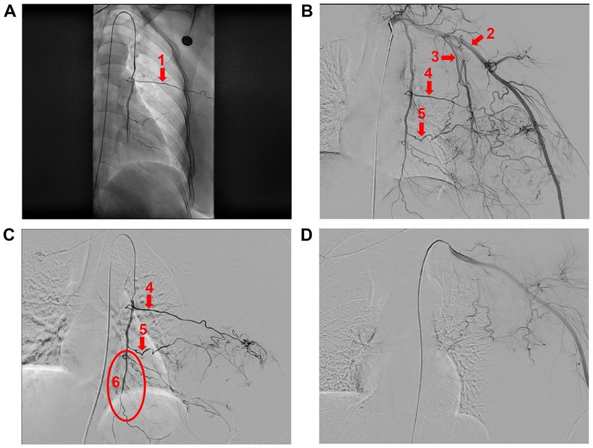
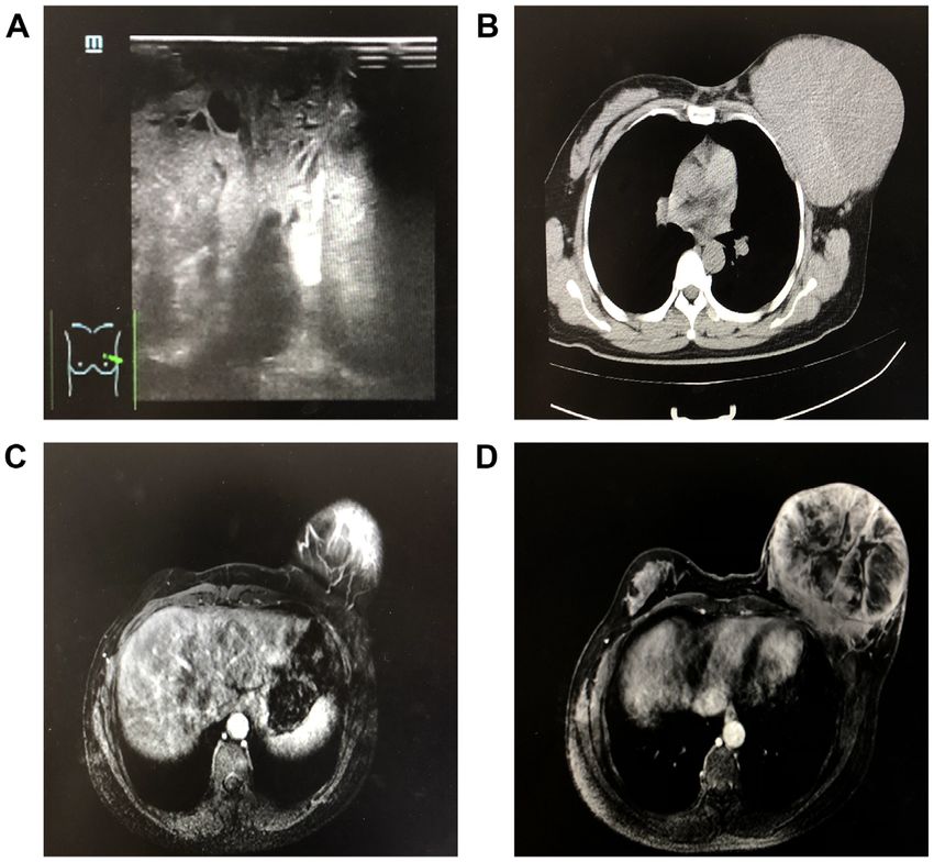
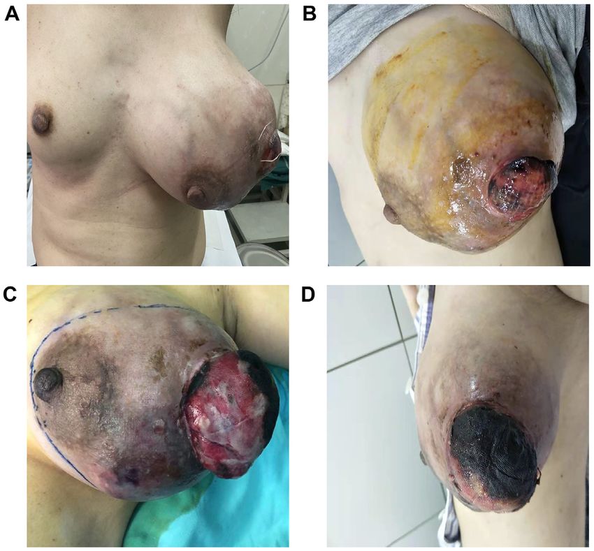
2                   TIAN et al: PREOPERATIVE INTERVENTIONAL EMBOLIZATION TO TREAT A GIANT MALIGNANT PT

Figure 1. Anteroposterior and lateral radiographs of the tumor before and after embolization. (A) Anteroposterior and (B) lateral film were acquired before and
after core needle biopsy. (C) Anteroposterior and (D) lateral film were taken 3 days after embolization. Following embolization, the growth rate of the tumor
decreased and the skin of the nipple and areola and tumor surface became ischemic and necrotic.

Figure 2. Imaging examination results of the tumor. (A) Color Doppler ultrasound and (B) T showed a giant circular tumor with a clear boundary occupying
the left breast. (C and D) MRI showed possible invasion of the major pectoralis muscle.

of breast cancer or childbearing. Upon admission to The First                    neous tissue (Fig. 1A and B). Palpation revealed that the tumor
Affiliated Hospital of Kunming Medical University, the tumor                     was slightly hard and elastic, and certain areas were fluctuant.
measured >20 cm in diameter and was larger than the right                        Because of hypermetabolism and chronic blood loss from
breast and the ulceration measured 2 cm in diameter; the                         the tumor, the patient’s heart rate increased to 128 beats per
engorgement of multiple veins was observed in the subcuta‑                       minute.

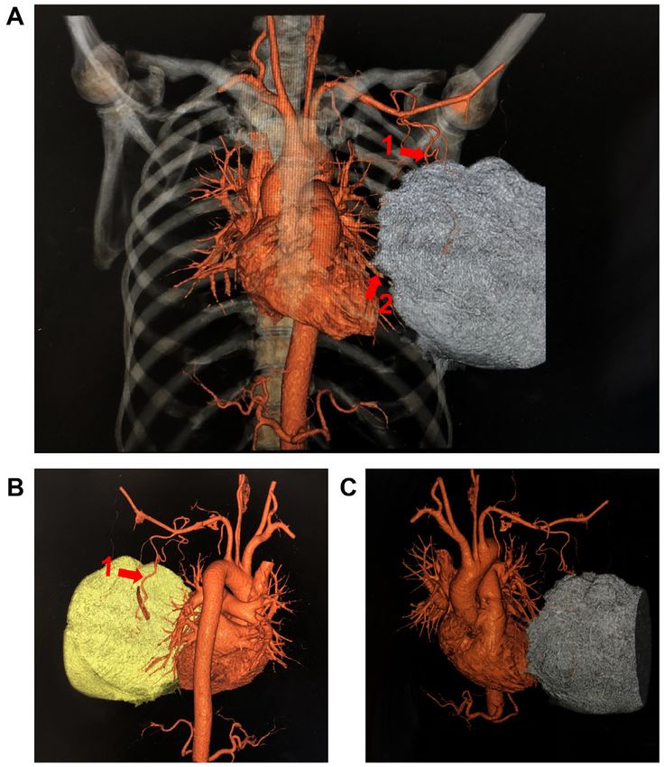
MOLECULAR AND CLINICAL ONCOLOGY 15: 133, 2021                                                                     3

Figure 3. Pathological examination of the tumor. (A) Preoperative core needle biopsy identified the tumor as malignant myxofibrosarcoma. (B and C) Postoperative
histology of the tumor. The malignant phyllodes tumor measured 17x16x11 cm. (D) Left axillary lymph node; 11 lymph nodes were found, with no tumor
metastasis.

Figure 4. CT angiography shows a large mass with a rich and powerful blood vessel supply. (A) Whole, (B) back and (C) front view of the tumor blood supply. Arrows 1
and 2 indicate the lateral and internal thoracic arteries, respectively.
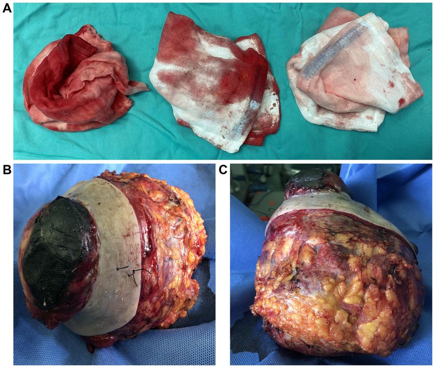
4                   TIAN et al: PREOPERATIVE INTERVENTIONAL EMBOLIZATION TO TREAT A GIANT MALIGNANT PT

Figure 5. Arteriography identifying the vascular blood supply to the tumor and performance of embolization. A single curved catheter was used to hook the
left subclavian artery for angiography, with superselective access to the internal and external thoracic arteries, and a gelatin sponge was used to protect the
distal end of the internal thoracic artery. (A) Superselective access to the internal thoracic artery (arrow 1). (B) Angiography showed that the primary blood
supply arteries of the tumor were the branches of the left internal thoracic artery and left subclavian artery (arrow 2). Arrow 3 indicates the external thoracic
artery; arrows 4 and 5 indicate the intercostal artery. (C) Branches of the internal thoracic artery were embolized using gelatin sponge particles and protective
embolization of the distal internal thoracic artery (arrow 6) was performed using a gelatin sponge. (D) Completed embolization.

Figure 6. Intraoperative bleeding and appearance of the tumor. (A) Amount of bleeding was ~50 ml, and
MOLECULAR AND CLINICAL ONCOLOGY 15: 133, 2021                                                               5

Figure 7. CT scan before postoperative radiotherapy and photos at the 14‑month follow‑up. (A) Srgical scar was signed by a galvanized wire on CT scan before
local radiotherapy. (B) Follow‑up image of the patient after 14 months.

rich blood supply inside. Multiple lymph nodes were enlarged                    thoracic arteries (Fig. 5A and B). Protective embolization
and rounded and hypoechoic nodules were observed in the left                    was performed distal to the internal thoracic artery using
axillary region (Fig. 2A). Computed tomography (CT) showed                      a gelatin sponge (Fig. 5C), and the 5F arterial sheath was
a giant tumor occupying the left breast and suspected invasion                  replaced with a 2.6F micropipe superselective to the domi‑
of the major pectoralis muscle, which indicated that the tumor                  nant blood‑supplying arteries guided by a 0.018‑inch micro
was potentially malignant (Fig. 2B). MRI showed a giant                         godet. Next, gelatin sponge granules (710‑1,000 µm) were
circular tumor with a clear boundary occupying the whole                        injected; imaging revealed that most of the staining in the
breast and a partially patchy unenhanced zone in the center                     tumor had disappeared (Fig. 5D). Following artery emboliza‑
measuring ~13x13x14 cm (upper and lower diameter x left and                     tion, fever and black necrotic tissue at the tumor site were
right diameter x before and after diameter). MRI also revealed                  observed. Fever resolved within 24 h, but the area of necrosis
possible invasion of the major pectoralis muscle and several                    on the tumor site expanded. No other serious complications
enlarged lymph nodes in the left axillary region, the largest of                were observed and the tumor stopped growing following
which measured ~1.6x0.8 cm. No abnormality was observed                         artery embolization (Fig. 1C and D).
in the entire right breast tissue (Fig. 2C and D). Preoperative                     Three days after artery embolization, the patient under‑
core needle biopsy (CNB) revealed that the biopsy specimen                      went surgery. Although axillary metastases of PTs are rare,
of the left breast was malignant myxofibrosarcoma. The                          mastectomy and level I axillary lymph node excision due to
immunohistochemical report was as follows: SMA (+), Des                         swelling were performed for safety after discussion. The tumor
(‑), KI‑67 (+)30%, CK5/6 (‑/+), P63 (‑), CK (‑) and VIM (+)                     measured 17x16x11 cm. The bleeding volume during the
(Fig. 3A). Additionally, the patient received comprehensive CT                  operation was
6                TIAN et al: PREOPERATIVE INTERVENTIONAL EMBOLIZATION TO TREAT A GIANT MALIGNANT PT

for antiangiogenic drugs. No recurrence was found at the            rare (14), and no complications were observed in the present
14‑month follow‑up (Fig. 7B).                                       case. However, potential complications associated with this
                                                                    type of operation, such as infection after tumor necrosis, distal
Discussion                                                          embolization and cerebral infarction (9), should be carefully
                                                                    monitored during the process.
PTs are rare but complex fibroepithelial lesions of the breast          The precise role of adjuvant radiotherapy remains debat‑
that are classified as benign, borderline or malignant (5). PTs     able, but it has been personalized for certain patients with
and malignant PTs present significant challenges in diagnosis,      malignant PT. Although radiotherapy has no significant effect
classification, predictive behavior and clinical treatment (10).    on survival rate, it can decrease local recurrence (5). When
From a diagnostic and management perspective, correctly             patients do not receive radiation and chemotherapy, they may
identifying malignant PTs is critical. They should be surgi‑        obtain passive results. For example, Kuo et al (6) described
cally eradicated and effectively treated at the time of diagnosis   patients who received preoperative embolization chemotherapy
because the risks of metastasis and death from such tumors,         and successful tumor suppression. However, chemotherapy is
although relatively small, are well‑established (5).                not an option for patients two months after recurrence (6). In
    PTs have non‑specific clinical and radiological manifesta‑      the present case, since the tumor invaded the muscle layer of
tions that can easily be confused with those of other similar       the chest wall, it was recommended that the patient receive
breast masses (11). Clinical and radiological diagnoses             chemotherapy and local radiotherapy to prevent recurrence;
are difficult, requiring joint confirmation from multiple           the 14‑month follow‑up after chemoradiotherapy was good.
assessments. In the present case, color Doppler ultrasound          This result suggests that, in the case of an MPTB, particularly
showed feature information that could assist in diagnosis.          an MPTB with chest wall invasion, chemoradiotherapy is
CT showed whether lung metastasis occurred and infiltration         necessary.
was assessed by MRI. However, these methods cannot help                 WGS and CTC detection lead to high‑resolution analysis
to make a definitive preoperative diagnosis. CNB is a widely        of cancers and reliably guided personalized therapy (17); the
used, highly sensitive method to obtain a preoperative diag‑        patient accepted CTC detection but rejected WGS. The CTC
nosis of breast cancer (12), although the tissue obtained in        detection results were negative.
CNB does not represent the whole tumor. Here, these results             Patients with large lesions are inclined to undergo
and clinical appearance were combined, and the diagnosis was        mastectomy. In cases of giant malignant PTs, preoperative
confirmed.                                                          intervention artery embolization can inhibit tumor growth,
    Surgical excision is the preferred procedure because it can     decrease bleeding during an operation, increase the operation
obtain a negative margin in the case of a final diagnosis of a      speed and shorten the period of postoperative convalescence.
malignant PT (5,11). Although mastectomy is the only treat‑         Preoperative intervention artery embolization was effective
ment method for large tumors, the extent of surgery (BCS            and safe for the present patient, and postoperative local radio‑
vs. mastectomy) for small tumors remains controversial (8).         therapy may be necessary for a good prognosis.
Though axillary lymph node metastasis is rare, some cases
of giant PT‑mixed breast cancer have been reported. In the          Acknowledgements
present case, the tumor grew rapidly and was very large. The
patient had metabolic consumption and risks were associated         Not applicable.
with surgery.
    Interventional embolization was developed to manage             Funding
vascular areas of bleeding, including in the brain, lung and
liver (13) and is an effective and safe choice (14). Vascular       The present report was supported by the National Natural
occlusion by embolization has also been used to control             Science Foundation of China (grant no. 81660438).
severe or recurrent bleeding in fungal breast cancer (15). In
the case of sternal erosion of breast cancer, embolization can      Availability of data and materials
effectively control bleeding and save lives (16). Interventional
embolization is used to decrease the preoperative breast tumor      The datasets used and/or analyzed during the current study are
volume and resection is performed 23 days after embolization.       available from the corresponding author on reasonable request.
Embolization effectively decreases intraoperative bleeding
and improves surgical results (16). In the present case, the left   Authors' contributions
breast tumor was primarily supplied by the left internal thoracic
artery branch and left internal thoracic artery perforator. The     FG, AJ and WC conceived and designed the study. YT, LC,
blood supply vessels were thickened, the blood supply was rich      SZ, RS, WZ and LL performed data acquisition and analysis.
and the tumor grew rapidly with bleeding. Therefore, catheter       YT, LL and FG drafted the manuscript. FG and LL reviewed
embolization was performed before surgery to slow tumor             the manuscript. All the authors read and approved the final
growth and decrease intraoperative bleeding. Three days             manuscript.
after embolization, resection was successfully performed. No
clear guidance exists to determine the operation time after         Ethics approval and consent to participate
embolization, and the operation should be performed carefully
according to the tumor growth rate, bleeding and ulceration for     Approval was obtained from the ethics committee of Kunming
each patient. Complications of interventional embolization are      Medical University. The procedures performed in this study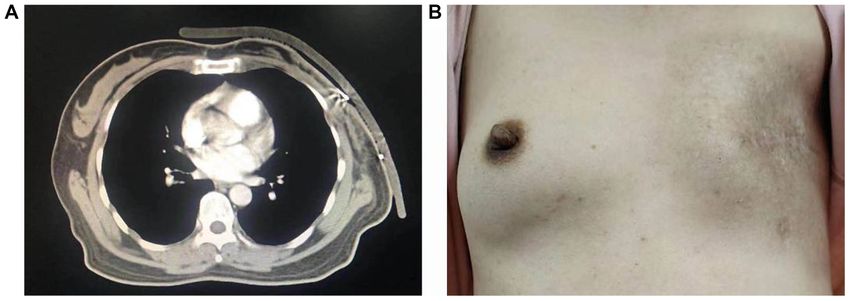
MOLECULAR AND CLINICAL ONCOLOGY 15: 133, 2021                                                     7

adhered to the tenets of the Declaration of Helsinki. Written           8. Mitus J, Reinfuss M, Mitus JW, Jakubowicz J, Blecharz P,
                                                                           Wysocki WW and Skotnicki P: Malignant phyllodes tumor
informed consent was obtained from the patient.                            of the breast: Treatment and prognosis. Breast J 20: 639‑644,
                                                                           2014.
Patient consent for publication                                         9. Hashimoto K, Mimura H, Arai Y, Doi M, Kojima Y,
                                                                           Tsugawa K and Nakajima Y: Successful preoperative
                                                                           chemoembolization in the treatment of a giant malignant
Written informed consent was obtained from the patient                     phyllodes tumor. Cardiovasc Intervent Radiol 39: 1070‑1075,
included in the study.                                                     2016.
                                                                       10. Moten AS and Goldberg AJ: Malignant phyllodes tumors of the
                                                                           breast: Association between race, clinical features, and outcomes.
Competing interests                                                        J Surg Res 239: 278‑283, 2019.
                                                                       11. Atalay C, Kınaş V and Çelebioğlu S: Analysis of patients with
The authors declare that they have no competing interests.                 phyllodes tumor of the breast. Ulus Cerrahi Derg 30: 129‑132,
                                                                           2014.
                                                                       12. Dillon MF, Quinn CM, McDermott EW, O’Doherty A,
References                                                                 O’Higgins N and Hill ADK: Needle core biopsy in the diagnosis
                                                                           of phyllodes neoplasm. Surgery 140: 779‑784, 2006.
 1. DeSantis C, Ma J, Bryan L and Jemal A: Breast cancer statistics,   13. Moriarty JM, Xing MZ and Loh CT: Particle embolization to
    2013. CA Cancer J Clin 64: 52‑62, 2014.                                control life‑threatening hemorrhage from a fungating locally
 2. Chang J, Denham L, Dong EK, Malek K and Lum SS: Trends                 advanced breast carcinoma: A case report. J Med Case Rep 6:
    in the diagnosis of phyllodes tumors and fibroadenomas before          186, 2012.
    and after release of WHO classification standards. Ann Surg        14. Corvino F, Giurazza F, Cangiano G, Cavaglià E, Amodio F,
    Oncol 25: 3088‑3095, 2018.                                             Magistris GD, Corvino A and Niola R: Safety and effectiveness of
 3. Krings G, Bean GR and Chen YY: Fibroepithelial lesions: The            transcatheter embolization in the treatment of internal mammary
    WHO spectrum. Semin Diagn Pathol 34: 438‑452, 2017.                    artery injuries. Radiol Med 123: 369‑377, 2018.
 4. Tan PH, Ellis I, Allison K, Brogi E, Fox SB, Lakhani S,            15. Aksoy Ş, Akçe B, Kiliçkesmez Ö, Gürsü RU, Çakır MS,
    Lazar AJ, Morris EA, Sahin A, Salgado R, et al: The 2019               Nazlı MA and Aren A: Transcatheter arterial embolization for
    WHO classification of tumours of the breast. Histopathology 77:        controlling severe bleeding from recurrent locally‑advanced
    181‑185, 2020.                                                         breast cancer. J Breast Health 12: 137‑140, 2016.
 5. Tan BY, Acs G, Apple SK, Badve S, Bleiweiss IJ, Brogi E,           16. Leung JWT, Gotway MB and Sickles EA: Preoperative
    Calvo JP, Dabbs DJ, Ellis IO, Eusebi V, et al: Phyllodes               embolization of vascular phyllodes tumor of the breast.
    tumours of the breast: A consensus review. Histopathology 68:          AJR Am J Roentgenol 184 (Suppl 3): S115‑S117, 2005.
    5‑21, 2016.                                                        17. Gulbahce N, Magbanua MJM, Chin R, Agarwal MR,
 6. Kuo CY, Lin SH, Lee KD, Cheng SJ, Chu JS and Tu SH:                    Luo XH, Liu J, Hayden DM, Mao Q, Ciotlos S, Li ZY, et al:
    Transcatheter arterial chemoembolization improves the resect‑          Quantitative whole genome sequencing of circulating
    ability of malignant breast phyllodes tumor with angiosarcoma          tumor cells enables personalized combination therapy of
    component: A case report. BMC Surg 19: 100, 2019.                      metastatic cancer. Cancer Res 77: 4530‑4541, 2017.
 7. Gradishar WJ, Anderson BO, Balassanian R, Blair SL,
    Burstein HJ, Cyr A, Elias AD, Farrar WB, Forero A,
    Giordano SH, et al: Breast cancer, version 4.2017, NCCN clinical                    This work is licensed under a Creative Commons
    practice guidelines in oncology. J Natl Compr Canc Netw 16:                         Attribution-NonCommercial-NoDerivatives 4.0
    310‑320, 2018.                                                                      International (CC BY-NC-ND 4.0) License.
You can also read


