Role of G2-S16 Polyanionic Carbosilane Dendrimer in the Prevention of Respiratory Syncytial Virus Infection In Vitro and In Vivo in Mice - MDPI
←
→
Page content transcription
If your browser does not render page correctly, please read the page content below
polymers
Article
Role of G2-S16 Polyanionic Carbosilane Dendrimer in the
Prevention of Respiratory Syncytial Virus Infection In Vitro
and In Vivo in Mice
Ignacio Rodriguez-Izquierdo 1,2 , Rafael Ceña-Diez 1 , Maria Jesús Serramia 1,2 , Rosa Rodriguez-Fernández 3 ,
Isidoro Martínez 4 and Mariángeles Muñoz-Fernández 1,2, *

                                          1   Immunology Section, Head Inmuno-Biology Molecular Laboratory, Gregorio Marañón University General
                                              Hospital (HGUGM), Gregorio Marañón Health Research Institute (IiSGM), 28007 Madrid, Spain;
                                              igna.iri.93@gmail.com (I.R.-I.); rcena48@gmail.com (R.C.-D.); mjserramia.hgugm@salud.madrid.org (M.J.S.)
                                          2   Spanish HIV HGM BioBank, C/Dr. Esquerdo 46, 28007 Madrid, Spain
                                          3   Hospital de Pediatría, Gregorio Marañón University General Hospital (HGUGM), Gregorio Marañón Health
                                              Research Institute (IiSGM), C/Dr. Esquerdo 46, 28007 Madrid, Spain; rrfernandez@salud.madrid.org
                                          4   Unidad de Infección Viral e Inmunidad, Centro Nacional de Microbiología, Instituto de Salud Carlos III,
                                              Majadahonda, 28007 Madrid, Spain; imago@isciii.es
                                          *   Correspondence: mmunoz.hgugm@gmail.com or mmunoz.hgugm@salud.madrid.org; Tel.: +34-91-462-4684

                                          Abstract: The respiratory syncytial virus (RSV) causes respiratory infection and bronchiolitis, requir-
                                          ing hospitalization mainly in infants. The interaction between RSV, envelope glycoproteins G and F,
                                and cell surface heparan sulfate proteoglycans (HSPG) is required for binding and entry into the host

                                          cells. A G2-S16 polyanionic carbosilane dendrimer was identified as a possible RSV inhibitor. We
Citation: Rodriguez-Izquierdo, I.;
                                          speculated that the G2-S16 dendrimer adheres to the host cell-surface HSPG, acts through binding
Ceña-Diez, R.; Serramia, M.J.;
                                          to HS receptors, and prevents further RSV infection. The G2-S16 dendrimer was non-toxic when
Rodriguez-Fernández, R.; Martínez, I.;
                                          applied intranasally to Balb/c mice, and interestingly enough, this G2-S16 dendrimer inhibits 85%
Muñoz-Fernández, M. Role of G2-S16
                                          RSV. Therefore, our G2-S16 dendrimer could be a candidate for developing a new possible therapy
Polyanionic Carbosilane Dendrimer
in the Prevention of Respiratory
                                          against RSV infection.
Syncytial Virus Infection In Vitro and
In Vivo in Mice. Polymers 2021, 13,       Keywords: RSV infection; G2-S16 dendrimer; syncytium; nanotechnology
2141. https://doi.org/10.3390/
polym13132141

Academic Editor: Diego Antonioli          1. Introduction
                                               Nanotechnology plays an important role in the treatment of broad viral infections.
Received: 9 June 2021
                                          Particularly, polyanionic carbosilane dendrimers, a class of hyper-branched polymers with
Accepted: 25 June 2021
                                          a nanoscale globular shape, well-defined functional groups at periphery, hydrophobic,
Published: 29 June 2021
                                          or hydrophilic cavities in the interior, and low polydispersity have proven to be broad-
                                          spectrum antivirals with promising results. Globally, RSV infection is estimated at 64
Publisher’s Note: MDPI stays neutral
                                          million cases and 160,000 deaths annually [1]. Current advances in the use of nanoparticle
with regard to jurisdictional claims in
                                          technology such as dendrimers have found new opportunities that address some limitations
published maps and institutional affil-
                                          in effective therapeutic agents. This RSV represents an important health problem in
iations.
                                          newborns, infants, young children, immunocompromised patients, and the elderly, due
                                          to the fact that there is no licensed vaccine for RSV and an effective therapeutic agent is
                                          needed [2–4]. RSV is the main cause of mortality and morbidity in infants until 2 years
                                          old [5].
Copyright: © 2021 by the authors.
                                               An early RSV infection treatment is mandatory to avoid lung tissue damage, the
Licensee MDPI, Basel, Switzerland.
                                          potential development of reactive airway disorders, and complications from sustained oxy-
This article is an open access article
                                          gen deprivation. Although different vaccines are being studied for respiratory viruses [6],
distributed under the terms and
                                          nanotechnology and different nanosystems such as gold nanomaterials have shown low
conditions of the Creative Commons
                                          toxicity and a wide range of biomedical applications, such as effective therapies [7]. Gold
Attribution (CC BY) license (https://
creativecommons.org/licenses/by/
                                          nanorods are being investigated as new possible treatments of RSV and for various biomed-
4.0/).
                                          ical applications [8]. Metallic nanoparticles show antiviral activity against respiratory

Polymers 2021, 13, 2141. https://doi.org/10.3390/polym13132141                                            https://www.mdpi.com/journal/polymers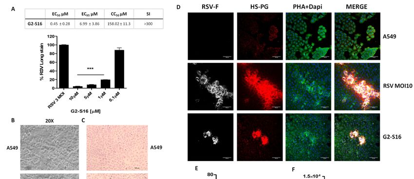
Polymers 2021, 13, x 2 of 14
Polymers 2021, 13, 2141 2 of 13
pies [7]. Gold nanorods are being investigated as new possible treatments of RSV and for
various biomedical applications [8]. Metallic nanoparticles show antiviral activity against
respiratory viruses [9]. Another study made with nanoparticles against RSV was shown
viruses
to have[9]. Another
very studydata
promising made as with nanoparticles
prophylactic against RSV
or therapeutic was shownplacing
approaches, to havenano-
very
promising
particles asdata as prophylactic
a very interesting or andtherapeutic approaches,
effective tool; meanwhile,placing
thenanoparticles as a very
vaccine is developed
interesting and effective tool; meanwhile, the vaccine is developed [10].
[10]. Additionally, mice models had improved the present airway epithelial cells culture Additionally,
mice models
as well as thehaddataimproved
obtained andthe present airway
mimic real epithelial
respiratory cells culture
infections as well as the data
[5,11,12].
obtained and mimic real respiratory infections [5,11,12].
The first infection step is mediated by the interactions of RSV envelope glycopro-
teinsThe firstF infection
G and stepsurface
and the cell is mediated
HSPGby the interactions
[13–20]. Indeed, toof RSV envelope
generate glycoproteins
a compound capable
G
of blocking the interaction between heparan sulfate (HS) and HBD, resulting capable
and F and the cell surface HSPG [13–20]. Indeed, to generate a compound of
in an inhi-
blocking the interaction between heparan sulfate (HS) and HBD, resulting in an inhibition of
bition of infection generated by different viruses, such as RSV, would be an interesting
infection generated by different viruses, such as RSV, would be an interesting therapeutic
therapeutic approach. This scenario makes the G2-S16 dendrimer an important tool for
approach. This scenario makes the G2-S16 dendrimer an important tool for antiviral
antiviral research, and the dendrimers could be a new possible effective therapeutic
research, and the dendrimers could be a new possible effective therapeutic agent against
agent against RSV. Our main objective has been to evaluate the G2-S16 dendrimer for its
RSV. Our main objective has been to evaluate the G2-S16 dendrimer for its possible anti-
possible anti-RSV activity and not only to research its mechanisms of action but also its
RSV activity and not only to research its mechanisms of action but also its biocompatibility
biocompatibility in human cells lines and in BALB/c mice.
in human cells lines and in BALB/c mice.
2. Materials
2. Materials and
and Methods
Methods
2.1. Dendrimers and Reagents
G2-S16 dendrimer (C112 112H
H244 NN
244 8Na
8 Na16O1648O S48
16Si 13;SiMw:
S16 3717.15
13 ; Mw: g/mol)
3717.15 and G2-S16-FITC
g/mol) and G2-S16-
dendrimer
FITC labeled
dendrimer labeled H249
(C129(C N H9 Na
129 249 9 N
14 ONa
47 S 15 Si
O 13 ;
14 47 15 13 3946.29
S Si ; g/mol)
3946.29 were
g/mol) synthesized
were and char-
synthesized and
acterized as previously
characterized described
as previously described [21–23]
[21–23] (Figure(Figure1A).1A).
Both dendrimers
Both dendrimers were dissolved
were in
dissolved
distillated
in water,
distillated and
water, dilution
and dilutiontotoµM µMrange
rangewas wasgenerated
generatedinindistillated
distillatedwater
water from
from stock.
stock.
Figure 1. (A)
Figure 1. (A) Schematic
Schematicrepresentation
representationofofG2-S16
G2-S16and
andG2-S16-FITC
G2-S16-FITC dendrimers.
dendrimers. (B–D)
(B–D)Viability of A549
Viability cellscells
of A549 waswas
evaluated
evalu-
by
atedMTT assayassay
by MTT after after
24 h 24(B),h 72 h 72
(B), (D), h or 5 days
(D), of exposure
or 5 days (C) in
of exposure (C)HEp-2 cellscells
in HEp-2 to a to
range of G2-S16
a range dendrimer
of G2-S16 dendrimerconcen-
con-
trations; 80%80%
centrations; of viability waswas
of viability set as
setthe
aslimit of toxicity
the limit for the
of toxicity forG2-S16 dendrimer.
the G2-S16 Dextran
dendrimer. 20 µM20and
Dextran µMDMSO
and DMSO10% were
10%
were as
used used as controls.
controls. Data Data
were were represented
represented as mean ± standard
as mean ± standard deviation
deviation of three
of three independent
independent experiments
experiments by tripli-
by triplicate.
cate.= NT
NT = non-treated
non-treated control;
control; DXT = DXT = dextran;
dextran; DMSO DMSO = dimethyl
= dimethyl sulfoxide.
sulfoxide.
2.2. Cells and Viruses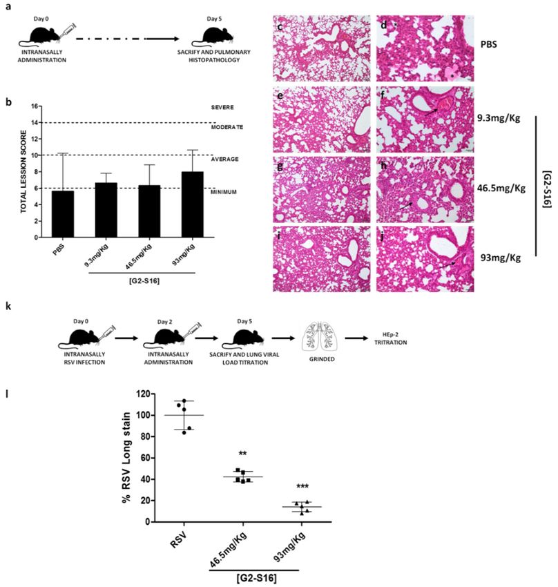
Polymers 2021, 13, 2141                                                                                          3 of 13

                          2.2. Cells and Viruses
                               The A549 human epithelial cell line (ATCC CCL-185, Manassas, VA, USA) and HEp-2
                          human epithelial cell line (ATCC CCL-23, Manassas, VA, USA) were grown and main-
                          tained in Dulbecco’s modified Eagle’s medium (DMEM; Biochrom AG, Berlin, Germany)
                          supplemented with 10% heat-inactivated fetal calf serum (FBS), a cocktail of antibiotics
                          (125 µg/mL ampicillin, 125 µg/mL cloxacillin, and 40 µg/mL gentamicin, Sigma, St Louis,
                          MO, USA), and 1% L-glutamine in 5% CO2 and 37 ◦ C.
                               This RSV (long Strain, ATCC VR-26) was obtained from the American Type Culture
                          Collection (ATCC, Manassas, VA, USA) and provided by Dr. Isidoro Martinez (Centro
                          Nacional de Microbiologia, Instituto de Salud Carlos III). This RSV was propagated in
                          HEp-2 cells as previously described [24,25] and G2-S16 dendrimer titers were determined
                          by plaque assay in HEp-2 cells.

                          2.3. Screening Procedure
                               G2-S16 dendrimer toxicity was screened by MTT assay (Sigma, St Louis, MO, USA)
                          according to manufacturer’s instructions. A range of concentrations between 0.1 and 20 µM
                          were evaluated for this G2-S16 dendrimer. Dextran 10 µM and DMSO 20 µM (Honeywell,
                          Charlotte, NC, USA) were used as control of cell viability and death, respectively, and
                          >80% cell viability was set as the minimum non-toxicity value. A549 or HEp-2 cells were
                          treated with G2-S16 dendrimer (0.1 to 20 µM), for 24 h in the case of A549 cells or 5 days in
                          the case of HEp-2 cells. MTT assay was added (0.5 mg/L), and generated crystals were
                          dissolved in DMSO. After 2 h, absorbance was read out in a Synergy 4 plate reader (BioTek,
                          Winooski, VT, USA) at 570/690 nm.

                          2.4. Inhibition Assay
                                To determine the antiviral capacity of G2-S16 dendrimer against RSV, the maximum
                          non-toxic concentration of the dendrimer was selected (10 µM). RSV infection was per-
                          formed on a monolayer of A549 cells in a 6-well plate. A549 cells (5 × 10⁵ ) were plated
                          and incubated for 24 h, washed with fresh DMEM medium at 2% SFB, infected with RSV
                          at 3 Multiplicity of Infection (MOI 3), and subsequently treated with G2-S16 dendrimer.
                          After 1 h at 37 ◦ C, the supernatant was removed, and the infection was left in 2 mL in fresh
                          medium at 2% FBS for 24 h at 37 ◦ C.
                                Viral titration was carried out on the HEp-2 cell line. HEp-2 cells were plated in a
                          p6, and the titration of each of the conditions was carried out, infecting with 400 µL of the
                          viral supernatants generated in the A549 cells serially diluted (10−1 , 10−2 and 10−3 ) and
                          the adsorption for 90 min at 37 ◦ C. After infection with the supernatants, 3 mL of DMEM
                          2% SFB with 0.7% low melting point agarose (LMP) were added per well and kept at 4 ◦ C
                          for 30 min so that the agarose solidified. The infection was maintained for 5 days at 37 ◦ C.
                                Plaques of lysis generated by RSV infection were revealed by immunostaining. Cells
                          were firstly fixed in 4% formaldehyde for 45 min at room temperature (RT); after removing
                          the semi-solid agarose medium, the monolayer was fixed with methanol for 5 min at RT.
                          Cells were washed twice and incubated with PBS-SAB 1% for 30 min at RT. Subsequently,
                          the PBS-SAB was removed, and primary antibody (1:50 in 1% PBS-SAB) was added for 1 h
                          at RT. Secondary antibody (anti-mouse-peroxidase 1:500 in PBS-SAB 1%) was added and
                          incubated for 1 h at RT. The secondary antibody was removed, and plates were washed
                          twice. Finally, the developing substrate was added ((citrate/phosphate 25/50 mM pH
                          5.0), 0.6 mL of 3-amino-9-ethylcarbazole (AEC) (Sigma, St Louis, MO, USA), 3.33 mg/mL
                          in DMSO, and 20 µL H2 O2 ) and after 20 min, the substrate was removed and the plates
                          were counted. The percentage of inhibition of each condition against RSV infection was
                          determined by comparing with controls.

                          2.5. Inhibitions of RSV Attachment to Host Cells
                               The mechanism of action of G2-S16 dendrimer was determined based on the results
                          obtained in the previous experiment, beginning to analyze the inhibition of RSV by means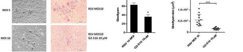
Polymers 2021, 13, 2141                                                                                            4 of 13

                          of the attachment mechanism, based on previously results obtained with G2-S16 dendrimer.
                          A549 cells were plated on a p6 as described above. After 24 h, the plate was pre-cooled at
                          4 ◦ C for 30 min. A549 cells were infected with RSV MOI 3 and simultaneously treated with
                          previously selected concentration (G2-S16 10 µM) for 1 h at 4 ◦ C. Subsequently, the cells
                          were washed with fresh DMEM medium with 2% FBS and incubated for 24 h at 37 ◦ C.
                                To titrate the RSV infection, HEp-2 cells were infected with the supernatants of the
                          cultures obtained in the A549 cell line, according to the procedure described in the previous
                          section, and 5 DPI, infection was revealed according to the protocol described above.

                          2.6. G2-S16 Dendrimer–Cell Interaction Assay
                                A549 cells were treated at maximum non-toxic concentrations of G2-S16 dendrimer
                          for 2 h to test the capability of the G2-S16 dendrimer to interact with the cellular receptors,
                          and it blocked the interaction of viral proteins with the host cell, inhibiting the infection.
                          A549 cells were washed to remove G2-S16 dendrimer that did not bind to the cell surface,
                          and MOI 3 RSV infection was performed. Then, 24 h post-infection, viral infection was
                          revealed in HEp-2 cells as mentioned before.

                          2.7. Binding of G2-S16 Dendrimer to RSV
                                 To test the percentage of inhibition due to the interaction between the G2-S16 den-
                          drimer and RSV, viral particles were pre-incubated at a MOI 3 with G2-S16 dendrimer
                          1 h at 4 ◦ C. HEp-2 cells were plated in a 96-well plate in DMEM medium at 2% FBS and
                          infected with the RSV-G2 inoculums for 1 h at 4 ◦ C. Cells were washed twice to remove
                          unbound virus and fixed with 4% formaldehyde and blocked with 5% SAB in PBS-Tween.
                          Cell-bound virus was determined by incubating a primary antibody (1:50 in PBS-Tween)
                          for 1 h at RT. Secondary antibody (anti-mouse–IgG peroxidase, 1:500 in PBS-Tween) was
                          added after washing the plates twice, and it was incubated for 2 h at 37 ◦ C. Then, the
                          secondary antibody was removed, plates were washed, and ABTS developing substrate
                          (2,20 -azino-bis- (3-ethylbenzothiazoline)-6-sulfonic acid)) (Thermo Scientific, Rock-ford, IL,
                          USA) was added and incubated for 20 min at RT. The reaction was stopped with 1% sodium
                          dodecyl sulfate (SDS) stop solution, and the absorbance at 405–410 nm was measured on
                          a microplate reader BioTek Synergy™ 4 Hybrid Microplate Reader (BioTek, Winooski,
                          VT, USA).

                          2.8. Syncytium Formation Assay
                                The ability of G2-S16 dendrimer to block cell-to-cell RSV spread was analyzed. A549
                          cell viability was determined by MTT assay at 72 h post-exposure as mentioned above. For
                          syncytium assay, A549 cells were infected with RSV at a MOI 10 and treated with a maxi-
                          mum non-toxic concentration of G2-S16 dendrimer or G2-S16-FITC dendrimer immediately
                          after RSV adsorption. Then, 72 h later, A549 cells were stained for hematoxylin/eosin
                          (H/E) or immune-stained. For H/E stain, A549 cells were washed with PBS and stained
                          10 min with hematoxylin and 10 min with eosin. Syncytium formation was evaluated
                          under light microscopy.
                                Syncytium inhibition was evaluated by the immunostaining of A549 cells. Cells were
                          infected and syncytium formation was determined by light microscopy. Once syncytia were
                          visualized, A549 monolayer were fixed (3.7–4% PFA; 10 min), permeabilized (0.1% Triton
                          X-100; 5 min), and incubated in PBS-BSA 5% 30 min. Monolayers were stained for RSV
                          fusion protein (anti-human RSV, Sino Biological Inc, Beijing, China) and cellular receptor
                          HSPG-2 (Sino Biological Inc, Beijing, China) (1:500 and 1:100 respectively). Phaloidin FITC
                          conjugated (1:100) was used for actin filaments visualization. Secondary antibody anti-
                          mouse IgG (Jackson InmunoResearch, Suffolk, UK) and anti-rabbit IgG (Thermo-Fisher
                          Scientific, Waltham, MA, USA) were incubated for 1 h, nuclei were stained with DAPI, and
                          preparations were observed in a Zeiss LSM710 confocal microscope by using Zen 2015
                          software (Carl Zeiss Microimaging Inc., Thornwood, NY, USA). Syncytium inhibition was
                          determined by counting number of syncytia in random files.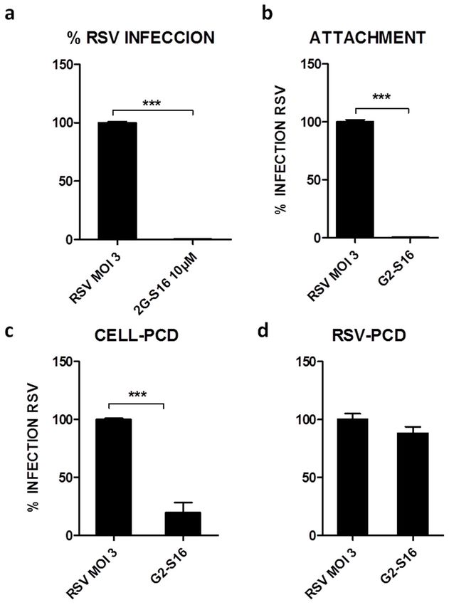
Polymers 2021, 13, 2141                                                                                          5 of 13

                          2.9. In Vivo Assays Statement
                                BALB/c mice studies were approved by the CBMSO Institutional Animal Care and
                          Use Committee (CEEA-CBMSO, Madrid, Spain) and carried out following CEEA-CBMSO,
                          National (Royal Decree 1201/2005), according to the Directive 2010/63/EU of the Euro-
                          pean Parliament guidelines and regulations. The ability of G2-S16 dendrimer to induce
                          irritation and damage to BALB/c mice lung when administrated intranasally was stud-
                          ied. Twelve BALB/c mice (Charles River Laboratories, Wilmington, MA) (22 ± 2 g and
                          8 weeks old) were purchased. G2-S16 dendrimer treatment was applied intranasally in
                          BALB/c mice previously anesthetized with isoflurane (Forane, Abbott, Madrid, Spain).
                          Therefore, BALB/c mice were randomized into four groups. The BALB/c mice control
                          group received 50 µL of PBS intranasally. The BALB/c mice G2-S16 dendrimer group
                          received 9.3 mg/kg, 46.5 mg/kg, or 93 mg/kg intranasally. Five days later, BALB/c mice
                          were sacrificed, and lungs were extracted and conserved in 3.7–4% PFA w/v (PanReac
                          AppliChem, Darmstadt, Germany).
                                The possible existence of inflammation, edema, alveolar congestion, bleeding, vascular
                          thrombosis, and congestion in pulmonary parenchyma and/or inflammatory infiltrate,
                          edema, bleeding, fibrosis, and neovascularization in pleura were evaluated in each sample.
                          Values (score) assigned for each of these lesions were 0–4 (no changes–very serious injuries)
                          as previously described [7].
                                Histological lesions in BALB/c mice lungs were evaluated with hematoxylin-eosin
                          staining as previously described [7]. The existence of injury in pulmonary parenchyma
                          and pleura was evaluated in each biological sample. These values were added, and the
                          determined level of pulmonary damage was determined as minimum (1–6), average (7–10),
                          moderate (10–14), or severe (>14).

                          2.10. In Vivo RSV Challenge Assay
                               BALB/c mice were randomized in the same four experimental groups as mentioned
                          above. All mice were intranasally infected with 2.5 × 10⁶ pfu of RSV while anesthetized
                          with isoflurane (Forane, Abbott, Madrid, Spain). G2-S16 dendrimer was applied in-
                          tranasally 2 DPI in anesthetized mice. BALB/c mice were sacrificed 5 DPI, and lungs
                          were extracted in order to extract RSV. The viral load of each lung was determined per-
                          forming serial dilutions of 5 mL of grinded RSV infected lungs and titrated on HEp-2 cells
                          as described.

                          2.11. Statistical Analysis
                               Statistical study was carried out with GraphPad Prism v7.0 software (GraphPad, San
                          Diego, CA, USA) using a t-Test of unpaired values and a non-parametric test, including
                          calculation of the mean, standard deviation, and p-values. The significance was solved
                          at p = 0.05. The data shown were analyzed from duplicate or triplicate in at least three
                          independent experiments, unless in vivo assays, where the n size and the pertinent analysis
                          were determined in its corresponding section.

                          3. Results
                          3.1. Biocompatibility of G2-S16 Polyanionic Carbosilane Dendrimer
                                The biocompatibility of G2-S16 and G2-S16-FITC dendrimers (Figure 1A) in A549
                          cells at 24 h (Figure 1B) or 72 h (Figure 1D) and in HEp-2 cells at 5 days (Figure 1C) were
                          evaluated by an MTT assay. The G2-S16 dendrimer concentration with viability > 80% in
                          comparison with the control group was regarded as non-toxic. The G2-S16 dendrimer was
                          not toxic at 10 µM. Biocompatibility at 72 h post-treatment in A549 cells showed that both
                          G2-S16 and G2-S16-FITC dendrimers were non-toxic at 5 µM.

                          3.2. G2-S16 Polyanionic Carbosilane Dendrimer against Respiratory Syncytial Virus
                               We studied the G2-S16 dendrimer at maximum non-toxic concentrations for MTT
                          assay. A549 cells were infected at an MOI 3 of RSV and treated at maximum non-toxic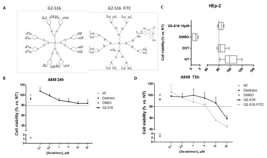
was not toxic at 10 µM. Biocompatibility at 72 h post-treatment in A549 cells showed that
                           both G2-S16 and G2-S16-FITC dendrimers were non-toxic at 5 µM.
Polymers 2021, 13, 2141                                                                                                                    6 of 13
                           3.2. G2-S16 Polyanionic Carbosilane Dendrimer against Respiratory Syncytial Virus
                                We studied the G2-S16 dendrimer at maximum non-toxic concentrations for MTT
                           assay. A549 cells were infected at an MOI 3 of RSV and treated at maximum non-toxic
                          concentrations
                           concentrations ofof G2-S16
                                               G2-S16 dendrimer in  in Hep-2
                                                                       Hep-2 cells.
                                                                              cells.The
                                                                                     TheG2-S16
                                                                                          G2-S16dendrimer
                                                                                                  dendrimer at at
                                                                                                               10 10
                                                                                                                   µMµM in-
                          inhibited  RSVinfection
                           hibited RSV     infectionbyby 99–100%
                                                         99–100% (Figure
                                                                   (Figure 2A). The G2-S16
                                                                                      G2-S16 dendrimer
                                                                                               dendrimer was
                                                                                                           was active
                                                                                                                 active atat
                          non-toxic
                           non-toxicconcentrations,
                                      concentrations,and
                                                       andthis
                                                            thisG2-S16
                                                                 G2-S16dendrimer
                                                                        dendrimerhalts
                                                                                     haltsRSV
                                                                                           RSVinfection
                                                                                               infection(p-value
                                                                                                         (p-value
Polymers 2021, 13, 2141 7 of 13
dendrimer also inhibits the attachment of RSV with A549 cells (99–100%; p-value < 0.0001)
(Figure 2B). This inhibition could be due to the interactions of the G2-S16 dendrimer either
with A549 cellular entry receptors or with viral proteins.
To test the first hypothesis, A549 cells were incubated 30 min with G2-S16 dendrimer
and we washed the unbound G2-S16 dendrimer prior to RSV infection, resulting in a
significant RSV inhibition of 83–84% (p < 0.01) (Figure 2C). The next step was to test the
other interaction of the G2-S16 dendrimer with the RSV virions hypothesis. In this case, not
significant inhibition was observed when the G2-S16 dendrimer was incubated with RSV
particles prior to infecting A549 cells (11–12%) (Figure 2D). These results are in line with
our previous experiments, demonstrating that the inhibitory action of G2-S16 dendrimer
was due to an interaction with host cell receptors.
3.4. Inhibition of Syncytium Formation
Syncytium formation is a well-known mechanism of cell-to-cell infection that con-
tributes significantly to virus spread in vivo. The RSV specifically uses syncytium formation
to spread infection. To determine whether G2-S16 dendrimer prevents cell-to-cell spread of
RSV after infection, a syncytium formation assay was performed on infected A549 cells
after 72 h. The G2-S16 dendrimer was not toxic at the range of concentrations evaluated on
A549 cells (Figure 1D). The inhibition range of the G2-S16 dendrimer was also measured
Polymers 2021, 13, x in A549 cells at a MOI 3 (Figure 3A) to determine EC50 and subsequently, therapeutic
8 of 14
index (SI).
Figure 3. Syncytium formation assay. (A) A549 cells were infected at a MOI 3 to evaluate the inhibition of G2-S16 den-
3. Syncytium
Figuredrimer. formation
EC50, EC95, assay.
CC50, and (A) A549
SI were cells were
calculated. infected
(B) A549 cells at a MOI
were 3 to at
infected evaluate the MOI
a different inhibition
of RSVoftoG2-S16
evaluate dendrimer.
the
EC50, syncytium
EC95, CC50, and SI were
formation (blackcalculated. (B) A549
asterisk). A549 cellsinfected
cells were were infected
at a MOIat 10
a different
(C–F) of MOI
RSV andof RSV to evaluate
treated the syncytium
with G2-S16 den-
drimer
formation in a range
(black of non-toxic
asterisk). concentrations.
A549 cells were infectedThen,
at a72MOI
h post-infection,
10 (C–F) of cells
RSVwereand (C) H/E stained
treated or immune-stained
with G2-S16 dendrimer infor a range
(D) RSV-F viral glycoprotein (white), HSPG (red), and actin filaments (phalloidin; green). Nuclei were stained with DAPI
of non-toxic concentrations. Then, 72 h post-infection, cells were (C) H/E stained or immune-stained for (D) RSV-F viral
(blue). (E) Media of syncytium formation/well after 72 h post-infection. (F) Media of syncytium size in four random
glycoprotein (white), HSPG
fields/experiment (red),
after 72 and actin filaments
h post-infection analyzed (phalloidin; green). Nuclei
with ImageJ software. Imageswere stained with of
are representative DAPI
three(blue). (E) Media
independ-
of syncytium formation/well
ent experiments. afterthe
Data showed 72 media
h post-infection.
± SD of three(F) Media of syncytium
independent experiments. size
*: pin four ***:
< 0.05; random fields/experiment after
p < 0.001.
72 h post-infection analyzed with ImageJ software. Images are representative of three independent experiments. Data
showed the media ± SD of three independentAs expected, in fields without
experiments. the presence
*: p < 0.05; of RSV, the G2-S16 dendrimer is located
***: p < 0.001.
inside A549 cells, mainly at the cellular surface (Figure 4; white arrows). These data are in
concordance with cellular protection assay results, where the G2-S16 dendrimer is able to
interact with A549 cells and protect RSV infection. In these fields where syncytium was
generated, the G2-S16-FITC dendrimer was visualized inside the syncytium (Figure 4),
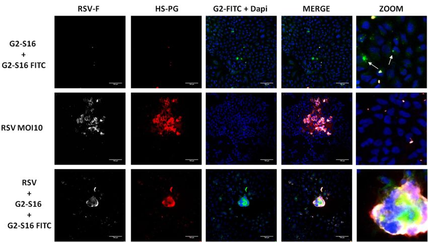
suggesting that it could interfere with adjacent cells, avoiding syncytium spread.Polymers 2021, 13, 2141 8 of 13
Firstly, A549 cells were infected with RSV at a different MOI to allow syncytium for-
mation and visualization. MOI 10 was selected for syncytium assays (Figure 3B). A549 cells
were infected with RSV, treated with G2-S16 dendrimer, and stained for H/E (Figure 3C).
Even the increase in RSV quantity, the G2-S16 dendrimer was able to reduce the number
and mainly the size of the syncytium generated (Figure 3E,F). Infected A549 cells were also
immune-stained to confirm inhibition observed in optical microscopy (Figure 3D). A549
cells were fixed 72 h post-infection and stained for RSV-F viral protein (white), HSPG (red),
phalloidin (green), and nuclei with DAPI (Figure 3D). Our data demonstrated that treat-
ment with G2-S16 dendrimer, even RSV infection, when performed at a MOI 10 reduced
significantly the syncytium quantity and mainly the size of the formed multinuclear cells.
Interestingly, when RSV infection occurs and syncytium is formed, the images reveal a
polarization in the main receptor of RSV, HSPG, favoring infection and co-locating with
RSV-F. We demonstrated that the G2-S16 dendrimer reduces syncytium formation, par-
ticularly the size of the syncytium, indicating a promising function against RSV infection.
Thus, the next step was to locate the G2-S16 dendrimer in syncytium formation.
The G2-S16-FITC labeled dendrimer was used to determine the position of the G2-S16
dendrimer in syncytium generation. A549 cells were infected at MOI 10 of RSV as men-
tioned above and treated with a mix of non-toxic concentrations (Figure 1) of G2-S16 den-
drimer (5 µM) and G2-S16-FITC (5 µM) to observe visualization of the dendrimers. Then,
Polymers 2021, 13, x 72 h post-infection, A549 cells were immune-stained for RSV-F glycoprotein and HSPG. 9 of 14
G2-S16-FITC was observed in green (Figure 4). DAPI was used for nuclei visualization.
Figure 4. Presence of G2-S16 dendrimer in syncytium formation. A549 cells were infected with RSV at MOI 10 and treated
Figure 4. Presence of G2-S16 dendrimer in syncytium formation. A549 cells were infected with RSV at MOI 10 and
with a mix of G2-S16 (5 µM) and G2-S16 FITC (5 µM) dendrimers. Then, 72 h post-infection, A549 cells were fixed and
treated with a mix
immune-stained forofRSV-F
G2-S16 (5 µM) and(white),
glycoprotein G2-S16 HSPG
FITC (5 dendrimers.
µM)and
(red), Then,
DAPI nuclei 72 h post-infection,
visualization A549 are
(blue). Images cellsrepre-
were
fixed and immune-stained for RSV-F glycoprotein
sentative of three independent experiments. (white), HSPG (red), and DAPI nuclei visualization (blue). Images are
representative of three independent experiments.
3.5 In Vivo Experiments: Murine Lung Model for Viability of G2-S16 Dendrimer and Respiratory
As expected,
Syncytial in fields without the presence of RSV, the G2-S16 dendrimer is located
Virus Challenge
inside A549 cells, mainly at the cellular surface (Figure 4; white arrows). These data are in
Once it was
concordance withdemonstrated that G2-S16
cellular protection dendrimer
assay results, inhibits
where RSV dendrimer
the G2-S16 and the underlying
is able to
mechanism of action by which the inhibition occurs, we studied if
interact with A549 cells and protect RSV infection. In these fields where G2-S16 dendrimer
syncytium was
induces irritation and/or damage in mice lungs when this G2-S16 dendrimer was ad-
ministrated intranasally in BALB/c mice. No damage or alteration to the lung epithelium
was shown when the G2-S16 dendrimer was applied intranasally at concentrations up to
46.5 mg/kg in mice lung. However, the G2-S16 dendrimer showed slight toxicity in lungs
(inflammation, vascular thrombosis, and congestion and atelectasis) at high concentra-Polymers 2021, 13, 2141 9 of 13
generated, the G2-S16-FITC dendrimer was visualized inside the syncytium (Figure 4),
suggesting that it could interfere with adjacent cells, avoiding syncytium spread.
3.5. In Vivo Experiments: Murine Lung Model for Viability of G2-S16 Dendrimer and Respiratory
Syncytial Virus Challenge
Once it was demonstrated that G2-S16 dendrimer inhibits RSV and the underlying
mechanism of action by which the inhibition occurs, we studied if G2-S16 dendrimer
induces irritation and/or damage in mice lungs when this G2-S16 dendrimer was admin-
istrated intranasally in BALB/c mice. No damage or alteration to the lung epithelium
was shown when the G2-S16 dendrimer was applied intranasally at concentrations up to
46.5 mg/kg in mice lung. However, the G2-S16 dendrimer showed slight toxicity in lungs
(inflammation, vascular thrombosis, and congestion and atelectasis) at high concentrations
of 93 mg/kg. However, those data suggest that the application method could be the reason
for the G2-S16 dendrimer toxicity in vivo (Table 1).
Table 1. Lung and pleura toxicity assay. The existence of injury in pulmonary parenchyma (inflammation, edema, alveolar
congestion, bleeding, and vascular thrombosis or congestion) was evaluated in each biological sample.
Pulmonary Parenchyma PBS 9.3 mg/kg 46.5 mg/kg 93 mg/kg
Inflammation 0 1 1 3 2 2 2 2 1 3 2 2 1
Edema 0 0 0 0 0 0 0 0 0 0 0 0
Alveolar congestion 0 0 3 1 1 2 1 2 3 2 1
Bleeding 1 0 1 1 0 1 0 0 0 0 0 0
Vascular thrombosis or
1 1 1 0 1 1 1 2 1 2 3 2
congestion
Atelectasis 1 1 3 2 2 2 2 1 3 3 2 1
Emphysema 0 0 0 0 0 0 0 0 0 0 0 0
Pleura PBS 9.3 mg/kg 46.5 mg/kg 93 mg/kg
Inflammatory infiltrate 0 0 0 0 0 0 0 0 0 0 0 0
Edema 0 0 0 0 0 0 0 0 0 0 0 0
Bleeding 0 0 0 0 0 0 0 0 0 0 0 0
Neovasculation 0 0 0 0 0 0 0 0 0 0 0 0
1 0 (no change); 1 (minimum); 2 (light); 3 (moderate); 4 (very serious). These values were added up to determine the level of vaginal
irritation as minimum 1–6, average 7–10, moderate 10–14, and severe 14+.
Histological analysis of mice lung clearly reveals the minimum or average damage in
all analyzed lungs. This is probably due to the application method, because PBS, which is
non-toxic control, also reveals average damage in all analyzed samples.
Due to the data obtained in an accumulated score, the presence of pulmonary injuries
in treatment with PBS showed that scores observed in G2-S16 dendrimer treatment are not
significantly related to PBS biocompatibility control, suggesting that the G2-S16 dendrimer
is safe for further in vivo assays (Figure 5). It is also important to note that no damage was
detected in pleura analysis in any of mice analyzed samples. Mice samples treated with
G2-S16 dendrimer present mainly an alveolar congestion and a moderate infiltrate, being
more serious in samples treated with 1 mM of G2-S16 dendrimer and minimum in those
BALB/c mice treated with 9.3 mg/Kg of G2-S16 dendrimer. Based on the data obtained,
the concentration of 93 mg/Kg is the best one as well as the concentration selected for RSV
in vivo challenge.tion selected for RSV in vivo challenge.
Finally, we also studied the antiviral activity of the G2-S16 dendrimer against RSV
challenge in BALB/C mice. Intranasal administration of the G2-S16 dendrimer reduced
RSV replication and infection in a significant manner. The administration of 50 µL of
Polymers 2021, 13, 2141 G2-S16 dendrimer 93 mg/Kg, after 4 h post-exposure to a high dose of RSV, abrogated 10 of 13
RSV infection in 86% (p < 0.0001 vs. placebo) (Figure 5). Interestingly and importantly, the
G2-S16 dendrimer at 46.5 mg/Kg inhibited RSV infection in 56% of BALB/C mice.
Figure 5.
Figure 5. In
In vivo
vivo experiments
experimentswith
withG2-S16
G2-S16dendrimer.
dendrimer.(a)(a)
Schematic
Schematicrepresentation of in
representation ofvivo via-
in vivo
bility procedure. (b) Media of the accumulate score for each treatment group. (c–j) Histological
viability procedure. (b) Media of the accumulate score for each treatment group. (c–j) Histological
studies of pulmonary damage. (b–d) Focal and minimal inflammatory infiltrate and atelectasis
studies of pulmonary damage. (b–d) Focal and minimal inflammatory infiltrate and atelectasis
around the presence of an acellular and eosinophilic substance (possibly PBS) (asterisk). (e,f) Alveolar
congestion of medium severity mainly around bronchioles and vessels in both cases (arrows). (g,h)
Thickening of the walls of the arterioles in the areas of inflammation (arrows). (i,j) More diffuse
lesions in the lung parenchyma with congestion and alveolar atelectasis. Presence of thrombi in
vessels (arrow). (k) Schematic representation of in vivo antiviral activity procedure. (l) Percentages
of inhibition of G2-S16 dendrimer corresponding to RSV infection of BALB/c mice and subsequent
titer of the lung infection HEp-2 cells for 5 days. The figure represents the relative mean of five mice
per group. RSV: BALB/c mice RSV infected and treated with PBS. **: p < 0.01; ***: p < 0.001.
Finally, we also studied the antiviral activity of the G2-S16 dendrimer against RSV
challenge in BALB/C mice. Intranasal administration of the G2-S16 dendrimer reduced
RSV replication and infection in a significant manner. The administration of 50 µL of
G2-S16 dendrimer 93 mg/Kg, after 4 h post-exposure to a high dose of RSV, abrogated
RSV infection in 86% (p < 0.0001 vs. placebo) (Figure 5). Interestingly and importantly, the
G2-S16 dendrimer at 46.5 mg/Kg inhibited RSV infection in 56% of BALB/C mice.
4. Discussion
RSV is the main cause of respiratory mortality and morbidity in persons of all
ages [28–30]. Particularly, RSV is the main cause of acute lower respiratory tract infec-
tions in children less than 5 years of age and responsible for substantial disease burden in
the elderly. The G2-S16 dendrimer presents different types of functionalized groups at their
periphery, including antiviral activities [31,32]. The biodistribution and biocompatibility of
the G2-S16 dendrimer have now been evaluated after administration at a systemic level,
and resistant mutation appearance was evaluated in HIV-1 infection [33].Polymers 2021, 13, 2141 11 of 13
Herein, we demonstrate that the G2-S16 dendrimer inhibits RSV infection not only
in an in vitro cellular model but also in a BALB/c mice in vivo model. Evaluation of the
cytotoxicity of the G2-S16 dendrimer has shown that this dendrimer is non-toxic in A549
cells at selected concentrations and that this G2-S16 dendrimer is capable of significantly
inhibiting RSV infection in the A549 cell line (>99%) (Figure 2). Studies of cytotoxicity
and inhibition curves made it possible to calculate the selectivity index (SI) of the G2-S16
dendrimer, enabling a value of >300 for RSV inhibition. Based on these previous results,
the mechanism of action by which the G2-S16 dendrimer inhibits RSV and the interaction
with A549 cells was studied.
Firstly, data obtained in attachment assay demonstrated that the inhibition by G2-S16
dendrimer (99–100%) (Figure 2) was due to the ability of the G2-S16 dendrimer to destabi-
lize or prevent the interactions of the RSV virions with the cell host receptors. This first step
confirms that G2-S16 dendrimer inhibition occurs in the entry early events of RSV infection.
Data also confirm that this inhibition is mediated by an interaction of G2-S16 dendrimer
with cellular receptors (Figure 2); meanwhile, the interaction of the G2-S16 dendrimer
with the RSV viral particles does not generate a significant effect in the RSV infection
(Figure 2). All these data demonstrate that the G2-S16 dendrimer is capable of interacting
with host cellular receptors implied in the RSV infection. We previously described that
G2-S16 dendrimer interacts with cellular HS and HBD in HSV infection [34–37]. We also
previously described that the G2-S16 dendrimer could interact electrostatically with the
viral glycoproteins of the viral surface as well as cellular receptors [21,22]. Interestingly, the
RSV cellular entry mediators in the viral infection are mainly the HSPG, in such a way that
it allows us to hypothesize that the RSV inhibition demonstrated by the G2-S16 dendrimer
could be due to the interaction of the dendrimer with HSPG of the cellular surface, blocking
this crucial initial step of the RSV infection.
In addition to the mechanism of action, we described the ability of the G2-S16 den-
drimer to halt RSV infection in vitro related to the syncytium formation and size in pul-
monary A549 cells (Figure 3). Our data suggest that the G2-S16 dendrimer halts syncytium
formation targeting cellular receptors, which are data that are in concordance with the
results obtained in mechanism of action assays.
Due to the potential use of the G2-S16 dendrimer as a possible treatment against RSV
in vivo, we studied its biocompatibility in BALB/C mice. Our results clearly showed that
the G2-S16 dendrimer applied intranasally at 93 mg/kg in BALB/c mice was well tolerated,
and no significant signs or damage to pulmonary parenchyma or pleura were observed
(Table 1). In addition, we also analyzed the antiviral activity of the G2-S16 dendrimer in
BALB/c mice, and we demonstrated that it decreased by 86% when RSV infected BALB/c
mice were treated with this G2-S16 dendrimer (Figure 5).
5. Conclusions
The G2-S16 dendrimer was demonstrated to be an effective treatment against RSV
infection. G2-S16 inhibits RSV infection in vitro and in vivo, and it presents good biocom-
patibility in vivo. In conclusion, G2-S16 dendrimer therapeutic application needs careful
design and treatment regimen to be considered effective for RSV treatment. G2-S16 den-
drimer in combination with biocompatible drugs and/or molecules could be a promising
therapeutic for that infection. The use of G2-S16 dendrimer could also contribute to finding
a solution to prevent RSV infection in infants and other target populations.
Author Contributions: Conceptualization, R.R.-F., I.M. and M.M.-F.; Data curation, I.R.-I., R.C.-D.
and M.J.S.; Formal analysis, R.R.-F. and I.M.; Funding acquisition, M.M.-F.; Investigation, I.R.-I.
and R.C.-D.; Methodology, I.R.-I., R.C.-D. and M.J.S.; Software, I.R.-I. and R.C.-D.; Supervision,
M.M.-F.; Validation, R.R.-F., I.M. and M.M.-F.; Writing—original draft, I.R.-I., R.C.-D. and R.R.-F.;
Writing—review & editing, I.M. and M.M.-F. All authors have read and agreed to the published
version of the manuscript.
Funding: This work has been (partially) funded by the RD16/0025/0019, projects as part of Acción
Estratégica en Salud, Plan Nacional de Investigación Científica, Desarrollo e Innovación TecnológicaPolymers 2021, 13, 2141 12 of 13
(2013–2016) and co-financed by Instituto de Salud Carlos III (Subdirección General de Evaluación) and
Fondo Europeo de Desarrollo Regional (FEDER), RETIC PT17/0015/0042, Fondo de Investigacion
Sanitaria (FIS) (grant number PI19/01638) and EPIICAL project. CIBER-BBN is an initiative funded
by the VI National R&D&i Plan 2008–2011, Iniciativa Ingenio 2010, the Consolider Program, and
CIBER Actions and financed by the Instituto de Salud Carlos III with assistance from the European
Regional Development Fund. This work has been supported partially by the EUROPARTNER:
Strengthening and spreading international partnership activities of the Faculty of Biology and
Environmental Protection for interdisciplinary research and innovation of the University of Lodz
Programme: NAWA International Academic Partnership Programme. This article/publication is
based upon work from COST Action CA 17140 “Cancer Nanomedicine from the Bench to the Bedside”
supported by COST (European Cooperation in Science and Technology).
Institutional Review Board Statement: The study was conducted according to the guidelines of the
Declaration of Helsinki. Animal studies were approved by Ethic Experimental Animals Committee of
the Centro de Biología Molecular “Severo Ochoa” (EEACCBMSO Institutional Animal Care and Use
Committee (EEAC-CBMSO, Madrid, Spain)). (PROES 136/14; Register number ES-280790000180).
All experiments were carried out following EECA-CBMSO, National (Royal Decree 1201/2005) and
the Directive 2010/63/EU of the European Parliament guidelines and regulations.
Informed Consent Statement: Not applicable.
Conflicts of Interest: The authors declare no conflict of interest. The funders had no role in the design
of the study; in the collection, analyses, or interpretation of data; in the writing of the manuscript, or
in the decision to publish the results.
References
1. Valdez, J.; Bawage, S.; Gomez, I.; Singh, S.R. Facile and rapid detection of respiratory syncytial virus using metallic nanoparticles.
J. Nanobiotechnol. 2016, 14, 13. [CrossRef]
2. Mazur, N.I.; Higgins, D.; Nunes, M.C.; Melero, J.A.; Langedijk, A.C.; Horsley, N.; Buchholz, U.J.; Openshaw, P.J.; McLellan, J.S.;
Englund, J.A.; et al. The respiratory syncytial virus vaccine landscape: Lessons from the graveyard and promising candidates.
Lancet Infect. Dis. 2018, 18, e295–e311. [CrossRef]
3. Mejias, A.; Rodriguez-Fernandez, R.; Peeples, M.E.; Ramilo, O. Respiratory syncytial virus vaccines: Are we making progress?
Pediatr. Infect. Dis. J. 2019, 38, e266–e269. [CrossRef]
4. Mejias, A.; Rodriguez-Fernandez, R.; Oliva, S.; Peeples, M.E.; Ramilo, O. The journey to a respiratory syncytial virus vaccine.
Ann. Allergy Asthma Immunol. 2020, 125, 36–46. [CrossRef]
5. Santos, L.D.; Antunes, K.H.; Muraro, S.P.; de Souza, G.F.; da Silva, A.G.; de Souza Felipe, J.; Zanetti, L.C.; Czepielewski, R.S.;
Magnus, K.; Scotta, M.; et al. Tnf-mediated alveolar macrophage necroptosis drives disease pathogenesis during respiratory
syncytial virus infection. Eur. Respir. J. 2020, 57, 2003764. [CrossRef] [PubMed]
6. Papadopoulos, N.G.; Megremis, S.; Kitsioulis, N.A.; Vangelatou, O.; West, P.; Xepapadaki, P. Promising approaches for the
treatment and prevention of viral respiratory illnesses. J. Allergy Clin. Immunol. 2017, 140, 921–932. [CrossRef] [PubMed]
7. Rodriguez-Izquierdo, I.; Seramia, M.J.; Gomez, R.; de la Mata, F.J.; Bullido, M.J.; Munoz-Fernandez, M.A. Gold nanoparticles
crossing blood-brain barrier prevent hsv-1 infection and reduce herpes associated amyloid-betasecretion. J. Clin. Med. 2020,
9, 155.
8. Pissuwan, D.; Niidome, T. Polyelectrolyte-coated gold nanorods and their biomedical applications. Nanoscale 2015, 7, 59–65.
[CrossRef]
9. Bawage, S.S.; Tiwari, P.M.; Singh, A.; Dixit, S.; Pillai, S.R.; Dennis, V.A.; Singh, S.R. Gold nanorods inhibit respiratory syncytial
virus by stimulating the innate immune response. Nanomedicine 2016, 12, 2299–2310. [CrossRef]
10. Markoutsa, E.; McGill, A.R.; Singer, A.; Jadhav, H.; Mohapatra, S.; Mohapatra, S.S. A multifunctional nanoparticle as a prophylactic
and therapeutic approach targeting respiratory syncytial virus. Nanomed. Nanotechnol. Biol. Med. 2020, 32, 102325. [CrossRef]
11. Nikonova, A.; Shilovskiy, I.; Galitskaya, M.; Sokolova, A.; Sundukova, M.; Dmitrieva-Posocco, O.; Mitin, A.; Komogorova,
V.; Litvina, M.; Sharova, N.; et al. Respiratory syncytial virus upregulates il-33 expression in mouse model of virus-induced
inflammation exacerbation in ova-sensitized mice and in asthmatic subjects. Cytokine 2020, 138, 155349. [CrossRef]
12. Kellar, G.G.; Barrow, K.A.; Rich, L.M.; Debley, J.S.; Wight, T.N.; Ziegler, S.F.; Reeves, S.R. Loss of versican and production of
hyaluronan in lung epithelial cells are associated with airway inflammation during rsv infection. J. Biol. Chem. 2020, 296, 100076.
[CrossRef]
13. Bourgeois, C.; Bour, J.B.; Lidholt, K.; Gauthray, C.; Pothier, P. Heparin-like structures on respiratory syncytial virus are involved
in its infectivity in vitro. J. Virol. 1998, 72, 7221–7227. [CrossRef] [PubMed]
14. Escribano-Romero, E.; Rawling, J.; Garcia-Barreno, B.; Melero, J.A. The soluble form of human respiratory syncytial virus
attachment protein differs from the membrane-bound form in its oligomeric state but is still capable of binding to cell surface
proteoglycans. J. Virol. 2004, 78, 3524–3532. [CrossRef]Polymers 2021, 13, 2141 13 of 13
15. Feldman, S.A.; Audet, S.; Beeler, J.A. The fusion glycoprotein of human respiratory syncytial virus facilitates virus attachment
and infectivity via an interaction with cellular heparan sulfate. J. Virol. 2000, 74, 6442–6447. [CrossRef]
16. Feldman, S.A.; Hendry, R.M.; Beeler, J.A. Identification of a linear heparin binding domain for human respiratory syncytial virus
attachment glycoprotein g. J. Virol. 1999, 73, 6610–6617. [CrossRef]
17. Harris, J.; Werling, D. Binding and entry of respiratory syncytial virus into host cells and initiation of the innate immune response.
Cell. Microbiol. 2003, 5, 671–680. [CrossRef]
18. Karger, A.; Schmidt, U.; Buchholz, U.J. Recombinant bovine respiratory syncytial virus with deletions of the g or sh genes: G and
f proteins bind heparin. J. Gen. Virol. 2001, 82, 631–640. [CrossRef]
19. Krusat, T.; Streckert, H.J. Heparin-dependent attachment of respiratory syncytial virus (rsv) to host cells. Arch. Virol. 1997, 142,
1247–1254. [CrossRef]
20. Martinez, I.; Melero, J.A. Binding of human respiratory syncytial virus to cells: Implication of sulfated cell surface proteoglycans.
J. Gen. Virol. 2000, 81, 2715–2722. [CrossRef]
21. Arnaiz, E.; Doucede, L.I.; Garcia-Gallego, S.; Urbiola, K.; Gomez, R.; Tros de Ilarduya, C.; de la Mata, F.J. Synthesis of cationic
carbosilane dendrimers via click chemistry and their use as effective carriers for DNA transfection into cancerous cells. Mol.
Pharm. 2012, 9, 433–447. [CrossRef]
22. Galan, M.; Sanchez Rodriguez, J.; Jimenez, J.L.; Relloso, M.; Maly, M.; de la Mata, F.J.; Munoz-Fernandez, M.A.; Gomez, R.
Synthesis of new anionic carbosilane dendrimers via thiol-ene chemistry and their antiviral behaviour. Org. Biomol. Chem. 2014,
12, 3222–3237. [CrossRef]
23. Rasines, B.; Sanchez-Nieves, J.; Maiolo, M.; Maly, M.; Chonco, L.; Jimenez, J.L.; Munoz-Fernandez, M.A.; de la Mata, F.J.; Gomez, R.
Synthesis, structure and molecular modelling of anionic carbosilane dendrimers. Dalton Trans. 2012, 41, 12733–12748. [CrossRef]
24. Martinez, I.; Dopazo, J.; Melero, J.A. Antigenic structure of the human respiratory syncytial virus g glycoprotein and relevance of
hypermutation events for the generation of antigenic variants. J. Gen. Virol. 1997, 78 Pt 10, 2419–2429. [CrossRef]
25. Mbiguino, A.; Menezes, J. Purification of human respiratory syncytial virus: Superiority of sucrose gradient over percoll,
renografin, and metrizamide gradients. J. Virol. Methods 1991, 31, 161–170. [CrossRef]
26. Guerrero-Beltran, C.; Rodriguez-Izquierdo, I.; Serramia, M.J.; Araya-Duran, I.; Marquez-Miranda, V.; Gomez, R.; de la Mata, F.J.;
Leal, M.; Gonzalez-Nilo, F.; Munoz-Fernandez, M.A. Anionic carbosilane dendrimers destabilize the gp120-cd4 complex blocking
hiv-1 entry and cell to cell fusion. Bioconjug. Chem. 2018, 29, 1584–1594. [CrossRef] [PubMed]
27. Rusnati, M.; Vicenzi, E.; Donalisio, M.; Oreste, P.; Landolfo, S.; Lembo, D. Sulfated k5 escherichia coli polysaccharide derivatives:
A novel class of candidate antiviral microbicides. Pharm. Ther. 2009, 123, 310–322. [CrossRef] [PubMed]
28. Thomas, E.; Mattila, J.M.; Lehtinen, P.; Vuorinen, T.; Waris, M.; Heikkinen, T. Burden of respiratory syncytial virus infection
during the first year of life. J. Infect. Dis. 2020, 223, 811–817. [CrossRef]
29. Pena, M.; Jara, C.; Flores, J.C.; Hoyos-Bachiloglu, R.; Iturriaga, C.; Medina, M.; Carcey, J.; Espinoza, J.; Bohmwald, K.; Kalergis,
A.M.; et al. Severe respiratory disease caused by human respiratory syncytial virus impairs language learning during early
infancy. Sci. Rep. 2020, 10, 22356. [CrossRef]
30. Behzadi, M.A.; Leyva-Grado, V.H. Overview of current therapeutics and novel candidates against influenza, respiratory syncytial
virus, and middle east respiratory syndrome coronavirus infections. Front. Microbiol. 2019, 10, 1327. [CrossRef]
31. Dzmitruk, V.; Szulc, A.; Shcharbin, D.; Janaszewska, A.; Shcharbina, N.; Lazniewska, J.; Novopashina, D.; Buyanova, M.;
Ionov, M.; Klajnert-Maculewicz, B.; et al. Anticancer sirna cocktails as a novel tool to treat cancer cells. Part (b). Efficiency of
pharmacological action. Int. J. Pharm. 2015, 485, 288–294. [CrossRef]
32. Perise-Barrios, A.J.; Gomez, R.; Corbi, A.L.; de la Mata, J.; Dominguez-Soto, A.; Munoz-Fernandez, M.A. Use of carbosilane
dendrimer to switch macrophage polarization for the acquisition of antitumor functions. Nanoscale 2015, 7, 3857–3866. [CrossRef]
[PubMed]
33. Rodriguez-Izquierdo, I.; Natalia, C.; Garcia, F.; los Ángeles Munoz-Fernandez, M. G2-s16 sulfonate dendrimer as new therapy for
treatment failure in hiv-1 entry inhibitors. Nanomedicine 2019, 14, 1095–1107. [CrossRef] [PubMed]
34. Cena-Diez, R.; Vacas-Cordoba, E.; Garcia-Broncano, P.; de la Mata, F.J.; Gomez, R.; Maly, M.; Munoz-Fernandez, M.A. Prevention
of vaginal and rectal herpes simplex virus type 2 transmission in mice: Mechanism of antiviral action. Int J. Nanomed. 2016, 11,
2147–2162.
35. Sepulveda-Crespo, D.; Sanchez-Rodriguez, J.; Serramia, M.J.; Gomez, R.; De La Mata, F.J.; Jimenez, J.L.; Munoz-Fernandez,
M.A. Triple combination of carbosilane dendrimers, tenofovir and maraviroc as potential microbicide to prevent hiv-1 sexual
transmission. Nanomedicine 2015, 10, 899–914. [CrossRef] [PubMed]
36. Rodriguez-Izquierdo, I.; Gasco, S.; Munoz-Fernandez, M.A. High preventive effect of g2-s16 anionic carbosilane dendrimer
against sexually transmitted hsv-2 infection. Molecules 2020, 25, 2965. [CrossRef] [PubMed]
37. Guerrero-Beltran, C.; Garcia-Heredia, I.; Cena-Diez, R.; Rodriguez-Izquierdo, I.; Serramia, M.J.; Martinez-Hernandez, F.; Lluesma-
Gomez, M.; Martinez-Garcia, M.; Munoz-Fernandez, M.A. Cationic dendrimer g2-s16 inhibits herpes simplex type 2 infection
and protects mice vaginal microbiome. Pharmaceutics 2020, 12, 515. [CrossRef]You can also read


