Milk Immune Cell Composition in Dromedary Camels With Subclinical Mastitis
←
→
Page content transcription
If your browser does not render page correctly, please read the page content below
ORIGINAL RESEARCH
                                                                                                                                                 published: 14 April 2022
                                                                                                                                         doi: 10.3389/fvets.2022.885523

                                             Milk Immune Cell Composition in
                                             Dromedary Camels With Subclinical
                                             Mastitis
                                             Gader Abdulaziz Alhafiz, Fatema Hassan Alghatam, Hams Almohammed and
                                             Jamal Hussen*
                                             Department of Microbiology, College of Veterinary Medicine, King Faisal University, Al-Ahsa, Saudi Arabia

                                             Mastitis represents one of the most important infectious diseases in camels with heavy
                                             economic losses due to reduced milk quantity and quality. Balanced immune cell
                                             composition and function in the mammary gland are essential for effective immune
                                             response to mastitis pathogens. The objective of the present study was to characterize
                                             the cellular immune response to subclinical mastitis in the mammary gland of dromedary
                                             camels. Therefore, immunostaining and flow cytometry were used to compare the cellular
                                             composition, leukocyte phenotype, and cell viability in camel milk from healthy she-
                                             camels (n = 8) and she-camels with subclinical mastitis (SCM; n = 6). In addition,
                                             the ex vivo phagocytic activity of milk phagocytes was compared between healthy and
                                             affected animals. The health status of the mammary gland was evaluated based on
                         Edited by:
       Abdelmalik Ibrahim Khalafalla,        the California Mastitis Test (CMT) score. SCM (CMT score of ≥3 in the absence of
 Independent Researcher, Abu Dhabi,          clinical signs of mastitis) was found in six of the 56 sampled quarters (10.7 %) with
               United Arab Emirates
                                             only one affected quarter per animal. In comparison to milk from healthy camels, milk
                       Reviewed by:
                      Maryam Dadar,
                                             from SCM animals showed higher somatic cell count (SCC), higher numbers of CD45+
    Razi Vaccine and Serum Research          leukocytes with an expanded fraction of CD172a+ myeloid cells. Within the myeloid cell
                         Institute, Iran     population, there was an increase in the percentage of granulocytes (CD172a+ CD14low )
            Mohanned Naif Alhussien,
                Technical University of      with a decreased percentage of macrophages (CD172a+ CD14high ) in milk from affected
                    Munich, Germany          animals compared to healthy animals. The decrease in lymphoid cells in SCM milk
                  *Correspondence:           was mainly due to the decreased fraction of CD4+ helper T cells. Camel SCM was
                       Jamal Hussen
                  jhussen@kfu.edu.sa
                                             also associated with a stimulated phenotype, increased cell viability, and enhanced
                                             phagocytic activity of the milk phagocytes, macrophages and granulocytes. Collectively,
                    Specialty section:       the present study identified significant changes in SCC, leukocyte count, phenotype,
         This article was submitted to
                                             viability, and function in association with subclinical mastitis in camels. The results of the
          Veterinary Experimental and
                 Diagnostic Pathology,       present study support a better understanding of host-pathogen interaction mechanisms
                a section of the journal     in the camel mammary gland.
        Frontiers in Veterinary Science
                                             Keywords: dromedary camel (Camelus dromedarius), subclinical mastitis (SCM), immune cells, flow cytometry,
         Received: 28 February 2022
                                             phagocytosis, bacteria
          Accepted: 23 March 2022
            Published: 14 April 2022

                               Citation:
             Alhafiz GA, Alghatam FH,
                                             INTRODUCTION
Almohammed H and Hussen J (2022)
     Milk Immune Cell Composition in
                                             Dromedary camels are well-adapted animals to the harsh environment of semiarid and arid zones
  Dromedary Camels With Subclinical          with the ability to produce milk of valuable quantity (1). The increased reports on the nutritional
   Mastitis. Front. Vet. Sci. 9:885523.      and health-promoting properties of camel milk resulted in a currently raised interest in camel milk
     doi: 10.3389/fvets.2022.885523          with growing market demand (2, 3).

Frontiers in Veterinary Science | www.frontiersin.org                               1                                               April 2022 | Volume 9 | Article 885523
Alhafiz et al.                                                                                                    Cellular Immunity to Camel Mastitis

   Like other dairy animals, camels may be affected by all types of          bacteria (38). The major histocompatibility complex (MHC) class
mastitis (4–7). Mastitis represents an inflammatory disease of the           II, an antigen-presentation receptor, and the scavenger receptor
mammary gland mainly caused by bacterial pathogens. Unlike                   CD163 are commonly used for the analysis of the functional
the clinical form of the disease, subclinical mastitis is difficult to       subtype of macrophages (39). CD4 and workshop cluster 1
be detected, because it does not cause any visible changes in milk           (WC1) antigen are cell markers that identify helper ab T cells and
or udder appearance, like swelling and redness (8). In addition              gd T cells, respectively (35).
to its effects on animal health and welfare, subclinical mastitis is             Milk phagocytes, including macrophages and neutrophils,
associated with huge economic losses due to the reduced milk                 are the primary effector cells of the mammary gland innate
yield and quality and high treatment costs. Moreover, it is a public         immune system with a key role during mammary gland
health concern for camel milk consumers (9).                                 infections (17). They contribute to the early elimination of
   The characterization of the innate and adaptive immune                    bacterial pathogens by several antimicrobial functions, including
response of the mammary gland to invading pathogens is                       phagocytosis, production of reactive oxygen and nitrogen species,
essential for the prevention and control of mastitis (10). In                and formation of extracellular traps (40).
addition to milk immunoglobulins and other humoral immune                        The mammary gland immune response associated with
factors, the mammary gland is equipped with several innate and               subclinical mastitis pathogens in camels is still somewhat
adaptive immune cells that orchestrate the immune response                   under-researched in comparison with other dairy animals. The
to mastitis pathogens (11–14). The cellular content of the                   present study was therefore aimed at the comparison of the
mammary gland secretions, which is called the somatic cell                   composition, phenotype, viability, and antimicrobial functions
count (SCC), consists of a complex network of cells including                of milk leukocytes from healthy camels and camels with
neutrophils, macrophages, lymphocytes, and epithelial cells                  subclinical mastitis.
(15, 16). The contribution of the different cell types to the
cellular compartment of milk differs according to species. While             MATERIALS AND METHODS
macrophages are the predominant cells in healthy human
and bovine milk (17, 18), the majority of cells in sow and                   Animals
goat milk have been identified as milk granulocytes (19–21).                 Investigations were conducted on milk samples collected from
The assessment of the cellular composition of milk, including                14 clinically healthy dromedary she-camels during their first 2
the absolute counting of somatic cells and the differential                  months of lactation. The animals were reared on a private camel
proportions of immune cell subpopulations is widely accepted                 farm in Al-Ahsa region in eastern Saudi Arabia. All camels were
as a valuable tool for the evaluation of the health status of the            from Al-majaheem camel breed and in their third and fourth
mammary gland (22–24). For milk samples from healthy camels,                 lactations. All animal procedures were approved by the Ethics
a broad SCC range has been reported in the literature (25) with              Committee of King Faisal University (Approval No. KFU-REC-
SCC values above 125 × 103 cells/ml milk being indicative of                 2021- DEC -EA000326).
mastitis in camel (8, 25).
   In contrast to the microscopic evaluation of the cellular                 Clinical Examination and Milk Sampling
composition of milk, which depends on the subjective evaluation              Milk samples were collected from all mammary gland quarters
of low numbers of milk cells (26, 27), flow cytometry has                    during the evening milking. Before sampling, the udder was
the advantage of measuring high cell numbers within a short                  palpated and checked for signs of clinical mastitis, such as
time, maximizing repeatability of test results. Flow cytometric              heat, swelling and pain in the infected quarters, and abnormal
protocols for the analysis of milk cell composition and viability            alteration in milk color and consistency (41). After discarding
have been described for several species including cattle (28), goats         the first milk jets, teat ends were cleaned and disinfected and
(19, 20), sheep (29), pigs (21), and humans (30).                            about 10 ml milk were collected into sterile glass tubes for the
   In the dromedary camel, several cell surface markers have                 California Mastitis Test (CMT), microbiological analysis, and
been recently identified and involved in phenotypic and                      SCC. Another 100 ml of milk were milked into sterile glass bottles
functional studies. Cluster of differentiation (CD) 45 is a pan-             for cell separation and flow cytometry. The health status of
leukocyte marker glycoprotein with tyrosine phosphatase activity             the animals was classified as healthy or mastitic based on the
involved in the maturation, activation, and differentiation of               results of the CMT (41, 42). For this, 3 ml of each quarter milk
several immune cells. The hyaluronan receptor CD44 is a type I               were added to an equal amount of CMT fluid and the mixture
transmembrane glycoprotein that is expressed on all leukocytes               was rotated by circular movement. The reactions were graded
and plays a role in cell–cell interactions and cell migration                according to the Scandinavian scoring system as previously
(31, 32). The surface molecules CD18 and CD11a represent                     described (41). A score of 1 was given if there was no visible
the alpha (α) and beta (β) chains that dimerize to form the                  thickening of the mixture; score 2 represented slight slime which
adhesion molecule lymphocyte function antigen-1 (LFA-1) (33–                 tends to disappear with continued swirling; score 3 indicated
35). The signal-regulatory protein alpha (SIRPa; CD172a) is a                distinct slime but without gel formation; score 4 represented the
myeloid cell marker with a regulatory role in several functional             immediate formation of gel that moves as a mass during swirling;
activities of myeloid cells (36, 37). CD14, which is mainly                  score 5 was given if the gel developed a convex surface and
expressed on monocytes and macrophages, plays an essential role              adhered to the bottom of the paddle. Animals with milk samples
in the recognition of LPS during infections with Gram-negative               of a test score equal to or more than 3 in the absence of clinical

Frontiers in Veterinary Science | www.frontiersin.org                    2                                      April 2022 | Volume 9 | Article 885523
Alhafiz et al.                                                                                                  Cellular Immunity to Camel Mastitis

signs of mastitis were classified as subclinical mastitis animals.       camel as previously described (48). Concisely, 1 ml blood was
For healthy she-camels (n = 8 animals) with a test score of
Alhafiz et al. Cellular Immunity to Camel Mastitis
TABLE 1 | List of monoclonal and polyclonal antibodies. of variance (ANOVA) with Bonferroni’s Multiple Comparison
Test. The results for each analyzed parameter were presented
Antigen Antibody clone Labeling Source Isotype
graphically as means ± standard error of the mean (SEM).
CD14 CAM36A - Kingfisher Mouse IgG1 Results were considered statistically significant if the p-value
CD14 Tuk4 APC Thermofisher Mouse IgG2a was 0.05)
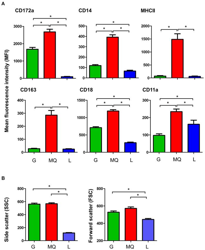
mean green fluorescence intensity (MFI), were calculated. cell size (as measured by the forward scatter; FSC)
and granularity (as measured by side scatter; SSC) for
Statistical Analyses milk granulocytes and macrophages (FSChigh SSChigh ),
Data were processed with the Microsoft office Excel R program while milk lymphocytes were identified as smaller
(version 2016 Microsoft) and statistical analysis was performed cells with lower granularity (FSClow SSClow ), in
using the software program Prism (GraphPad software version comparison to granulocytes and macrophages (p < 0.05)
5, GraphPad Software, San Diego, USA). The Kolmogorov- (Figure 2B).
Smirnov test (with the Dallal-Wilkinson-Lilliefor P-value) was
performed to check the normal distribution of data. For normal- Subclinical Mastitis
distributed data, the unpaired student’s t-test was used to Subclinical mastitis (SCM) was diagnosed based on the
compare the mean of the two groups. For the data that failed California Mastitis Test (CMT) score and the absence of
to pass the normality test, the non-parametric Mann-Whitney signs of clinical mastitis. SCM (CMT score of ≥3 in the
test was used to compare the means. The comparison between absence of clinical signs of mastitis) was found in six of the
milk granulocytes, macrophages, and lymphocytes regarding 56 sampled quarters (10.7%). All affected camels had only
their phenotype was performed using a one-factorial analysis 1 quarter with SCM. The bacteriological analysis identified
Frontiers in Veterinary Science | www.frontiersin.org 4 April 2022 | Volume 9 | Article 885523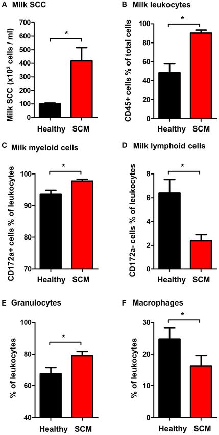
Alhafiz et al.                                                                                                                   Cellular Immunity to Camel Mastitis

  FIGURE 1 | Flow cytometric analysis of leukocytes in milk and blood from dromedary camel. Separated milk and blood leukocytes were labeled with monoclonal
  antibodies to CD45, CD172a, and CD14, and stained cells were analyzed by flow cytometry. (A) Using an SSC/CD45 gate, milk, and blood leukocytes were identified
  as CD45-positive cells. (B) Cell debris were excluded in an FSC-H/SSC-H dot plot. (C) Milk and blood granulocytes (G), macrophages (MQ), and lymphocytes (L)
  were identified based on their staining with monoclonal antibodies to CD172a, and CD14. (D) Cells stained with mouse IgG1 and IgM isotype controls.

bacterial cultures in all milk samples collected from the six                       Somatic Cell Count, Total and Differential
SCM quarters and only in one sample collected from healthy                          Leukocyte Composition in Milk Samples
animals. While two milk samples yielded single bacterial cultures                   From Healthy Camels and Camels With
with staphylococcus or streptococcus species, the other five                        Subclinical Mastitis
samples yielded mixed bacterial cultures with streptococcus and                     The SCC, the fraction of milk leukocytes (CD45+ cells), and
staphylococcus (three samples) or streptococcus and coliform                        the differential leukocyte composition were compared between
bacteria (two samples).                                                             the healthy and SCM animals. The SCC was significantly (p =

Frontiers in Veterinary Science | www.frontiersin.org                           5                                             April 2022 | Volume 9 | Article 885523
Alhafiz et al.                                                                                                                     Cellular Immunity to Camel Mastitis

  FIGURE 2 | (A) The expression levels of the cell surface antigens, CD172a, CD14, MHCII, CD163, CD18, and CD11a on milk granulocyte (G), macrophages (MQ),
  and lymphocytes (L) as measured by flow cytometry. (B) Forward and side scatter characteristics of milk granulocytes (G), macrophages (MQ), and lymphocytes (L) as
  measured by flow cytometry. *indicates statistically-significant difference between the means.

0.001) higher in milk samples from SCM camels (418.3 × 10³                            was decreased (16.2 ± 3.4% of leukocytes vs. 24.7 ± 3.6% of
cell/ ml) than healthy animals (103.8 × 10³ cell/ml) (Figure 3A).                     leukocytes in healthy milk; p = 0.04) (Figure 3F).
Milk samples with SCM contained higher percentages (p =
0.001) of total leukocytes (90.3 ± 3.1% of total cells) than
healthy (48.5 ± 9.3% of total cells) milk samples (Figure 3B).                        Impact of Subclinical Mastitis on Milk
The fraction of myeloid cells within the leukocyte population                         Leukocyte Viability
was also significantly elevated (p = 0.01) in SCM milk samples                        The percentage of viable, PI-negative (Figure 4A) milk
(97.8 ± 0.5% of leukocytes) compared to healthy milk samples                          leukocytes was significantly (p = 0.01) higher in milk samples
(93.5 ± 1.3% of leukocytes) (Figure 3C), while the fraction of                        from SCM animals (91.9 ± 1.9% of CD45+ cells) than healthy
lymphoid cells was significantly (p = 0.01) lower in SCM than                         animals (85.8 ± 1.3% of CD45+ cells). In milk from SCM
in healthy milk samples (Figure 3D). Similarly, the fraction of                       animals, the myeloid cell population contained a higher (p <
milk granulocytes was significantly (p = 0.02) expanded in SCM                        0.05) percentage of viable cells (91.9 ± 1.7% vs. 77.0 ± 3.3% of
samples (79.1 ± 2.7% of leukocytes vs. 67.8 ± 3.7% of leukocytes                      CD172a+ myeloid cells), while the percentage of viable cells
in healthy milk) (Figure 3E), while the fraction of macrophages                       under lymphoid cells was significantly lower (p < 0.05) in SCM

Frontiers in Veterinary Science | www.frontiersin.org                             6                                             April 2022 | Volume 9 | Article 885523
Alhafiz et al. Cellular Immunity to Camel Mastitis
milk (85.2 ± 4.3% vs. 91.0 ± 2.2% of CD172a-lymphoid cells)
compared to healthy milk (Figure 4B).
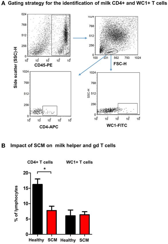
Milk Lymphocytes From Animals With SCM
Contained a Lower Percentage of Helper T
Cells
Using monoclonal antibodies to camel CD4 and WC1, the
percentages of helper T cells and γδ T cells were analyzed in
milk from healthy and SCM animals (Figure 5A). While the
fraction of WC1+ γδ T cells was comparable (p > 0.05) between
milk samples from healthy and SCM animals, the percentage of
CD4+ T helper cells was significantly (p = 0.002) lower in SCM
(7.8 ± 1.4% of lymphocytes) than healthy milk (16.3 ± 1.8% of
lymphocytes) (Figure 5B).
Granulocytes and Macrophages Shape
Change in SCM Milk
The analysis of forward scatter (FSC) and side scatter (SSC),
which are indicators for cell size and granularity, respectively,
revealed higher (p < 0.05) FSC values for granulocytes and
macrophages from SCM milk compared to healthy milk
(Figure 6A). Only for granulocytes, the SSC values were lower
(p < 0.05) in SCM milk compared to healthy milk (Figure 6B).
Impact of SCM on the Phenotype of Milk
Macrophages
The comparison between macrophages from SCM and healthy
milk regarding their expression levels of several cell surface
molecules revealed significant changes in their phenotype. While
the abundance of cell surface CD172a, CD163, and CD18 did
not differ (p < 0.05) between SCM and healthy milk samples,
macrophages from SCM milk showed higher levels of MHCII but
lower levels of CD14 and CD11a when compared (p < 0.05) to
macrophages from healthy milk (Figure 7).
Impact of SCM on the Antimicrobial
Function of Milk Phagocytes
The antimicrobial function of milk phagocytes (granulocytes
and macrophages) was analyzed by the evaluation of bacterial
phagocytosis by flow cytometry (Figure 8A). The percentage of
phagocytosis-positive cells was higher (p < 0.05) for phagocytes
from SCM milk than healthy milk. The phagocytic capacity (the
number of bacteria ingested by each cell as measured by MFI of
FIGURE 3 | (A) Somatic cell count (SCC) in milk samples from healthy camels phagocytosis-positive cells), however, did not differ (p > 0.05)
and camels with subclinical mastitis (SCM). Milk SCC was counted using the between cells from SCM and healthy milk (Figure 8B).
direct microscopic method after fat globule removal by a spin-wash followed
by staining with Turk solution and microscopic counting on a Neubauer cell
counter. The percentage of (B) leukocytes (CD45+ cells % of total milk cells),
DISCUSSION
(C) myeloid cells (CD172a+ cells % of total leukocytes), (D) lymphoid cells
(CD172a- cells % of total leukocytes), (E) granulocytes (CD172a+ CD14low The immune cell composition of the mammary gland secretions
cells % of total leukocytes), and (F) macrophages (CD172a+ CD14high cells has been investigated for several animal species in health
% of total leukocytes) in milk samples from healthy and SCM camels as and disease (19, 20, 55–58). However, studies on the cellular
identified by flow cytometry after labeling milk cells with antibodies to CD45,
composition of camel milk are limited. The present study
CD172a, and CD14. *indicates significant differences between the means with
p-values < 0.05. employed flow cytometry and monoclonal antibody staining to
investigate the differential composition, phenotype, vitality, and
Frontiers in Veterinary Science | www.frontiersin.org 7 April 2022 | Volume 9 | Article 885523Alhafiz et al. Cellular Immunity to Camel Mastitis FIGURE 4 | Separated milk cells were labeled with CD45 antibodies and labeled cells were loaded with the nucleic acid stain propidium iodide and analyzed by flow cytometry. (A) After gating on leukocytes (CD45 + cells) and the exclusion of cell debris in a SSC-H against FSC-H dot plot, milk phagocytes and lymphocytes were gated based on their FSC and SSC properties. (B) The percentage of viable PI-negative cells were calculated in a SSC-H against FL-3 dot plot and the values were presented for total leukocytes, myeloid cells, and lymphoid cells, and presented graphically. *indicates significant differences between the means with p-values < 0.05. some functional aspects of milk leukocytes in clinically healthy CD172a+ /CD14lhigh /SSChigh macrophages and a minor camels. The evaluation of the health status of the mammary gland CD172a− /CD14− /SSClow lymphocytes population. The higher and the classification of the camels was based on the results proportion of granulocytes in camel milk, compared to of the California Mastitis Test (CMT) with a test score of ≥3 other species like bovine (17, 28), could be a result of their in the absence of signs of clinical mastitis being indicative for dominance in peripheral blood (49). The results of the present subclinical mastitis (41). study, however, largely correspond to data reported for goats The present study divided camel milk leukocytes based regarding the differential composition of milk cells (19, 20). The on their differential expression of the cell surface molecules expression pattern of the cell surface antigens, CD172a, CD14, CD172a and CD14 into a dominant CD172a+ /CD14low /SSChigh CD163, MHCII, CD11a, and CD18 on milk granulocytes and granulocyte population followed by a smaller fraction of macrophages indicates similarities with the immunophenotype Frontiers in Veterinary Science | www.frontiersin.org 8 April 2022 | Volume 9 | Article 885523
Alhafiz et al.                                                                                                                       Cellular Immunity to Camel Mastitis

                                                                                          FIGURE 6 | Shape change in milk granulocytes. Milk granulocytes and
                                                                                          macrophages were identified based on their staining with CD172a and CD14
  FIGURE 5 | Flow cytometric analysis of milk lymphocyte subsets. Separated               antibodies in flow cytometry. Mean FSC (A) and SSC (B) were calculated and
  milk cells were labeled with monoclonal antibodies to CD45, CD4, and WC1                presented for healthy and SCM animals. *indicates significant differences
  and analyzed by flow cytometry. (A) After setting a gate on milk leukocytes in a        between the means with p-values < 0.05.
  CD45 against SSC-H dot plot, lymphocytes were identified based on their
  SSC and FSC properties. Milk helper T cells and gd T cells were identified
  within the lymphocyte population based on their staining with anti-CD4 and
  anti WC1 antibodies, respectively. (B) The percentage of T helper cells and gd
                                                                                            The two-times increase in whole milk leukocytes with more
  T cells within the milk lymphocyte population were calculated for healthy and
  SCM camels and presented graphically. *indicates significant differences
                                                                                         myeloid cells (CD172a+ cells) in the SCM milk suggests a
  between the means with p-values < 0.05.                                                significant role of the innate immune phagocytes, granulocytes
                                                                                         and macrophages, in the immune response to bacterial infections
                                                                                         of the camel mammary gland. The increase in the proportion
                                                                                         of granulocytes with the decrease in macrophages in SCM milk
of peripheral blood granulocytes and monocytes, respectively,                            could be a result of enhanced recruitment of blood neutrophils to
with CD172a, CD18, and CD11a being highest expressed on                                  the infected mammary gland.
camel monocytes/macrophages, while MHCII and CD163 being                                    According to reports in dairy cows with bacterial mastitis,
exclusively expressed on monocytes/macrophages (53).                                     the survival of neutrophils was higher in infected than healthy
   The milk somatic cell count and the CMT are widely accepted                           mammary glands (59). The same study identified a link between
tools for the evaluation of the mammary gland health status                              the higher viability of milk phagocytes with their enhanced
(17, 28). In camels, elevated SCC values were observed in milk                           antibacterial function. In the present study, the viability of
samples from infected mammary glands with values ranging                                 granulocytes and macrophages, as well as the fraction of
from 1 × 10⁵ to 10 × 106 cells/ml milk (8, 25). In the present                           phagocytosis-positive cells, were higher in SCM milk compared
study, the four-times elevated SCC with the identification of                            to healthy milk, suggesting similarity in the host-pathogen
bacterial cultures in milk samples with a CMT test score ≥3                              interaction mechanisms in the mammary gland of cattle
confirm the results from previous reports regarding the efficiency                       and camel.
of CMT and SCC as diagnostic tools for monitoring mammary                                   The lipopolysaccharide (LPS) receptor CD14, the antigen
gland infections in camels.                                                              presentation receptor MHCII, and the hemoglobin-haptoglobin

Frontiers in Veterinary Science | www.frontiersin.org                                9                                            April 2022 | Volume 9 | Article 885523
Alhafiz et al. Cellular Immunity to Camel Mastitis
FIGURE 8 | (A) Separated milk cells were incubated with FITC-conjugated S.
aureus and the fraction of phagocytosis-positive cells within the myeloid cell
population (including granulocytes and macrophages defined based on
SCG/SSC properties) was estimated by flow cytometry based on their
enhanced fluorescence in the green fluorescence channel. (B) The percentage
of phagocytosis-positive cells and their mean fluorescence intensity (MFI),
which is a metric of phagocytic capacity of the cells, were calculated and
presented for healthy and SCM animals.
FIGURE 7 | Impact of SCM on the phenotype of milk macrophages.
Separated milk cells were stained with monoclonal antibodies to CD172a,
CD14, CD163, MHCII, CD18, and CD11a and stained cells were analyzed by
flow cytometry. After setting a gate on milk macrophages (based on their
higher expression of CD14), the mean fluorescence intensity of the monocytic indicate the activation status of these phagocytes in the infected
cell markers and the cell adhesion molecules was calculated and presented for mammary gland. This is also supported by the higher fraction of
healthy and SCM animals. *indicates significant differences between the
means with p-values < 0.05.
phagocytosis-positive cells in SCM milk.
The lower percentage of helper T cells in SCM milk with
no difference in the percentage of γδ T cells suggests a
selective impact of bacterial mammary gland infections on T cell
receptor CD163 are well-established markers of monocyte and subpopulations. As we did not analyze all lymphocyte subsets
macrophage phenotype (53, 60–62). The differences in the due to the lack of specific monoclonal antibodies (49), we cannot
expression levels of MHCII and CD14 on milk macrophages exclude changes in other milk lymphocyte subsets like CD8+
indicate a significant modulatory effect of subclinical mastitis cytotoxic T cells, B cells, or NK cells in SCM animals.
on the functional type of macrophages. Reduced expression of Collectively, the present study identified significant
CD14, which plays a key role in the binding of LPS, a cell- differences between healthy camels and camels with SCM
wall component of gram-negative bacteria (63–65), may have an regarding the cellular composition of their milk. Milk from SCM
impact on the innate recognition function of macrophages in camels had higher SCC with higher fractions of total leukocytes,
SCM milk. myeloid cells, and granulocytes, but reduced fractions of
Macrophages and neutrophils are key effector innate immune lymphoid cells and macrophages. Within the lymphoid cell
cells of the mammary gland with an essential role during the population, the percentage of CD4+ T helper cells was reduced
early immune response to mastitis pathogens (17, 40). In the in milk from SCM camels. In addition, SCM was associated
present work, the observed shape-change of granulocytes and with improved cell viability and phagocytic activity of milk
macrophages from SCM milk with higher forward scatter values, phagocytes. The results of the present study pave the way
which correlates with the cell size, and lower side scatter values for the characterization of the camel immune response to
of granulocytes, which is indicative of cell degranulation (66, 67), mammary gland infections and support a better understanding
Frontiers in Veterinary Science | www.frontiersin.org 10 April 2022 | Volume 9 | Article 885523Alhafiz et al. Cellular Immunity to Camel Mastitis
of host-pathogen interaction mechanisms on mucosal surfaces analysis. HA: flow cytometric analysis and writing the original
in camels. manuscript. JH: funding acquisition, conceptualization, flow
cytometric analysis, and writing the manuscript. All authors have
DATA AVAILABILITY STATEMENT read and approved the final manuscript.
The raw data supporting the conclusions of this article will be FUNDING
made available by the authors, without undue reservation.
This work was supported through the Annual Funding track by
ETHICS STATEMENT the Deanship of Scientific Research, Vice Presidency for Graduate
Studies and Scientific Research, King Faisal University, Saudi
The animal study was reviewed and approved by Ethics Arabia (Project No. AN000337).
Committee of King Faisal University.
ACKNOWLEDGMENTS
AUTHOR CONTRIBUTIONS
The authors are thankful to Prof. Ali Fadlelmula, the Bacteriology
GA: sample collection and sample preparation for flow Unit, College of Veterinary medicine, king Faisal University for
cytometric analysis. FA: sample collection and flow cytometric his support in the bacteriological evaluation of milk samples.
REFERENCES 13. Chang CC, Winter AJ, Norcross NL. Immune response in the bovine
mammary gland after intestinal, local, and systemic immunization. Infect
1. Musaad A, Faye B, Nikhela AA. Lactation curves of dairy Immun. (1981) 31:650-9. doi: 10.1128/iai.31.2.650-659.1981
camels in an intensive system. Trop Anim Health Prod. (2013) 14. Newby TJ, Bourne J. The nature of the local immune system of the bovine
45:1039-46. doi: 10.1007/s11250-012-0331-x mammary gland. J Immunol. (1977) 118:461-5.
2. Nagy P, Faigl V, Reiczigel J, Juhasz J. Effect of pregnancy and embryonic 15. Kimura K, Goff JP, Schmerr MJ, Stabel JR, Inumaru S, Yokomizo Y. Activation
mortality on milk production in dromedary camels (Camelus dromedarius). J of immune cells in bovine mammary gland secretions by zymosan-treated
Dairy Sci. (2015) 98:975-86. doi: 10.3168/jds.2014-8546 bovine serum. J Dairy Sci. (2008) 91:1852-64. doi: 10.3168/jds.2007-0895
3. Nagy P, Juhasz J. Review of present knowledge on machine milking and 16. Riollet C, Rainard P, Poutrel B. Kinetics of cells and cytokines during
intensive milk production in dromedary camels and future challenges. Trop immune-mediated inflammation in the mammary gland of cows systemically
Anim Health Prod. (2016) 48:915-26. doi: 10.1007/s11250-016-1036-3 immunized with Staphylococcus aureus alpha-toxin. Inflamm Res. (2000)
4. Ranjan R, Narnaware SD, Prakash V. Incidence, risk factors and economic 49:486-96. doi: 10.1007/s000110050621
impact of clinical mastitis in dromedary camel (Camelus dromedarius). Trop 17. Alhussien MN, Panda BSK, Dang AK. A comparative study on changes
Anim Health Prod. (2021) 54:31. doi: 10.1007/s11250-021-03035-0 in total and differential milk cell counts, activity, and expression of milk
5. Geresu MA, Abera Leliso S, Liben GW. Camel mastitis: prevalence, phagocytes of healthy and mastitic indigenous sahiwal cows. Front Vet Sci.
risk factors, and isolation of major bacterial pathogens in gomole (2021) 8:670811. doi: 10.3389/fvets.2021.670811
district of Borena Zone, Southern Ethiopia. Vet Med Int. (2021) 18. Cacho NT, Lawrence RM. Innate immunity and breast milk. Front Immunol.
2021:9993571. doi: 10.1155/2021/9993571 (2017) 8:584. doi: 10.3389/fimmu.2017.00584
6. Alebie A, Molla A, Adugna W, Tesfaye A, Ejo M. Prevalence, isolation, 19. Guiguen F, Greenland T, Pardo E, Panaye G, Mornex JF. Flow
identification, and risk factors of major bacterial cause of camel subclinical cytometric analysis of goat milk lymphocytes: subpopulations and
mastitis. Biomed Res Int. (2021) 2021:5522331. doi: 10.1155/2021/5522331 adhesion molecule expression. Vet Immunol Immunopathol. (1996)
7. Al-Ashqar RA, Al-Mohammad Salem KM, Al Herz AK, Al-Haroon AI, 53:173-8. doi: 10.1016/0165-2427(96)05553-5
Alluwaimi AM. The CD markers of camel (Camelus dromedarius) milk 20. Winnicka A, Klucinski W, Hoser G, Sikora J, Kawiak J.
cells during mastitis: the LPAM-1 expression is an indication of possible Flow cytometry analysis of milk and peripheral blood cells
mucosal nature of the cellular trafficking. Res Vet Sci. (2015) 99:77- from goats during lactation. Zentralbl Veterinarmed A. (1999)
81. doi: 10.1016/j.rvsc.2015.01.011 46:459-64. doi: 10.1046/j.1439-0442.1999.00234.x
8. Abdurahman OA. The detection of subclinical mastitis in the bactrian camel 21. Forner R, Bombassaro G, Bellaver FV, Maciag S, Fonseca FN, Gava D, et al.
(Camelus bactrianus) by somatic cell count and California mastitis test. Vet Distribution difference of colostrum-derived B and T cells subsets in gilts and
Res Commun. (1996) 20:9-14. doi: 10.1007/BF00346570 sows. PLoS ONE. (2021) 16:e0249366. doi: 10.1371/journal.pone.0249366
9. Osman KM, Samir A, Orabi A, Zolnikov TR. Confirmed low 22. McDougall S, Williamson J, Lacy-Hulbert J. Bacteriological outcomes
prevalence of Listeria mastitis in she-camel milk delivers a safe, following random allocation to quarter-level selection based on California
alternative milk for human consumption. Acta Trop. (2014) Mastitis Test score or cow-level allocation based on somatic cell count for dry
130:1-6. doi: 10.1016/j.actatropica.2013.10.001 cow therapy. J Dairy Sci. (2022) 105:2453-72. doi: 10.3168/jds.2021-21020
10. Petzl W, Zerbe H, Günther J, Seyfert H-M, Hussen J, Schuberth H-J. Pathogen- 23. Halasa T, Kirkeby C. Differential somatic cell count: value for udder health
specific responses in the bovine udder. Models and immunoprophylactic management. Front Vet Sci. (2020) 7:609055. doi: 10.3389/fvets.2020.609055
concepts. Res Vet Sci. (2018) 116:55-61. doi: 10.1016/j.rvsc.2017.12.012 24. Gussmann M, Kirkeby C, Schwarz D, Farre M, Halasa T. A simulation study
11. Wellnitz O, Bruckmaier RM. The innate immune response of the to investigate the added value in using differential somatic cell count as an
bovine mammary gland to bacterial infection. Vet J. (2012) 192:148- additional indicator for udder health management in dairy herds. Prev Vet
52. doi: 10.1016/j.tvjl.2011.09.013 Med. (2020) 182:105090. doi: 10.1016/j.prevetmed.2020.105090
12. Oviedo-Boyso J, Valdez-Alarcon JJ, Cajero-Juarez M, Ochoa-Zarzosa A, 25. Guliye AY, Van Creveld C, Yagil R. Detection of subclinical mastitis in
Lopez-Meza JE, Bravo-Patino A, et al. Innate immune response of bovine dromedary camels (Camelus dromedarius) using somatic cell counts and
mammary gland to pathogenic bacteria responsible for mastitis. J Infect. the N-acetyl-beta-D-glucosaminidase test. Trop Anim Health Prod. (2002)
(2007) 54:399-409. doi: 10.1016/j.jinf.2006.06.010 34:95-104. doi: 10.1023/a:1014324421258
Frontiers in Veterinary Science | www.frontiersin.org 11 April 2022 | Volume 9 | Article 885523Alhafiz et al. Cellular Immunity to Camel Mastitis
26. Schwarz D, Diesterbeck US, Konig S, Brugemann K, Schlez K, 45. Wehr HM, Frank JF. Standard Methods for the Examination of Dairy Products.
Zschock M, et al. Microscopic differential cell counts in milk for 17 ed. Washington, D.C.: Apha Press (2004).
the evaluation of inflammatory reactions in clinically healthy and 46. Koneman E, Schreckenberger P, Janda W, Allen S, Winn W. Koneman’s Color
subclinically infected bovine mammary glands. J Dairy Res. (2011) Atlas and Textbook of Diagnostic Microbiology. 6 ed. New York: Lippincott
78:448-55. doi: 10.1017/S0022029911000574 Williams & Wilkins (2005).
27. Pilla R, Schwarz D, Konig S, Piccinini R. Microscopic differential cell counting 47. Boulaaba A, Grabowski N, Klein G. Differential cell count of caprine
to identify inflammatory reactions in dairy cow quarter milk samples. J Dairy milk by flow cytometry and microscopy. Small Rumin Res. (2010) 97:117-
Sci. (2012) 95:4410-20. doi: 10.3168/jds.2012-5331 23. doi: 10.1016/j.smallrumres.2011.02.002
28. De Matteis G, Grandoni F, Scata MC, Catillo G, Moioli B, Buttazzoni L. Flow 48. Hussen J, Shawaf T, Alhojaily SM. The impact of anticoagulation agent on the
cytometry-detected immunological markers and on farm recorded parameters composition and phenotype of blood leukocytes in dromedary camels. Vet Sci.
in composite cow milk as related to udder health status. Vet Sci. (2020) (2022) 9:78. doi: 10.3390/vetsci9020078
7:114. doi: 10.3390/vetsci7030114 49. Hussen J, Schuberth HJ. Recent advances in camel immunology. Front
29. Albenzio M, Santillo A, Caroprese M, Ruggieri D, Ciliberti M, Sevi A. Immunol. (2020) 11:614150. doi: 10.3389/fimmu.2020.614150
Immune competence of the mammary gland as affected by somatic cell and 50. Mosaad AA, Elbagory AR, Khalid AM, Waters W, Tibary A, Hamilton MJ,
pathogenic bacteria in ewes with subclinical mastitis. J Dairy Sci. (2012) et al. Identification of monoclonal antibody reagents for use in the study of
95:3877-87. doi: 10.3168/jds.2012-5357 the immune response to infectious agents in camel and water buffalo. J Camel
30. Trend S, de Jong E, Lloyd ML, Kok CH, Richmond P, Doherty DA, Pract Res. (2006) 13:91-101.
et al. Leukocyte populations in human preterm and term breast 51. Al-Mubarak AIA.D ifferential expression of the coronavirus (Mers-
milk identified by multicolour flow cytometry. PLoS ONE. (2015) cov) Receptor, Dipeptidyl Peptidase 4, on normal and stimulated
10:e0135580. doi: 10.1371/journal.pone.0135580 leukocytes of dromedary camels. J Camel Pract Res. (2018)
31. Senbanjo LT, Chellaiah MA. CD44: a multifunctional cell surface adhesion 25:249. doi: 10.5958/2277-8934.2018.00033.4
receptor is a regulator of progression and metastasis of cancer cells. Front Cell 52. Hussen J, Shawaf T, Al-herz AI, Alturaifi HR, Alluwaimi AM. Reactivity of
Dev Biol. (2017) 5:18. doi: 10.3389/fcell.2017.00018 commercially available monoclonal antibodies to human CD antigens with
32. Wang Q, Teder P, Judd NP, Noble PW, Doerschuk CM. CD44 peripheral blood leucocytes of dromedary camels (Camelus dromedarius.
deficiency leads to enhanced neutrophil migration and lung injury Open Vet J. (2017) 7:150-3. doi: 10.4314/ovj.v7i2.12
in Escherichia coli pneumonia in mice. Am J Pathol. (2002) 53. Hussen J, Shawaf T, Al-Mubarak AIA, Al Humam NA, Almathen F,
161:2219-28. doi: 10.1016/S0002-9440(10)64498-7 Schuberth HJ. Dromedary camel CD14(high) MHCII(high) monocytes
33. van de Vijver E, Maddalena A, Sanal O, Holland SM, Uzel G, display inflammatory properties and are reduced in newborn camel calves.
Madkaikar M, et al. Hematologically important mutations: leukocyte BMC Vet Res. (2020) 16:62. doi: 10.1186/s12917-020-02285-8
adhesion deficiency (first update). Blood Cells Mol Dis. (2012) 54. Silva VM, Souza MT, Blagitz MG, Souza FN, Batista CF, Alves AJ, et al.
48:53-61. doi: 10.1016/j.bcmd.2011.10.004 Milk lymphocyte profile and macrophage functions: new insights into the
34. Roos D, Law SK. Hematologically important mutations: leukocyte immunity of the mammary gland in quarters infected with Corynebacterium
adhesion deficiency. Blood Cells Mol Dis. (2001) 27:1000- bovis. BMC Vet Res. (2021) 17:282. doi: 10.1186/s12917-021-02989-5
4. doi: 10.1006/bcmd.2001.0473 55. Zecconi A, Zanini L, Cipolla M, Stefanon B.F actors affecting the patterns of
35. Hussen J, Shawaf T, Al-herz AI, Alturaifi HR, Al khamees M, Alluwaimi total amount and proportions of leukocytes in bovine milk. Animals. (2020)
AM. Expression patterns of cell adhesion molecules on CD4+ T Cells and 10:992. doi: 10.3390/ani10060992
WC1+ T Cells in the peripheral blood of dromedary camels. Pak Vet J. (2018) 56. De UK, Mukherjee R. Dynamics of milk leukocytes in response to a biological
38:231-6. doi: 10.29261/pakvetj/2018.055 response modifier during bovine subclinical mastitis. Res Vet Sci. (2013)
36. Verjan Garcia N, Umemoto E, Saito Y, Yamasaki M, Hata E, Matozaki T, et 95:352-7. doi: 10.1016/j.rvsc.2013.06.010
al. SIRPalpha/CD172a regulates eosinophil homeostasis. J Immunol. (2011) 57. Blagitz MG, Souza FN, Santos BP, Batista CF, Parra AC, Azevedo LF, et
187:2268-77. doi: 10.4049/jimmunol.1101008 al. Function of milk polymorphonuclear neutrophil leukocytes in bovine
37. Ring NG, Herndler-Brandstetter D, Weiskopf K, Shan L, Volkmer JP, George mammary glands infected with Corynebacterium bovis. J Dairy Sci. (2013)
BM, et al. Anti-SIRPalpha antibody immunotherapy enhances neutrophil and 96:3750-7. doi: 10.3168/jds.2012-6370
macrophage antitumor activity. Proc Natl Acad Sci USA. (2017) 114:E10578- 58. Leitner G, Merin U, Krifucks O, Blum S, Rivas AL, Silanikove N. Effects
85. doi: 10.1073/pnas.1710877114 of intra-mammary bacterial infection with coagulase negative staphylococci
38. Triantafilou M, Triantafilou K. Lipopolysaccharide recognition: CD14, and stage of lactation on shedding of epithelial cells and infiltration of
TLRs and the LPS-activation cluster. Trends Immunol. (2002) 23:301- leukocytes into milk: comparison among cows, goats and sheep. Vet Immunol
4. doi: 10.1016/S1471-4906(02)02233-0 Immunopathol. (2012) 147:202-10. doi: 10.1016/j.vetimm.2012.04.019
39. Yao Y, Xu XH, Jin L. Macrophage polarization in physiological 59. Mehrzad J, Duchateau L, Burvenich C. Viability of milk neutrophils
and pathological pregnancy. Front Immunol. (2019) and severity of bovine coliform mastitis. J Dairy Sci. (2004) 87:4150-
10:792. doi: 10.3389/fimmu.2019.00792 62. doi: 10.3168/jds.S0022-0302(04)73558-4
40. Swain DK, Kushwah MS, Kaur M, Patbandha TK, Mohanty AK, Dang 60. Hu JM, Liu K, Liu JH, Jiang XL, Wang XL, Chen YZ, et al. CD163 as a marker
AK. Formation of NET, phagocytic activity, surface architecture, apoptosis of M2 macrophage, contribute to predicte aggressiveness and prognosis of
and expression of toll like receptors 2 and 4 (TLR2 and TLR4) Kazakh esophageal squamous cell carcinoma. Oncotarget. (2017) 8:21526-
in neutrophils of mastitic cows. Vet Res Commun. (2014) 38:209- 38. doi: 10.18632/oncotarget.15630
19. doi: 10.1007/s11259-014-9606-1 61. Svendsen P, Etzerodt A, Deleuran BW, Moestrup SK. Mouse CD163 deficiency
41. Seligsohn D, Nyman AK, Younan M, Sake W, Persson Y, Bornstein S, et strongly enhances experimental collagen-induced arthritis. Sci Rep. (2020)
al. Subclinical mastitis in pastoralist dairy camel herds in Isiolo, Kenya: 10:12447. doi: 10.1038/s41598-020-69018-7
Prevalence, risk factors, and antimicrobial susceptibility. J Dairy Sci. (2020) 62. Hussen J, Schuberth HJ. Heterogeneity of bovine peripheral blood monocytes.
103:4717-31. doi: 10.3168/jds.2019-17701 Front Immunol. (2017) 8:1875. doi: 10.3389/fimmu.2017.01875
42. Schalm OW, Noorlander DO. Experiments and observations leading 63. Zanoni I, Granucci F. Role of CD14 in host protection
to development of the California mastitis test. J Am Vet Med Assoc. against infections and in metabolism regulation. Front
(1957) 130:199-204. Cell Infect Microbiol. (2013) 3:32. doi: 10.3389/fcimb.2013.
43. Schroder AC, Hamann J. The influence of technical factors on differential cell 00032
count in milk. J Dairy Res. (2005) 72:153-8. doi: 10.1017/S0022029905000804 64. Schumann RR. Function of lipopolysaccharide (LPS)-binding protein
44. Hussen J, Al-Sukruwah MA. The impact of the animal housing system on (LBP) and CD14, the receptor for LPS/LBP complexes: a short
immune cell composition and function in the blood of dromedary camels. review. Res Immunol. (1992) 143:11-5. doi: 10.1016/0923-2494(92)8
Animals. (2022) 12:317. doi: 10.3390/ani12030317 0074-U
Frontiers in Veterinary Science | www.frontiersin.org 12 April 2022 | Volume 9 | Article 885523Alhafiz et al. Cellular Immunity to Camel Mastitis
65. Wright SD, Ramos RA, Tobias PS, Ulevitch RJ, Mathison JC. CD14, a receptor Publisher’s Note: All claims expressed in this article are solely those of the authors
for complexes of lipopolysaccharide (LPS) and LPS binding protein. Science. and do not necessarily represent those of their affiliated organizations, or those of
(1990) 249:1431-3. doi: 10.1126/science.1698311 the publisher, the editors and the reviewers. Any product that may be evaluated in
66. MacDonald HR, Zaech P. Light scatter analysis and sorting of this article, or claim that may be made by its manufacturer, is not guaranteed or
cells activated in mixed leukocyte culture. Cytometry. (1982)
endorsed by the publisher.
3:55-8. doi: 10.1002/cyto.990030112
67. Stern AD, Rahman AH, Birtwistle MR. Cell size assays for mass Copyright © 2022 Alhafiz, Alghatam, Almohammed and Hussen. This is an open-
cytometry. Cytometry A. (2017) 91:14-24. doi: 10.1002/cyto.a. access article distributed under the terms of the Creative Commons Attribution
23000 License (CC BY). The use, distribution or reproduction in other forums is permitted,
provided the original author(s) and the copyright owner(s) are credited and that the
Conflict of Interest: The authors declare that the research was conducted in the original publication in this journal is cited, in accordance with accepted academic
absence of any commercial or financial relationships that could be construed as a practice. No use, distribution or reproduction is permitted which does not comply
potential conflict of interest. with these terms.
Frontiers in Veterinary Science | www.frontiersin.org 13 April 2022 | Volume 9 | Article 885523You can also read