Knockdown of CCL28 inhibits endometriosis stromal cell proliferation and invasion via ERK signaling pathway inactivation
←
→
Page content transcription
If your browser does not render page correctly, please read the page content below
Molecular Medicine REPORTS 25: 56, 2022

     Knockdown of CCL28 inhibits endometriosis stromal cell
proliferation and invasion via ERK signaling pathway inactivation
      YINGTING WU1, FEILONG ZHU1, WENQIN SUN1, WEIWEI SHEN1, QIN ZHANG2 and HUIFEN CHEN1

           1
               Department of Laboratory Medicine, Shanghai First Maternity and Infant Hospital, School of Medicine,
                       Tongji University, Shanghai 200092; 2Phase I Clinical Trial Unit, Tongren Hospital,
                        Shanghai Jiao Tong University School of Medicine, Shanghai 200336, P.R. China

                                     Received March 19, 2021; Accepted September 15, 2021

                                                  DOI: 10.3892/mmr.2021.12573

Abstract. Endometriosis (EM), the presence of functional          Introduction
endometrial glands and stroma outside the uterine cavity, is
a common gynecological disorder. At present, the pathogen‑        Endometriosis (EM), the presence of functional endometrial
esis of EM has not been fully elucidated, so there is still a     glands and stroma outside the uterine cavity, is a common
lack of effective therapy. The present study aimed to explore     gynecological disorder characterized by dysmenorrhea,
the role of C‑C motif chemokine ligand 28 (CCL28) and its         chronic pelvic pain, menstrual abnormalities and infer‑
underlying mechanism in endometrial stromal cells to propose      tility (1‑4). Previous study demonstrated that EM affects ~10%
a novel therapy for EM treatment. The expression of CCL28         of individuals who have a uterus, and that the probability
and CC chemokine receptor 10 (CCR10) were examined. After         of symptomatic perimenopause increases to 30‑50% (5).
CCL28 knockdown or overexpression by lentivirus infection,        Currently, the most common primary diagnostic method for
cell proliferation and invasion were measured. It was revealed    EM is laparoscopy, supplemented with the screening for cancer
that compared with normal, the expression levels of CCL28         antigen 125 (CA125) and endometrium antibody, as well as
and CCR10 were significantly elevated in endometrial tissues      B‑ultrasounds, x‑ray and magnetic resonance imaging (6).
of patients with EM. Knockdown of CCL28 in endometrial            Although EM is a benign disease, it has certain characteristics
stromal cells significantly suppressed cell proliferation and     of malignant tumors, including cell invasion, new blood vessel
invasion, and this was accompanied by significantly reduced       generation, unlimited growth, reduced numbers of apoptotic
expression levels of CCR10, MMP2, MMP9, integrin β1               cells, infiltration and destruction of surrounding tissues and
(ITGB1) and phosphorylated (p)‑ERK/ERK ratio. The addi‑           metastasis (7). Therefore, inhibition of the growth and inva‑
tion of the CCL28 recombinant protein had an opposite effect      sion of EM may be a possible treatment strategy for EM.
to CCL28 downregulation. Furthermore, the ERK inhibitor,               Chemokines are selective mediators of leukocyte migra‑
PD98059, reduced CCL28‑induced cell proliferation and inva‑       tion to inflammatory sites (8). It has been demonstrated
sion, as well as the expression levels of MMP2, MMP9, ITGB1       that chemokines are key players in a variety of physi‑
and p‑ERK. Therefore, the present study indicated that CCL28      ological and pathological events, including chemotaxis, cell
may contribute to the progression of EM by regulating MMP2,       proliferation, apoptosis, angiogenesis and inflammatory
MMP9 and ITGB1 expression and function via the activation         processes/diseases (9,10). C‑C motif chemokine ligand (CCL)28
of the ERK signaling pathway.                                     is a mucosa‑associated epithelial chemokine that is selectively
                                                                  expressed in certain mucosal tissue, such as epithelial mucosal
                                                                  tissues (11,12). CCL28 is a functional ligand for CC chemokine
                                                                  receptor (CCR)10, a member of the chemokine receptor family,
                                                                  which belongs to the G protein‑coupled receptor superfamily,
Correspondence to: Dr Huifen Chen, Department of Laboratory
Medicine, Shanghai First Maternity and Infant Hospital, School
                                                                  and is normally expressed by melanocytes, plasma cells and
of Medicine, Tongji University, 536 Changle Road, Jingan,         skin‑homing T cells (13). Upregulation of CCR10 can facili‑
Shanghai 200092, P.R. China                                       tate cell proliferation and invasion in glioma, contributing to
E‑mail: chf51mch@126.com                                          gliomagenesis (14). Furthermore, CCR10 can stimulate breast
                                                                  cancer cell invasion and migration by increasing MMP7 expres‑
Dr Qin Zhang, Phase I Clinical Trial Unit, Tongren Hospital,
Shanghai Jiao Tong University School of Medicine, 1111 Xianxia
                                                                  sion via ERK1/2 activation (15,16). In ectopic endometrial
Road, Changning, Shanghai 200336, P.R. China                      stromal cells, depletion of CCL27 can suppress cell prolifera‑
E‑mail: zq1980@shtrhospital.com                                   tion, metastasis and adhesion (17). CCL28 induces apoptosis of
                                                                  decidual stromal cells via binding of CCR3/CCR10 in human
Key words: endometriosis, C‑C motif chemokine ligand 28,          spontaneous abortion (18). Furthermore, estrogen may serve a
endometriosis stromal cells, MMPs, integrin beta 1, ERK pathway   crucial role in the protection against genital infection by regu‑
                                                                  lating mucosa‑associated epithelial chemokine (MEC)/CCL28
                                                                  expression in the uterus (19). There are also several studies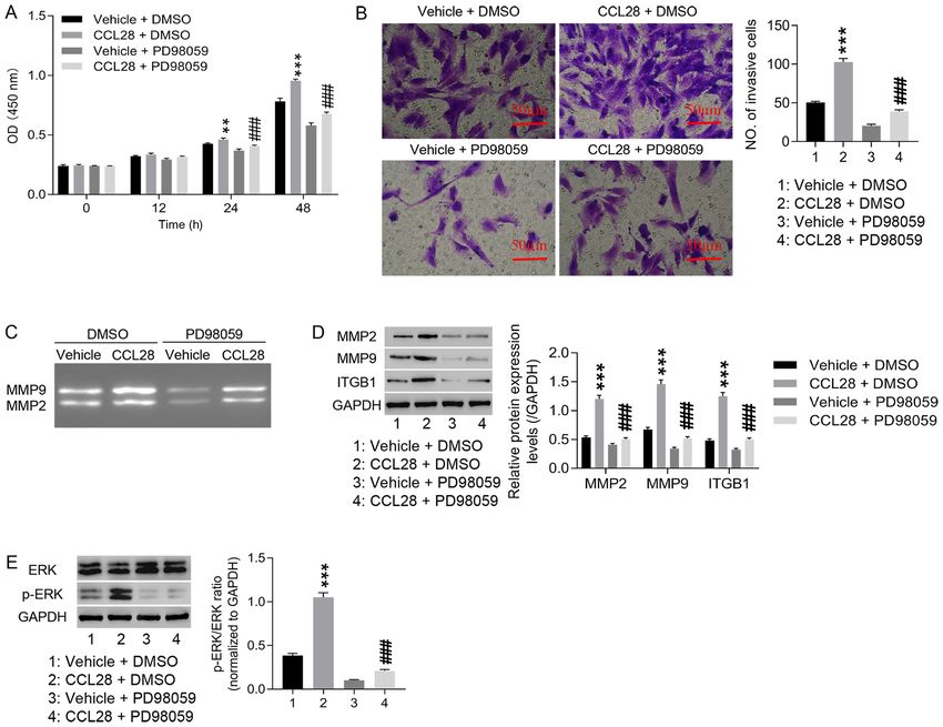
2                                 WU et al: CCL28 REGULATES ENDOMETRIOSIS STROMAL CELLS

demonstrating that the ERK1/2 signaling pathway is associ‑          Plasmid construction and lentivirus packaging. Targeting
ated with migration and apoptosis of endometrial stromal            different sites of CCL28, three interference sequences were
cells (20‑24). Another study has reported that CCL28 can            synthesized (Table II). Short hairpin RNA (sh) constructs were
promote cell proliferation and metastasis in breast cancer via      created using double chain annealing and inserted into the
the MAPK signaling pathway (25). However, the function of           pLKO.1‑Puro vector (Addgene, Inc.) at AgeI‑EcoRI restriction
CCL28 in endometrial stromal cells and its underlying mecha‑        sites, while a negative control shRNA (shNC) as a negative
nisms are still unknown.                                            control. Subsequently, plasmids of pLKO.1‑Puro‑shCCL28‑1,
    The present study aimed to explore the role of CCL28            ‑2 and ‑3 (1,000 ng) were co‑transfected with viral packaging
and its underlying mechanism in endometrial stromal cells to        plasmids psPAX2 (900 ng) and pMD2.G (100 ng; Addgene,
propose a novel therapy for EM treatment. It provided impor‑        Inc.; packaging vector:envelope vector, 1:9) into 3rd generation
tant leads for designing studies in the future to understand        293T cells (ATCC) using Lipofectamine® 2000 (Invitrogen;
the mechanism of EM and aid in the development of novel             Thermo Fisher Scientific, Inc.) for 48 h at 37˚C in a 5% CO2
therapeutic strategies.                                             incubator. Following 48 h of incubation, viral particles were
                                                                    collected via ultracentrifugation at 55,000 x g, 4˚C for 2.5 h,
Materials and methods                                               and then the viral supernatant (MOI, 10) was used to infect
                                                                    EM stromal cells (1x10⁶). After 24 h infection, the cells were
Patient samples. Patients who met the EM diagnostic criteria        cultured for 24 h with serum‑free transfer solution before
(visual inspection/laparoscopy/laparotomy) participated in the      further experiments were performed.
study. EM is usually diagnosed by visual inspection of the pelvis
during laparoscopy or laparotomy (6). After informed consent        ELISA. An ELISA was employed to detect CCL28 levels in the
was obtained, the EM tissues from 15 patients (female, age          serum of patients with EM or in the supernatant of endome‑
range from 25 years to 55 years, mean age: 37 years, Shanghai,      trial stromal cells (1x10⁶ cells/ml). The Human MEC/CCL28
China) with deep‑infiltrating EM who underwent laparoscopic         ELISA Kit (cat. no. RAB0072; Sigma‑Aldrich; Merck KGaA)
treatment at the Shanghai First Maternity and Infant Hospital,      was used according to the manufacturer's protocol.
Tongji University School of Medicine (Shanghai, China)
between February 2020 and December 2020 were removed                Immunohistochemical (IHC) detection. Tissue sections (4 µm)
via biopsy. Deep‑infiltrating EM is located 5 mm below the          were washed with 0.02 M PBS and fixed with 4% formalde‑
surface of the peritoneum. Control endometrial samples were         hyde for 30 min at room temperature. After three washes
collected from 15 patients (female, age range from 23 years to      with 0.02 M PBS, samples were incubated in 0.3% H 2O2
51 years, mean age: 35 years, Shanghai, China) without EM           in a wet‑box for 10 min and then blocked in 1% BSA (cat.
who underwent laparoscopy and hysteroscopy surgery for              no. A8010; Beijing Solarbio Science & Technology Co., Ltd.)
benign gynecological diseases. CCL28 and CCR10 expression           for 1 h at room temperature. Subsequently, samples were
was detected in these tissues by IHC. Furthermore, 80 serum         incubated with primary antibodies against CCL28 (1:200;
samples (healthy individuals: 40; EM patients, 40) were             cat. no. 18214‑1‑AP; ProteinTech Group, Inc.) and CCR10
collected to detect CCL28 levels by ELISA. Tissue samples           (1:100; cat. no. 22071‑1‑AP; ProteinTech Group, Inc.) for 1
were collected independent of menstrual cycle stage. The            h at room temperature. The primary incubation was then
following inclusion criteria were used: i) EM was confirmed         followed by a 30‑min incubation with HRP‑labeled secondary
by two pathologists following laparoscopic biopsy; and ii) No       antibodies (cat. no. D‑3004; Shanghai Changdao Biological
preoperative chemotherapy or radiotherapy was received.             Technology Co., Ltd.) at room temperature. Samples were
Patients who had received hormonal treatment and birth              then subjected to 3, 3'‑diaminobenzidine (DAB) staining (cat.
control prior to enrollment were excluded from the study. The       no. FL‑6001; Shanghai Changdao Biological Technology Co.,
clinical characteristics of the patients and controls are shown     Ltd.), 3 min of hematoxylin staining (cat. no. 714094; Zhuhai
in Table I. The Ethics Committee of Shanghai First Maternity        Besso Biotechnology Co., Ltd.) and alcohol differentiation
and Infant Hospital, Tongji University School of Medicine           with 1% hydrochloric acid at room temperature, followed
(Shanghai, China) approved all experiments involving patients.      by washing with tap water for 10 min and drying in a 65˚C
                                                                    oven for 15 min. Finally, samples were made transparent in
Cell culture. Primary endometrial stromal cells derived             xylene for 3 min and sealed with neutral gum (cat. no. G8590;
from ectopic endometria of female patients with EM, or              Solarbio) at room temperature. After drying in a 65˚C oven for
from normal endometria of female patients without EM,               15 min, samples were imaged using an upright fluorescence
were isolated and cultured as previously described (26,27).         microscope (ECLIPSE Ni; Nikon Corporation). CCL28 and
Immunocytochemistry using anti‑cytokeratin 19 and                   CCR10 expression was analyzed using an image analysis
anti‑Vimentin antibodies was performed to identify cell purity.     system version 11.0 (IMS; Beijing Changheng Rongchuang
The cells were cultured in DMEM/F12 (cat.no. SH30023.01B;           Technology).
Hyclone; Cytiva) containing 10% FBS (cat. no. 16000‑044;
Gibco; Thermo Fisher Scientific, Inc.), 1% double antibiotics       Immunocytochemical detection. Endometrial cells were
(penicillin and streptomycin mixture), 2 mM L‑glutamine             cultured on coverslips for 24 h. The cells were washed with
and 1 ng/ml fibroblast growth factor‑2 at 37˚C in a 5% CO2          0.02 M PBS to remove the medium, fixed with 4% formaldehyde
incubator. 293T cells were cultured with DMEM containing            for 30 min at room temperature and washed with 0.02 M PBS.
10% FBS (cat. no. 16000‑044; Gibco; Thermo Fisher Scientific,       Cells were permeated with 0.5% Triton X‑100 (cat. no. T8200;
Inc.), 1% double antibiotics in a 37˚C, 5% CO2 incubator.           Beijing Solarbio Science & Technology Co., Ltd.) for 10 min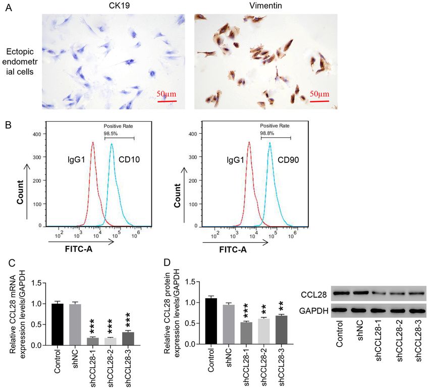
Molecular Medicine REPORTS 25: 56, 2022                                                 3

Table I. Clinical characteristics of patients.

                                                     Patients with                        Healthy patients
Characteristics                                  endometriosis (n=40)                         (n=40)                         P-value

Age, year                  35.55±3.64  31.32±5.87                                                                              0.092
BMI, kg/m   2
                           19.10±2.53  21.32±2.12                                                                              0.061
CA125, IU/ml               23.74±2.56  14.57±2.04                                                                              0.013
EM stage, n (%)
 III	                       21 (52.5)	NA
 IV                         19 (47.5)	NA
Benign conditions, n (%)
 Uterine myoma	NA	                       8 (20.0)
 Endometrial hyperplasia	NA	            13 (32.5)
 Others	NA	                             19 (47.5)
Menstrual phase, n (%)			                                                                                                      0.171
 Proliferative              21 (52.5)   27 (67.5)
 Secretory                  19 (47.5)   13 (32.5)

Data are presented as the mean ± SD or n (%). CA125, cancer antigen 125; NA, not applicable.

Table II. CCL28 sequences for gene silencing.                         detected using a Flow cytometer (CytoFLEX; Beckman
                                                                      Coulter, Inc.) and analyzed using BD Accuri™ C6 Software
Target site name                        Sequence (5'-3')              (Version 1.0.264.21; BD Biosciences).

CCL28‑1 (site: 167-185) GCACGGAGGTTTCACATCA                           Cell proliferation assay. Endometrial stromal cells in the loga‑
CCL28‑2 (site: 260-278)	CTGTCATCCTTCATGTCAA                           rithmic growth phase were digested with trypsin and cultured
CCL28‑3 (site: 321-339) GCAGTGGATGAAAGTGCAA                           overnight in 96‑well plates (cat. no. TR4001; TrueLine) at
                                                                      a density of 3,000 cells/well in a 37˚C, 5% CO2 incubator.
CCL28, C-C motif chemokine ligand 28.                                 At 0, 12, 24 and 48 h of treatment of shNC, shCCL28‑1and
                                                                      shCCL28‑2, or different concentrations of CCL28 recombinant
                                                                      protein (0, 5, 10, 20 and 40 ng/ml), or vehicle + DMSO, CCL28
                                                                      + DMSO, vehicle + PD98059 and CCL28 + PD98059, Cell
at room temperature and then blocked with 1% BSA for 1 h              Counting Kit‑8 (CCK‑8; cat. no. CP002; SAB Biotherapeutics,
at room temperature. Subsequently, cells were incubated with          Inc.) reagent and serum‑free medium were mixed at a volume
primary antibodies against CK19 (1:200; cat. no. ab52625;             ratio of 1:10. Subsequently, 100 µl CCK‑8 mixture was added
Abcam) and vimentin (1:500; cat. no. ab92547; Abcam) at               to the aforementioned groups. After 1 h incubation at 37˚C,
4˚C overnight. Following primary incubation, cells were               the optical density at 450 nm was measured using a microplate
incubated for 30 min with HRP‑labeled secondary antibodies            reader.
(cat. no. D‑3004; Shanghai Changdao Biological Technology
Co., Ltd.). Cells were then subjected to DAB staining. Finally,       Cell invasion assay. Endometrial stromal cells in the loga‑
cells were imaged using an upright fluorescence microscope            rithmic growth phase were digested with trypsin and seeded
and the expression levels of CK19 and vimentin were analyzed          into 6‑well plates at a density of 300,000 cells/well. After
using an image analysis system version 11.0 (IMS; Beijing             24 h of culture at 37˚C, the stromal cells were transduced
Changheng Rongchuang Technology).                                     with shCCL28 (shCCL28‑1, shCCL28‑2) lentivirus for 48 h,
                                                                      or pre‑treated with PD98059 (an ERK inhibitor; 10 µmol/l;
Flow cytometry analysis. The EM markers of CD10 and CD90              S1177; Selleck) for 30 min at 37˚C. Subsequently, cells were
have been detected to verify the purity of endometrial stromal        treated with CCL28 recombinant protein for 48 h at 37˚C and
cells. Endometrial stromal cells in the logarithmic growth            then collected for Transwell detection. For the cell invasion
phase were digested, resuspended and counted. Resuspended             assay, a 24‑well Transwell plate was used (pore size, 8 µm;
cells (5,000,000‑10,000,000) were centrifuged at 1,000 x g            MilliporeSigma; Merck KGaA). The upper chamber of the
for 5 min to obtain the cell precipitants, and then incubated         Transwell plate was coated with 30 µl Matrigel at 37˚C for
with the following antibodies: FITC Mouse Anti‑Human                  30 min and 2x10⁵ cells in 200 µl DMEM/F12 were added.
CD10 (1:50; cat. no. 340925; BD Biosciences); FITC Mouse              DMEM/F12 containing 10% FBS was added to the lower
Anti‑Human CD90 (1:100; cat. no. 561969; BD Biosciences);             chamber. After a 48‑h incubation at37˚C, the membrane was
and FITC Mouse IgG1 (1:100; cat. no. 555748; BD Biosciences).         fixed with 4% formaldehyde and stained with 0.5% crystal
After 30 min of incubation at 4˚C in the dark, the cells were         violet (1 ml) for 30 min at room temperature. The number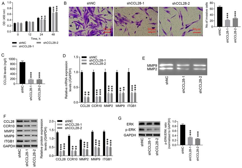
4                                 WU et al: CCL28 REGULATES ENDOMETRIOSIS STROMAL CELLS

Table III. Sequences of primers for reverse transcription-          antibodies against CCL28 (dilution, 1:500; cat. no. ab196567;
quantitative PCR.                                                   Abcam), CCR10 (dilution, 1:250; cat. no. ab3904; Abcam),
                                                                    MMP2 (dilution, 1:5,000; cat. no. ab37150; Abcam), MMP9
Gene                             Sequence (5'-3’)                   (dilution, 1:1,000; cat. no. ab194316; Abcam), ITGB1 (1:1,000;
                                                                    cat. no. ab24693; Abcam), phosphorylated (p)‑ERK (dilution,
CCL28 F:	CTGATGGGGATTGTGACTTG                                       1:1,000; cat. no. ab214362; Abcam), ERK (dilution, 1:10,000;
	R: TGGTGTTTCTTCCTGTGGC                                             cat. no. ab184699; Abcam) and GAPDH (dilution, 1:2,000; cat.
CCR10 F:	AGGGCTGGAGTCTGGGAAGTG                                      no. 5174; Cell Signaling Technology, Inc.). The membranes
	R:	CACGATGACGGAGACCAAGTGT                                          were washed three times with TBS‑0.05% Tween‑20 (TBST),
                                                                    followed by a 2‑h incubation at room temperature with
MMP2  F: GGGAGTACTGCAAGTTCCCCTTCTT
                                                                    HRP‑conjugated goat anti‑rabbit (cat. no. A0208) and goat
	R: TGGAAGCGGAATGGAAAC
                                                                    anti‑mouse (catalog no. A0216) secondary antibody (dilu‑
MMP9  F:	AGGACGGCAATGCTGATG                                         tion, 1:1,000; Beyotime Institute of Biotechnology). Membranes
	R: TCGTAGTTGGCGGTGGTG                                              were washed with TBST and visualized using a chemilu‑
ITGB1 F:	AATGTAACCAACCGTAGC                                         minescent reagent (cat. no. WBKLS0100; MilliporeSigma;
	R: GGTCAATGGGATAGTCTTC                                             Merck KGaA) and ECL imaging system (Tanon‑5200; Tanon
                                                                    Science and Technology Co., Ltd.). ImageJ version 1.47
GAPDH F:	AATCCCATCACCATCTTC
                                                                    (National Institutes of Health) was used to semi‑quantify
	R:	AGGCTGTTGTCATACTTC
                                                                    protein expression levels using GAPDH as a loading control.
CCL28, C-C motif chemokine ligand 28; CCR10, C chemokine
receptor 10; ITGB1, integrin β1; F, forward; R, reverse.            Gelatinase zymography. Total protein from cells was isolated
                                                                    using RIPA buffer, quantified by a BCA kit (cat. no. 23223;
                                                                    Thermo Fisher Scientific, Inc.) and 25 µl protein per lane
                                                                    was separated via 10% SDS‑PAGE containing 1% gelatin.
of invasive cells was counted at a magnification of x200 via        The gels were then washed with eluent (2.5% Triton X‑100,
a light microscope (XDS‑500C; Shanghai Caikang Optical              50 mM Tris‑HCl, 5 mM CaCl2; pH 7.6) twice for 30 min,
Instrument Co., Ltd.).                                              and rinsed with rinsing solution (eluent without Triton X‑100)
                                                                    twice for 20 min. The gels were subsequently incubated in
Reverse transcription‑quantitative PCR (RT‑qPCR). Total             incubation solution (50 mM Tris‑HCL, 5 mM CaCl2, 0.02%
RNA was extracted from endometrial tissues or stromal               brij‑35; pH 7.6) for 20 h at 37˚C. The gels were stained using
cells using TRIzol® reagent (cat. no. 1596‑026; Invitrogen;         Coomassie brilliant blue staining solution for 3 h at room
Thermo Fisher Scientific, Inc.). Total RNA was quantified and       temperature in a low‑speed shaker, and the staining solution
underwent RNA integrity confirmation. Total RNA (1 µg) was          was recovered. A decolorizing solution (30% methanol and
reverse transcribed into complementary DNA using a Reverse          10% acetic acid) was added to highlight clear bands on a blue
Transcription Kit (cat. no. K1622; Fermentas; Thermo Fisher         background for 30 min at room temperature. Images of the
Scientific, Inc.) according to the manufacturer's instructions as   gels were then captured for observation using a gel imager.
follows: 37˚C for 60 min; 85˚C for 5 min; 4˚C for 5 min. qPCR
was subsequently performed using an ABI‑7300 (Applied               Statistical analysis. Statistical analysis was performed using
Biosystems; Thermo Fisher Scientific, Inc.) and a SYBR‑Green        GraphPad Prism version 7.0 software (GraphPad Software,
PCR Kit (cat. no. K0223; Thermo Fisher Scientific, Inc.). The       Inc.). One‑way ANOVA followed by Tukey's post hoc test
following thermocycling conditions were used for qPCR:              was used for statistical comparisons among more than two
Initial denaturation: 10 min at 95˚C; followed by 40 cycles of      groups, whereas unpaired Student's t‑tests were used for statis‑
denaturation, elongation and annealing at 15 sec at 95˚C and        tical comparisons between two groups. For Table I, the t‑test
45 sec at 60˚C. CCL28, CCR10, MMP2, MMP9 and ITGB1                  was used for age, BMI and cancer antigen‑125 comparisons
mRNA expression levels were quantified using the 2 ‑ΔΔCq            between the patient and control groups. The χ2 test was used
method (28) and normalized to the internal reference gene           for comparisons between menstrual phases of the patient and
GAPDH. The primers are listed in Table III.                         control groups. Data are presented as the mean ± standard
                                                                    deviation of ≥3 independent experimental repeats. P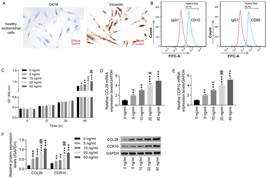
Molecular Medicine REPORTS 25: 56, 2022 5
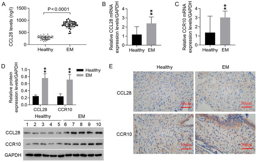
Figure 1. CCL28 and CCR10 are highly expressed in the serum and endometrial tissues of patients with EM. Serum samples of 40 patients with EM and
40 healthy patients were collected to detect CCL28 levels, and 15 endometrial tissue samples from patients with EM and 15 endometrial tissues from healthy
patients were also collected. (A) CCL28 levels in the serum of patients with EM were detected using ELISA. mRNA expression levels of (B) CCL28 and
(C) CCR10 in endometrial tissues of patients with EM were detected by reverse transcription‑quantitative PCR. (D) Protein expression levels of CCL28
and CCR10 in endometrial tissues of patients with EM were detected using western blotting. (E) Protein expression levels of CCL28 and CCR10 in endo‑
metrial tissues of patients with EM were detected by immunohistochemistry (x200, 50 µm). **P95% of the isolated cells were EM results suggested that knockdown of CCL28 attenuated EM
stromal cells (Fig. 2B). To understand the function of CCL28 progression by inhibiting cell proliferation and invasion via
in EM, lentiviral transduction was used to downregulate the regulation of CCR10, MMP2, MMP9 and ITGB1 expres‑
CCL28 expression in ectopic endometrial stromal cells. Both sion, and this may involve the ERK signaling pathway.
mRNA and protein expression levels of CCL28 were signifi‑
cantly downregulated by shCCL28‑1, ‑2 and ‑3 compared with CCL28 recombinant proteins significantly increase CCL28
negative control (shNC; Fig. 2C and D). Among the three, and CCR10 expression in healthy endometrial stromal cells.
shCCL28‑1 and shCCL28‑2 were more efficient and selected The results demonstrated that positive vimentin expression was
for use in subsequent experiments. observed in endometrial stromal cells from healthy controls,
whereas CK19 expression was negative (Fig. 4A). Detection of
Knockdown of CCL28 in endometrial stromal cells signifi‑ CD10 and CD90 by flow cytometry verified that >95% of the
cantly suppresses cell proliferation and invasion. Following isolated cells were healthy endometrial stromal cells (Fig. 4B).
CCL28 knockdown, cell proliferation and invasion were evalu‑ A series of CCL28 recombinant protein concentrations (0, 5,
ated. Cell proliferation (0‑48 h) and cell invasion in shCCL28 10, 20 and 40 ng/ml) were used to treat healthy endometrial
endometrial stromal cells were significantly decreased stromal cells. At 48 h, CCL28 could significantly promote
compared with those in the shNC group (Fig. 3A and B). healthy endometrial stromal cell proliferation in a dose‑depen‑
Compared with the shNC group, a significant decrease in dent manner compared with 0 ng/ml, whereas at 0, 12 and 24 h6 WU et al: CCL28 REGULATES ENDOMETRIOSIS STROMAL CELLS Figure 2. Knockdown of CCL28 expression in endometrial stromal cells by lentiviral transduction. (A) endometrial stromal cells were identified via immuno‑ cytochemistry, which was used to analyze CK19 and vimentin expression (x200, 50 µm). (B) CD10 and CD90 were detected using flow cytometry to identify the percentage of EM stromal cells. shCCL28‑1, ‑2 and ‑3 were constructed to transduce endometrial stromal cells, CCL28 (C) mRNA and (D) protein expression levels were detected to determine the knockdown efficiency of the constructs. **P
Molecular Medicine REPORTS 25: 56, 2022                                                                   7

Figure 3. Knockdown of CCL28 in endometrial stromal cells significantly suppresses cell proliferation and invasion. (A) Following CCL28 knockdown in
endometrial stromal cells, cell proliferation was detected using a Cell Counting Kit‑8 assay at 0, 12, 24 and 48 h. (B) Cell invasion at 48 h was detected using
a Transwell invasion assay (x200, 50 µm). (C) CCL28 levels in endometrial stromal cell supernatants were detected using an ELISA. (D) mRNA expression
levels of CCL28, CCR10, MMP2, MMP9 and ITGB1 were examined using reverse transcription‑quantitative PCR. (E) Activities of MMP2 and MMP9
were detected using gelatinase zymography. (F) Protein expression levels of CCL28, CCR10, MMP2, MMP9 and ITGB1 were detected via western blotting.
(G) Protein expression levels of p‑ERK/ERK ratio were detected via western blotting. **P
8                                       WU et al: CCL28 REGULATES ENDOMETRIOSIS STROMAL CELLS

Figure 4. Treatment with CCL28 recombinant protein increases CCL28 and CCR10 expression in healthy human endometrial stromal cells. (A) Healthy human
endometrial stromal cells were identified using immunocytochemistry to analyze CK19 and vimentin expression (x200, 50 µm). (B) CD10 and CD90 were
detected via flow cytometry to identify the percentage of healthy endometrial stromal cells. Healthy human endometrial stromal cells were then treated with
CCL28 recombinant protein at concentrations of 0, 5, 10, 20 and 40 ng/ml. (C) Cell proliferation was detected using a Cell Counting Kit‑8 assay to determine
the effect of CCL28 recombinant protein. Subsequently, at 48 h after CCL28 recombinant protein treatment, the mRNA expression levels of (D) CCL28
and (E) CCR10 were detected using reverse transcription‑quantitative PCR. (F) Relative protein expression levels of CCL28 and CCR10 were analyzed via
western blotting at 48 h after CCL28 recombinant protein treatment. *P
Molecular Medicine REPORTS 25: 56, 2022                                                                 9

Figure 5. CCL28 may contribute to endometriosis progression by regulating MMP2, MMP9 and ITGB1 expression via activating the ERK signaling pathway.
Healthy endometrial stromal cells were pre‑treated with 10 µmol/l PD98059 (ERK inhibitor) for 30 min, and then treated with 20 ng/ml CCL28 recom‑
binant protein for 48 h. (A) Cell proliferation was detected using the Cell Counting Kit‑8 assay at 0, 12, 24 and 48 h. (B) Cell invasion was detected using
the Transwell invasion assay at 48 h (x200, 50 µm). (C) MMP2 and MMP9 activity was detected via gelatinase zymography. Relative protein expression
levels of (D) MMP2, MMP9 and ITGB1 and (E) the p‑ERK/ERK ratio were analyzed via western blotting. **P
10                                     WU et al: CCL28 REGULATES ENDOMETRIOSIS STROMAL CELLS

References                                                                  21. Tong JS, Zhang QH, Huang X, Fu XQ, Qi ST, Wang YP, Hou Y,
                                                                                Sheng J and Sun QY: Icaritin causes sustained ERK1/2 activation
                                                                                and induces apoptosis in human endometrial cancer cells. PLoS
 1. Kennedy S, Bergqvist A, Chapron C, D'Hooghe T, Dunselman G,                 One 6: e16781, 2011.
    Greb R, Hummelshoj L, Prentice A and Saridogan E; ESHRE                 22. Banu SK, Lee J, Speights VO Jr, Starzinski‑Powitz A and
    Special Interest Group for Endometriosis and Endometrium                    Arosh JA: Selective inhibition of prostaglandin E2 receptors
    Guideline Development Group: ESHRE guideline for the diagnosis              EP2 and EP4 induces apoptosis of human endometriotic cells
    and treatment of endometriosis. Hum Reprod 20: 2698‑2704, 2005.             through suppression of ERK1/2, AKT, NFkappaB, and β‑catenin
 2. Eskenazi B and Warner ML: Epidemiology of endometriosis.                    pathways and activation of intrinsic apoptotic mechanisms. Mol
    Obstet Gynecol Clin North Am 24: 235‑258, 1997.                             Endocrinol 23: 1291‑1305, 2009.
 3. Zondervan KT, Yudkin PL, Vessey MP, Jenkinson CP, Dawes MG,             23. Li MQ, Li HP, Meng YH, Wang, Zhu XY, Mei J and Li DJ:
    Barlow DH and Kennedy SH: The community prevalence of                       Chemokine CCL2 enhances survival and invasiveness of endo‑
    chronic pelvic pain in women and associated illness behaviour.              metrial stromal cells in an autocrine manner by activating Akt and
    Br J Gen Pract 51: 541‑547, 2001.                                           MAPK/Erk1/2 signal pathway. Fertil Steril 97: 919‑929, 2012.
 4. Zondervan KT, Cardon LR and Kennedy SH: What makes a                    24. Li MQ, Shao J, Meng YH, Mei J, Wang Y, Li H, Zhang L,
    good case‑control study? Design issues for complex traits such              Chang KK, Wang XQ, Zhu XY, et al: NME1 suppression
    as endometriosis. Hum Reprod 17: 1415‑1423, 2002.                           promotes growth, adhesion and implantation of endometrial
 5. Rogers PA, D'Hooghe TM, Fazleabas A, Gargett CE,                            stromal cells via Akt and MAPK/Erk1/2 signal pathways in the
    Giudice LC, Montgomery GW, Rombauts L, Salamonsen LA                        endometriotic milieu. Hum Reprod 28: 2822‑2831, 2013.
    and Zondervan KT: Priorities for endometriosis research:                25. Yang XL, Liu KY, Lin FJ, Shi HM and Ou ZL: CCL28 promotes
    Recommendations from an international consensus workshop.                   breast cancer growth and metastasis through MAPK‑mediated
    Reprod Sci 16: 335‑346, 2009.                                               cellular anti‑apoptosis and pro‑metastasis. Oncol Rep 38:
 6. Rolla E: Endometriosis: Advances and controversies in classifi‑             1393‑1401, 2017.
    cation, pathogenesis, diagnosis, and treatment. F1000 Res 8: 8, 2019.   26. Liu J, Zhang Z, Liu J and Wang D: LIM Kinase 1 Mediates
 7. Liu J, Wang Y, Chen P, Ma Y, Wang S, Tian Y, Wang A and Wang D:             estradiol effects on the phosphorylation of Cofilin1 in eutopic
    AC002454.1 and CDK6 synergistically promote endometrial                     endometrial stromal cells during the invasion and proliferation
    cell migration and invasion in endometriosis. Reproduction 157:             of endometriosis. Reprod Sci 26: 1499‑1505, 2019.
    535‑543, 2019.                                                          27. Chen Q, Hang Y, Zhang T, Tan L, Li S and Jin Y: USP10
 8. Bakogiannis C, Sachse M, Stamatelopoulos K and Stellos K:                   promotes proliferation and migration and inhibits apoptosis of
    Platelet‑derived chemokines in inflammation and atherosclerosis.            endometrial stromal cells in endometriosis through activating
    Cytokine 122: 154157, 2019.                                                 the Raf‑1/MEK/ERK pathway. Am J Physiol Cell Physiol 315:
 9. Korbecki J, Grochans S, Gutowska I, Barczak K and                           C863‑C872, 2018.
    Baranowska‑Bosiacka I: CC Chemokines in a tumor: A review of            28. Livak KJ and Schmittgen TD: Analysis of relative gene
    pro‑cancer and anti‑cancer properties of receptors CCR5, CCR6,              expression data using real‑time quantitative PCR and the 2‑ΔΔCT
    CCR7, CCR8, CCR9, and CCR10 ligands. Int J Mol Sci 21: 21,                  method. Methods 25: 402‑408, 2001.
    2020.                                                                   29. Minkwitz C, Schoon HA, Zhang Q and Schöniger S: Plasticity of
10. Bian X, Xiao YT, Wu T, Yao M, Du L, Ren S and Wang J:                       endometrial epithelial and stromal cells‑A new approach towards
    Microvesicles and chemokines in tumor microenvironment:                     the pathogenesis of equine endometrosis. Reprod Domest
    Mediators of intercellular communications in tumor progression.             Anim 54: 835‑845, 2019.
    Mol Cancer 18: 50, 2019.                                                30. Deng X, Zhang X, Li W, Feng RX, Li L, Yi GR, Zhang XN,
11. Hieshima K, Ohtani H, Shibano M, Izawa D, Nakayama T,                       Yin C, Yu HY, Zhang JP, et al: Chronic Liver Injury Induces
    Kawasaki Y, Shiba F, Shiota M, Katou F, Saito T, et al: CCL28 has           Conversion of Biliary Epithelial Cells into Hepatocytes. Cell
    dual roles in mucosal immunity as a chemokine with broad‑spectrum           Stem Cell 23: 114‑122.e3, 2018.
    antimicrobial activity. J Immunol 170: 1452‑1461, 2003.                 31. Gou Y, Li X, Li P, Zhang H, Xu T, Wang H, Wang B, Ma X,
12. Berri M, Virlogeux‑Payant I, Chevaleyre C, Melo S, Zanello G,               Jiang X and Zhang Z: Estrogen receptor β upregulates CCL2
    Salmon H and Meurens F: CCL28 involvement in mucosal                        via NF‑κ B signaling in endometriotic stromal cells and recruits
    tissues protection as a chemokine and as an antibacterial peptide.          macrophages to promote the pathogenesis of endometriosis. Hum
    Dev Comp Immunol 44: 286‑290, 2014.                                         Reprod 34: 646‑658, 2019.
13. Moed H, Boorsma DM, Tensen CP, Flier J, Jonker MJ,                      32. Mei J, Zhou WJ, Li SY, Li MQ and Sun HX: Interleukin‑22
    Stoof TJ, von Blomberg BM, Bruynzeel DP, Scheper RJ,                        secreted by ectopic endometrial stromal cells and natural killer
    Rustemeyer T, et al: Increased CCL27‑CCR10 expression in                    cells promotes the recruitment of macrophages through promoting
    allergic contact dermatitis: implications for local skin memory.            CCL2 secretion. Am J Reprod Immunol 82: e13166, 2019.
    Journal Pathol 204: 39‑46, 2004.                                        33. Hirata T, Osuga Y, Takamura M, Kodama A, Hirota Y, Koga K,
14. Chen L, Liu X, Zhang H‑Y, Du W, Qin Z, Yao Y, Mao Y                         Yoshino O, Harada M, Takemura Y, Yano T, et al: Recruitment
    and Zhou L: Upregulation of chemokine receptor CCR10 is                     of CCR6‑expressing Th17 cells by CCL20 secreted from IL‑1
    essential for glioma proliferation, invasion and patient survival.          β‑, TNF‑α‑, and IL‑17A‑stimulated endometriotic stromal cells.
    Oncotarget 5: 6576‑6583, 2014.                                              Endocrinology 151: 5468‑5476, 2010.
15. Xiong N, Fu Y, Hu S, Xia M and Yang J: CCR10 and its ligands            34. Guo P, Bi K, Lu Z, Wang K, Xu Y, Wu H, Cao Y and Jiang H:
    in regulation of epithelial immunity and diseases. Protein Cell 3:          CCR5/CCR5 ligand‑induced myeloid‑derived suppressor cells
    571‑580, 2012.                                                              are related to the progression of endometriosis. Reprod Biomed
16. Lin HY, Sun SM, Lu XF, Chen PY, Chen CF, Liang WQ and                       Online 39: 704‑711, 2019.
    Peng CY: CCR10 activation stimulates the invasion and migration         35. Jaiswal U, Yadav RK, Bhat MA, Kriplani A, Roy KK and
    of breast cancer cells through the ERK1/2/MMP‑7 signaling                   Netam RK: Cytokine and growth factor profile in endometriosis:
    pathway. Int Immunopharmacol 51: 124‑130, 2017.                             a multiplex analysis of peritoneal fluid to assess diagnostic utility.
17. Ruan F, Ma J and Zhou J: Depletion of CCL27 inhibits cell                   Gynecol Endocrinol 36: 718‑722, 2020.
    proliferation, metastasis and adhesion in ectopic endometrial           36. Rashidiani S, Jalili A, Babaei E, Sheikhesmaeili F, Fakhari S,
    stromal cells. Int J Clin Exp Med 9: 19074‑19083, 2016.                     Ataee P and Parhizkar B: The chemokine CCL28 is elevated
18. Sun C, Zhang YY, Tang CL, Wang SC, Piao HL, Tao Y, Zhu R,                   in the serum of patients with celiac disease and decreased after
    Du MR and Li DJ: Chemokine CCL28 induces apoptosis of                       treatment. Am J Clin Exp Immunol 6: 60‑65, 2017.
    decidual stromal cells via binding CCR3/CCR10 in human spon‑            37. Sanaei MJ, Shirzad H, Soltani A, Abdollahpour‑Alitappeh M,
    taneous abortion. Mol Hum Reprod 19: 676‑686, 2013.                         Shafigh MH, Rahimian G, Mirzaei Y and Bagheri N: Up‑regulated
19. Cha HR, Ko HJ, Kim ED, Chang SY, Seo SU, Cuburu N,                          CCL18, CCL28 and CXCL13 expression is associated with the
    Ryu S, Kim S and Kweon MN: Mucosa‑associated epithelial                     risk of gastritis and peptic ulcer disease in Helicobacter pylori
    chemokine/CCL28 expression in the uterus attracts CCR10+ IgA                infection. Am J Med Sci361: 43‑54, 2021.
    plasma cells following mucosal vaccination via estrogen control.        38. Lv Y, Zhao X, Zhu L, Li S, Xiao Q, He W and Yin L: Targeting
    J Immunol 187: 3044‑3052, 2011.                                             intracellular MMPs efficiently inhibits tumor metastasis and
20. Gentilini D, Busacca M, Di Francesco S, Vignali M, Viganò P and             angiogenesis. Theranostics 8: 2830‑2845, 2018.
    Di Blasio AM: PI3K/Akt and ERK1/2 signalling pathways are               39. Wang X, Yang B, She Y and Ye Y: The lncRNA TP73‑AS1 promotes
    involved in endometrial cell migration induced by 17β‑estradiol             ovarian cancer cell proliferation and metastasis via modulation of
    and growth factors. Mol Hum Reprod 13: 317‑322, 2007.                       MMP2 and MMP9. J Cell Biochem 119: 7790‑7799, 2018.
Molecular Medicine REPORTS 25: 56, 2022 11
40. Farina P, Tabouret E, Lehmann P, Barrie M, Petrirena G, 47. Kodarahmian M, Amidi F, Moini A, Kashani L, Nashtaei MS,
Campello C, Boucard C, Graillon T, Girard N and Chinot O: Pazhohan A, Bahramrezai M, Berenjian S and Sobhani A: The
Relationship between magnetic resonance imaging character‑ modulating effects of resveratrol on the expression of MMP‑2
istics and plasmatic levels of MMP2 and MMP9 in patients and MMP‑9 in endometriosis women: A randomized exploratory
with recurrent high‑grade gliomas treated by Bevacizumab and trial. Gynecol Endocrinol 35: 719‑726, 2019.
Irinotecan. J Neurooncol 132: 433‑437, 2017. 48. Shan B, Li W, Yang SY and Li ZR: Estrogen up‑regulates MMP2/9
41. Pino M, Galleguillos C, Torres M, Sovino H, Fuentes A, expression in endometrial epithelial cell via VEGF‑ERK1/2
Boric MA and Johnson MC: Association between MMP1 and pathway. Asian Pac J Trop Med 6: 826‑830, 2013.
MMP9 activities and ICAM1 cleavage induced by TNF in 49. Wang D, Wang D, Wang N, Long Z and Ren X: Long non‑coding
stromal cell cultures from eutopic endometria of women with RNA BANCR promotes endometrial cancer cell proliferation
endometriosis. Reproduction 138: 837-847, 2009. and invasion by regulating MMP2 and MMP1 via ERK/MAPK
42. Aglund K, Rauvala M, Puistola U, Angström T, Turpeenniemi- signaling pathway. Cell Physiol Biochem 40: 644‑656, 2016.
Hujanen T, Zackrisson B and Stendahl U: Gelatinases A and B 50. Lei Y, Huang K, Gao C, Lau QC, Pan H, Xie K, Li J, Liu R,
(MMP‑2 and MMP‑9) in endometrial cancer‑MMP‑9 correlates Zhang T, Xie N, et al: Proteomics identification of ITGB3 as a
to the grade and the stage. Gynecol Oncol 94: 699‑704, 2004. key regulator in reactive oxygen species‑induced migration and
43. Honkavuori M, Talvensaari‑Mattila A, Soini Y, Turpeenniemi- invasion of colorectal cancer cells. Mol Cell Proteomics: May 27,
Hujanen T and Santala M: MMP‑2 expression associates with 2011 (Epub ahead of print). doi: 10.1074/mcp.M110.005397.
CA 125 and clinical course in endometrial carcinoma. Gynecol 51. Chen J, Gu L, Ni J, Hu P, Hu K and Shi YL: MiR‑183 regulates
Oncol 104: 217‑221, 2007. ITGB1P expression and promotes invasion of endometrial
44. Barbe AM, Berbets AM, Davydenko IS, Koval HD, Yuzko VO stromal cells. Biomed Res Int 2015: 340218, 2015.
and Yuzko OM: Expression and significance of matrix metal‑
loproteinase‑2 and matrix metalloproteinas‑9 in Endometriosis.
J Med Life 13: 314‑320, 2020. This work is licensed under a Creative Commons
45. Freitas S, Meduri G, Le Nestour E, Bausero P and Perrot- Attribution-NonCommercial-NoDerivatives 4.0
Applanat M: Expression of metalloproteinases and their inhibitors International (CC BY-NC-ND 4.0) License.
in blood vessels in human endometrium. Biol Reprod 61:
1070‑1082, 1999.
46. Yoshiji H, Harris SR, Raso E, Gomez DE, Lindsay CK, Shibuya M,
Sinha CC and Thorgeirsson UP: Mammary carcinoma cells
over‑expressing tissue inhibitor of metalloproteinases‑1 show
enhanced vascular endothelial growth factor expression. Int J
Cancer 75: 81‑87, 1998.You can also read