RADY 403 Case Presentation - Susana Bracewell, MS4 May 21, 2021 - UNC Radiology Medical Student Website
←
→
Page content transcription
If your browser does not render page correctly, please read the page content below
RADY 403 Case Presentation

   Susana Bracewell, MS4
       May 21, 2021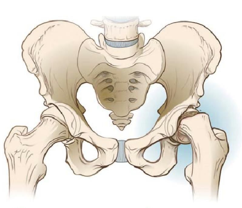
Focused History

• 9-year-old male with past medical history significant for obesity
• Presented to clinic with left hip pain over the past two months
   • Describes the pain as non-radiating, dull, and aching
   • No history of preceding trauma
• Furthermore, the patient recently began limping
• On exam, he was afebrile
• CBC, ESR, and CRP were unremarkable
Causes of Hip Pain in a Child

                                                           Mechanical or
    Infectious         Inflammatory                         Orthopedic
                                                                                            Neoplastic                    Other
-   Septic arthritis   -   Transient synovitis         -    Slipped capital femoral     -   Osteoid osteoma        -   Sickle cell disease
-   Lyme disease       -   Spondyloarthropathy              epiphysis                   -   Leukemia               -   Gaucher disease
                       -   Juvenile idiopathic         -    Legg-Calvé-Perthes
-   Osteomyelitis                                                                       -   Solid tumor (primary   -   Neuromuscular
                           arthritis                        disease
-   Psoas abscess      -   Infectious/post-                                                 or metastatic)             disorders (e.g.,
                                                       -    Secondary avascular
-   Pyomyositis            infectious arthritis             necrosis                    -   Pigmented                  muscular dystrophy)
-   Spinal epidural    -   Other rheumatologic         -    Femoral stress fracture         villonodular
    abscess                conditions (e.g., SLE,      -    Muscular strain                 synovitis
-   Appendicitis           MCTD, vasculitis,           -    Iliac apophysitis
                           dermatomyositis,            -    Snapping iliopsoas
-   Abdominal/pelvic
                           CRMO, localized                  tendon
    abscess                scleroderma, FMF)           -    Trochanteric bursitis
                       -   Arthritis associated with   -    Acetabular labral tear
                           gastrointestinal            -    Femoroacetabular
                           conditions (e.g., IBD,           impingement
                           celiac disease)             -    Apophyseal avulsion
                       -   Idiopathic chondrolysis          fracture with apophysitis
                           of the hip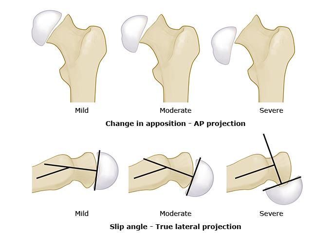
ACR Appropriateness Criteria

Therefore, two views of the pelvis were obtained.
List of Imaging Studies

• AP Radiograph of Pelvis
• Lateral Radiograph of Left Hip
AP Radiograph of Pelvis

Normal Klein’s line (a line
along the superior edge
of the neck of the femur
that intersects the lateral                                        Abnormal Klein’s line where
part of the superior                                               the epiphysis is malaligned
femoral epiphysis).                                                with no intersection of the
                                                                   capital femoral epiphysis.

                              Proximal femoral capital epiphysis
Lateral Radiograph of Left Hip
Widening and
malalignment of
the left capital
femoral epiphysis
consistent with a
mild slipped capital
femoral epiphysis.
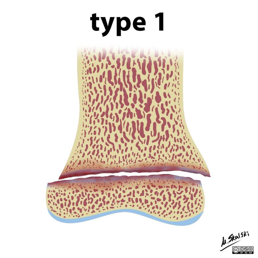
Proximal femoral capital epiphysisSlipped Capital Femoral Epiphysis (SCFE) – Definition
• Also called slipped upper femoral
epiphysis (SUFE)
• Type I Salter-Harris fracture through
the proximal femoral physis
• Fracture that is completely contained
within the physis
• Loss of structural integrity along the
physis results in displacement of the
femoral neck and the appearance of
a posteriorly and inferiorly displaced
epiphysis
Case courtesy of Dr Matt Skalski, Radiopaedia.org, rID: 27144Clinical Features
• Affects 1 to 6 in 20,000 patients with a 2:1 male
predominance
• Bilateral in up to one third of patients
• Most common in patients 10 to 15 years of age
• Due to the combination of increased biomechanical stress
and a weakened perichondrial ring during puberty from
increased growth and hormonal changes
• Risk factors include endinocrinopathies (such as
hypothyroidism, renal disease, and hypogonadism),
biochemical stress (such as trauma and obesity),
and African or Hispanic ancestry
• Acute SCFE is associated with an unstable joint and
severe pain of less than 3 weeks’ duration whereas Image via Reid, J, Davros, W, Paladin, A, Lee, E, & Carrico, C (Eds.). (2014). Pediatric radiology.
ProQuest Ebook Central https://ebookcentral-proquest-com.libproxy.lib.unc.edu
pain is mild and may subside with rest in a chronic
SCFE
• In stable SCFE, the patient maintains the ability to
bear weight, which is lost in unstable SCFECorrect interpretation of
the radiographs is critical.
Radiographic Findings In as many as 25 percent
of patients whose SCFEs
are missed, radiographs
were misinterpreted, or
• Widening, lucency, and irregularity the diagnosis could not be
of the physis established with the
radiographs that were
• Sclerosis of the femoral neck obtained.
• Bone remodeling
• Displacement of the epiphysis from
the metaphysis anteriorly or
posteriorly on lateral view Case courtesy of Dr Hani Makky Al Salam, Radiopaedia.org, rID: 9298
• Osteonecrosis, chrondrolysis, and
osteoarthritis in the hip joint
• Klein’s line does not intersect with
the epiphysis on frontal view
• In the normal AP view, a line drawn
along the superior femoral neck
intersects the lateral portion of the Case courtesy of Assoc Prof Frank Gaillard, Radiopaedia.org,
femoral head rID: 8004Severity based upon radiographic
Severity Grading findings determines prognosis.
• Mild: Displacement of epiphysis 50 degrees on true
lateral projection Image via Kienstra, AJ, & Macias, CG (2021). Evaluation and management of slipped capital femoral
epiphysis (SCFE). Phillips, WA, & Singer, JI (Eds.), UptoDate. Available
from https://www.uptodate.com/contents/evaluation-and-management-of-slipped-capital-femoral-
epiphysis-scfe?csi=6870df90-0491-4ddf-850c-d60bb93ab3f2&source=contentShareManagement
• SCFE should be treated as an emergency upon diagnosis and
confirmation by imaging
• Children with SCFE should be made non-weightbearing and promptly referred
to an orthopedic surgeon
• Surgery is the treatment of choice to prevent further slippage and
promote physeal closure
• Screw fixation is the most commonly used and widely accepted treatment
• Prophylactic pinning of the opposite hip is considered an option by
some surgeons but is controversialAP Radiograph of Pelvis
Back to our case…
Right hip is normally Interval pinning of
aligned. left hip. Fully
threaded screw
transversing the left
femoral neck.Lateral Radiograph of Left Hip
No adverse
hardware features of
left hip.AP Radiograph of Pelvis
Two years later…
Interval removal of pin
in left hip due to
fixation failure.
Patient has since
developed right-sided
SCFE. Interval pinning
of right hip. Fragmentation and
sclerosis of left
proximal femoral
epiphysis.
Fully threaded screw
transversing the
right femoral neck.Lateral Radiograph of Hips
Stable alignment of
left hip with
elements of healing
avascular necrosis.
No adverse features
of right hip.Complications Sclerosis
• Avascular necrosis
• The most serious complication and has the
worst prognosis
• May be a complication from an acute slip
or surgical fixation
• Chondrolysis
• Defined as narrowing of the joint space Lucency
and loss of articular cartilage Spurring
• Femoroacetabular impingement
• Abnormal contact between the proximal
femoral metaphysis and the acetabular rim
• Osteoarthritis
Image via Reid, J, Davros, W, Paladin, A, Lee, E, & Carrico, C (Eds.). (2014). Pediatric
radiology. ProQuest Ebook Central https://ebookcentral-proquest-
com.libproxy.lib.unc.eduKey Points • Approximately 2:1 male predominance • Type I Salter-Harris fracture through physis • Displacement of the metaphysis while the epiphysis remains within the acetabulum • Risk factors include obesity, trauma, and endincrinopathies • Common complications include avascular necrosis, chrondrolysis, and osteoarthritis • Always obtain both AP and lateral views of the hips when suspicious for SCPE
References

• Nigrovic, PA. (2019). Approach to hip pain in childhood. JE Drutz, WA Phillips, & SC
  Li (Eds.), UptoDate. Available
  from https://www.uptodate.com/contents/approach-to-hip-pain-in-
  childhood?csi=b26d3ae2-95b3-4753-b567-2e8743048219&source=contentShare
• ACR Appropriateness Criteria. (2021). Retrieved May 18, 2021, from
  https://www.acr.org/Clinical-Resources/ACR-Appropriateness-Criteria
• Kienstra, AJ, & Macias, CG (2021). Evaluation and management of slipped capital
  femoral epiphysis (SCFE). Phillips, WA, & Singer, JI (Eds.), UptoDate. Available
  from https://www.uptodate.com/contents/evaluation-and-management-of-
  slipped-capital-femoral-epiphysis-scfe?csi=6870df90-0491-4ddf-850c-
  d60bb93ab3f2&source=contentShare
• Reid, J, Davros, W, Paladin, A, Lee, E, & Carrico, C (Eds.). (2014). Pediatric radiology.
  ProQuest Ebook Central https://ebookcentral-proquest-com.libproxy.lib.unc.edu
You can also read