Molecular Modeling 2021 lecture 3 - Tues Feb 2 - Protein classification SCOP TOPS
←
→
Page content transcription
If your browser does not render page correctly, please read the page content below
Molecular Modeling 2021
           lecture 3 -- Tues Feb 2

Protein classification
  SCOP
  TOPS
  Contact maps

                         domains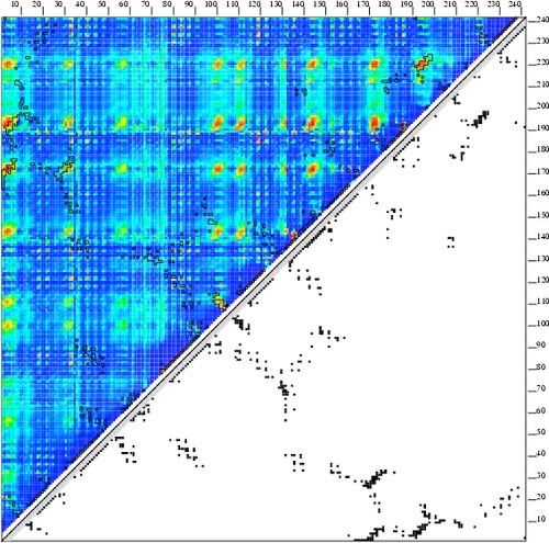
Domains

To a cell biologist a domain is a sequential unit
within a gene, usually with a specific function.    To a structural biologist a domain is a compact
                                                    globular unit within a protein, classified by its 3D
                                                    structure.
                                                                                                           2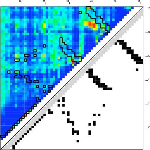
A domain is...
• ... an autonomously-folding substructure of a
  protein.
• ... > 30 residues, but typically < 200. May be
  bigger.
• ...usually has a single hydrophobic core
• ... usually composed of one chain (occasionally composed
  of multiple chains)

• ...is usually composed on one contiguous segment
  (occasionally made of discontiguous segments of the same chain)

                                                                    3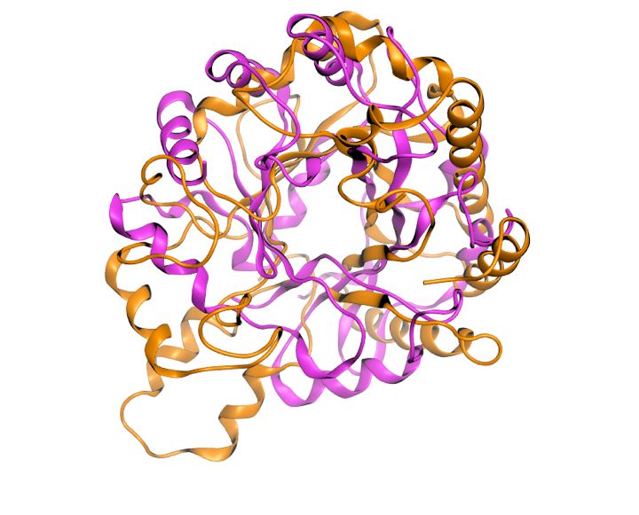
SARS-CoV-2 spike protein — a
   multi domain protein

                         4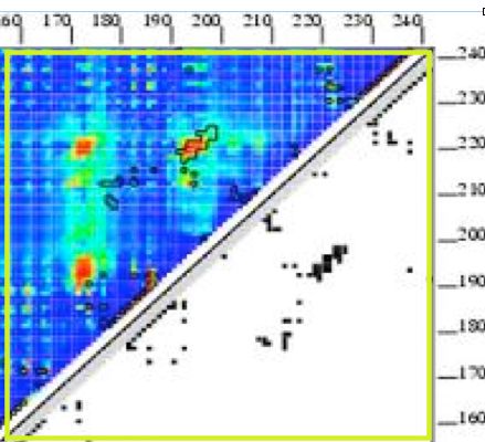
SCOPe -- classification of domains
                                              !http://scop.berkeley.edu

                                                               similar secondary structure
                                   (1) class                   content
increasing structural similarity

                                   (2) fold                   vague structural homology
                                   (3) superfamily
                                                                Clear structural homology
                                   (4) family
                                   (5) protein                Clear sequence homology

                                   (6) species          nearly identical sequences

                                                     individual structures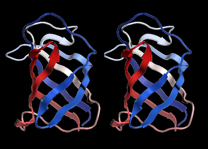
SCOPe -- class

 1. all α (289)
                               classes of domains
 2. all β (178)
 3. α/β (148)
 4. α+β (388)
 5. multidomain (71)
 6. membrane (60)
 7. small (98)                 Not true classes of globular
 8. coiled coil (7)            protein domains
 9. low-resolution (25)
 10. peptides (148)
 11. designed proteins (44)
 12. artifacts (1)

Proteins of the same class conserve secondary structure content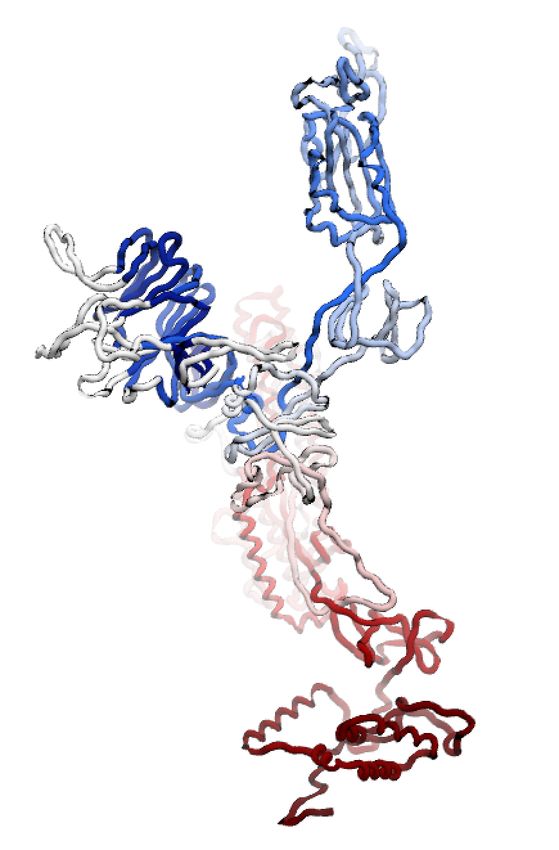
SCOPe -- fold level
within α/β proteins -- Mainly parallel beta sheets (beta-alpha-beta units)

TIM-barrel (22)
swivelling beta/beta/alpha domain (5)
                                            Many folds have historical
spoIIaa-like (2)                            names. “TIM” barrel was
flavodoxin-like (10)                        first seen in TIM. These
restriction endonuclease-like (2)           classifications are done by
ribokinase-like (2)
                                            eye, by experts.
chelatase-like (2)

       Proteins of the same Fold conserve topology.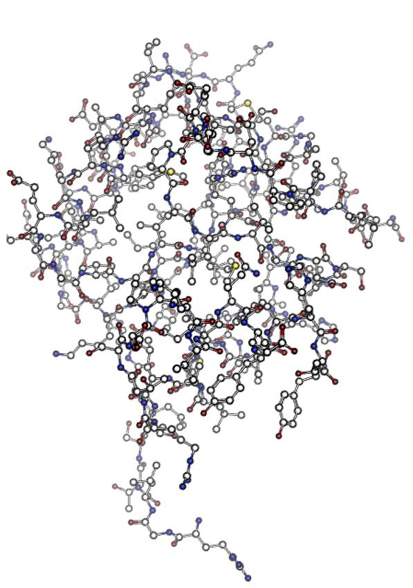
fold level jargon
               example: α/β proteins: flavodoxin-like
 SCOP Description: 3 layers, α/β/α; parallel beta-sheet of 5 strand, order 21345

                         “layers”
           Rough arrangements of
           secondary structure elements.

             “order”
The sequential order of beta
strands in a beta sheet.

                                                   α layer             α layer
        2 1 3 4                                              β layer
                5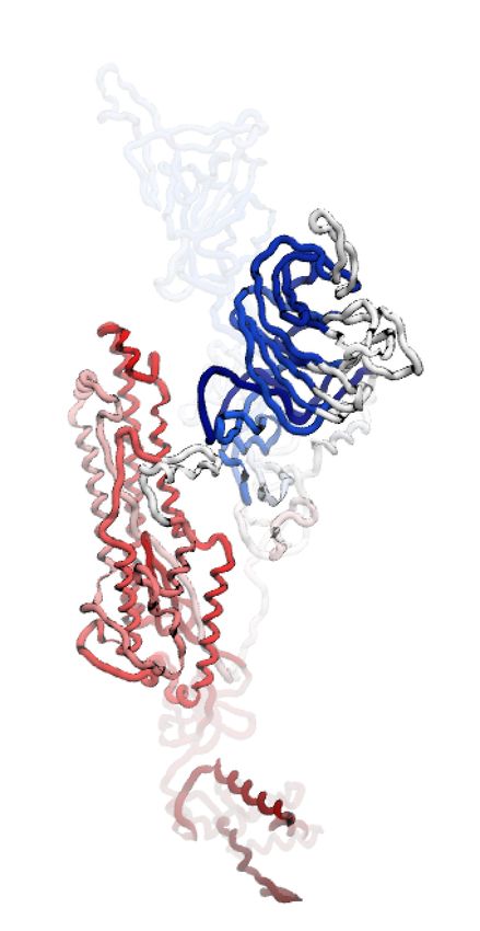
Fold-level similarity
           7-stranded alpha/beta barrel

                         SSE are in the same order along the
                         chain, and trace roughly the same path
                         through space. Similarity is evident
                         when viewed side-by-side

2bod

1m65
                         But the SSE do not superpose. Some
                         superposition algorithms fail to
                         superpose proteins of the same fold.
                                                      9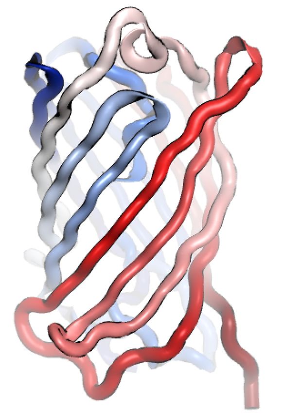
Superfamily level similarity
                                         is hard to see.

       Members of the same superfamily cannot usually be found in a BLAST search. But can be
       identified by structural superposition.

1djq                                                                                           1h6v

                                      FAD-linked reductases

 Proteins in the same superfamily may look completely different, but upon close
 inspection they contains a superposable domain of secondary structure elements.
Family level similarity
is obvious.
FAD/NAD-linked reductases, N-terminal and central domains [51943]
1h6v
1fec
Different members of the same family superimpose well. At this level, a structure may be
used as a molecular replacement model for Xray crystallography.
A BLAST search using one family member finds all other family members.Definition of SCOP Family, Superfamily, Fold
family family
family family
family sequence difference family
superfamily superfamily
Fold
A Family is the set of homologs we can find by BLAST sequence database search.
A Superfamily is a set of distant homologs that cannot be easily found by BLAST search, but can be
recognized by sophisticated fold recognition algorithms
A Fold is an even more distant homologous relationship, recognizable only when the structure is
known
A Class is not a homologous relationship but just a statement of the gross secondary structure content.Contact maps and TOPS
diagrams
13TOPS topology cartoons
Secondary structure elements (SSE)
beta strand beta strand alpha helix connections
pointing up pointing
down
A parallel beta sheet
An anti- parallel beta sheetTOPS topology cartoons
connection in middle
means on top.
connection on side
means on bottom.
A right-handed βαβ unit
A left-handed βαβ unit
(rare)How to draw TOPS To do this on your own, find the link "TOPS practice" (tops_practice.moe) on the course web site. Download. Open it in moe. Or just follow along as I guide you through it. Get pen and paper.
How to draw TOPS
Line up the molecule along the beta sheet, if present.
Otherwise choose a direction so that secondary structures are
mostly perpendicular to the screen.
TOPS diagram
• Draw secondary structures first.
TOPS diagram
• number them and connect

                     2        1

        4        3        2        1

            C                             N
                  4       3

Be careful to draw connections to the center or side,
when it is in front or in back, respectively.
Name it. SCOP-style.
• 3 layers, 2-4-2 αβα, all parallel, 1234
2 1
4 3 2 1
C N
4 3Exercise 16.2: contact map and TOPS cartoon
Open MOE
File | Open: RCSB PDB: codes: 2ptl
Ribbon | Style: oval
Ribbon | Color : structure β4
Identify SSEs. Draw triangles and circles β3
α
Ribbon | Color : terminus
β2
Number and connect SSEs. β1
β1 β2 α β3 β42ptl contact map
H-bonds Distance cutoff
22TOPS diagram of a beta barrel
• all anti-parallel barrel, closed; n=6, S=10; greek-key
loops are N loops
ignored in drawn as
naming 4 simple lines
1 3 or curves
6 2
5
C
5 32 3 4 1 6
To draw a barrel, determine strand neighbors, up or it’s a greek-key barrel!
down, arrange triangles in a circle. Draw connector lines
in front, or in back, of triangles.Exercise 3.3: TOPS cartoon of beta barrel Open MOE. Open Green Fluorescent Protein File | Open: RCSB PDB: code: 2b3p Ribbon | Style: oval Ribbon | Color : structure Identify SSEs. Draw triangles and circles Ribbon | Color : terminus Number SSEs. Draw connections. Label termini.
• Mostly anti-parallel barrel, closed, containg a helix; n=11
• strand order 1 2 3 11 10 7 8 9 4 5 6

                                                            7
                                                   10               8
                                      N
                                          11
                                                                        9
                                          3                                 4
                                               2                        5
                                                        1       6
                                                   C

                  GFP-like fluorescent proteins
TOPS and contact maps
β α β
β
helix contacts.
α
β
parallel sheet.
A "contact map" for a βαβ unit.Contact map for a small protein
beta-hairpins A contact map contains enough
alpha-helix information to build the 3D structure
contacts between
within ~2Å RMSD.
helix and sheetA simplified contact map based on SSEs
(1) Arrange the SSEs along the sequence (a line) in both directions
(2) Draw a line parallel to the diagonal for each helix
(3) For any two SSEs that touch, draw a line parallel to the diagonal if the
contacts are parallel, draw a line perpendicular to the diagonal if the
contacts are anti-parallel. Draw a dotted line if a helix is involved.
contact map TOPS simplified contact map
Structure
28Simplified contact map to TOPS diagram
alpha i-
3 2 >i+4
α4
5 4 1 3 2
β5 beta-
beta
4 1
β4
α3
alpha-beta
β3
α2
β2
α1 alpha-
alpha
β1Simplified contact map to TOPS diagram
alpha i-
3 2 >i+4
α4
5 4 1 3 2
β5 beta-
beta
4 1
β4
α3
alpha-beta
β3
α2
β2
α1 alpha-
alpha
β1Exercise 4.4: TOPS from contact map
Do this on paper.
β4
α3
α3
β3
α2
α2
β2
α1
α1
β1
β1 α1 β2 α2 β3 α3 β4Many genes represent multidomain proteins
~40% of known structures (crystal, NMR) are multidomain
proteins, but
Most of all proteins are multidomain.(~60% in uncellular
organisms, ~90% in eukaryotes).
Domain boundaries can be seen as
"weak" connections in the
structure.
"Weak" means few contacts and
few chain cross-overs.
Domain boundaries can be seen in multiple sequence alignments if the
alignments are of whole genes.Seeing two domains within a contact map
(research)
Contacts are mostly within domains,
not between domains. One domain consis
of N and C-terminal partsC/N-Terminal domain, cut-and-pasted
(research)
β5
β4
α3
β3
α2
β2
α1
β1in-class
Exercise 3.1: Superimpose by hand
Do this pair: 1WFA.A vs 1WFA.B (2 chains of the same PDB structure)
File | Open: RCSB PDB: code: 1WFA
Ribbon | Style: oval, Color: chain or terminus
Select | synchronize (check if not already checked)
In SEQ window (cntl-Q)
Double-click on chain label to select one molecule.
In MOE window (cntl-M) practice these moves. Superpose the chains.
Rotate selected : meta-middlemouse-drag.
Translate selected : shift-meta-middlemouse-drag
Rotate all: middlemouse-drag
Translate all: shift-middlemouse-drag
Share screen to show me your superposition.in-class
Exercise 3.2: Superimpose automatically
Same chains: 1WFA.A vs 1WFA.B
Do these steps.
1. SEQ | Alignment|Align/Superpose
2. Open setup chains. Select waters (click on chain name), set to “i” (ignore)
Ignore selected chains Align
Group chains
Align individual chains
3. Align (sequence and structural)
4. Inspect by showing straight-line trace ribbon.
5. Superpose. (explore options). Try selecting the C-terminal half (either MOE | left-
mouse drag or SEQ | left-mouse drag along “ruler”), in menu set Selected Residues, then
Superpose again. Do same after selecting N-terminal half. What is happening?
Share screen to show me your superposition.Exercise 3.3: domain boundaries 6vsb — Coronavirus spike protein, a multi domain protein. File | Open | PDB: 6vsb Double-click 1st chain. Select | invert. Delete. Display ribbon, colored by Terminus. Hide all atoms. Where are the domains? What kind are they? Select atoms of each domain. Color domains differently.
Homework 1 -- domains in
    coronavirus spike protein
• Align and superpose the three protein chains of SARS-2
  spike (6vsb)
• Why doesn't the whole molecule superpose well?
• Superpose based on the receptor domain only ACE2
  binding domain, residues 330-440
• Draw a TOPS diagram.
• Some loops are missing!
• Do homework1.pdf
• Turn in on LMS as PDF file.

                                                 38
Review questions
• What is a domain?
• What is a sequence “family” according to SCOP?
• What does “strand order” mean w/respect to SCOP naming?
• What defines a sequence “superfamily”?
• What characterizes a “fold”?
• Draw a beta-alpha-beta unit using TOPS.
• Draw a simplified contact maps based on a TOPS diagram.
• Find domain boundaries using a contact map.
• How can we infer domain boundaries using a multiple sequence alignment?
• In a TOPS diagram, what does a triangle pointing up mean?
39Supplementary slides
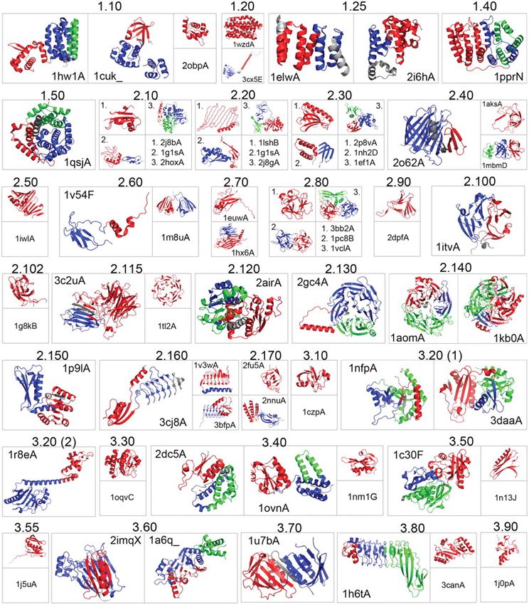
40• Class
• Architecture
CATH
• Topology
• Homology
Class = like SCOPe "Class"
Architecture = conserves arrangement of
SSE (secondary structural elements) but
not sequential order.
Topology = like SCOPe "Fold".
Homology = like SCOPe "Superfamily".
https://www.cathdb.info/protein structure and representation - a
hierarchy or a continuum?
Structure -- representation.
Secondary structure-- 1D, three states
Local structure -- motifs, backbone angles.
Super-secondary structure -- TOPS.
Inter-residue distances -- 2D contact maps
Tertiary structure -- 3D backbone
Side chain conformation -- rotamers
Domain-domain interactions -- interface maps
Quaternary structure -- poses, interaction maps.You can also read