Middle Turbinate mucocele: An unusual location - Journal ...
←
→
Page content transcription
If your browser does not render page correctly, please read the page content below
CAS CLINIQUE

   Middle Turbinate mucocele: An unusual location
             Mucocèle du cornet moyen: une localisation rare
                D. Chiboub, N. Romdhane, H. Belaid, A. Ouerghi, S. Nefzaoui, I. Hariga, Ch. Mbarek
                                  ENT department of Habib Thameur hospital
                        Recieved:22/12/2021, Revised: 28/01/2022, Accepted: 20/02/2022

                                                   ABSTRACT
Objective: Nasal mucoceles are rare. The symptoms depend on the extension of lesion. They are especially related
to nasal obstruction. It causes progressive distension inducing organ compression and bone erosion. Most com-
monly seen in the frontoethmoidal area, it may also develop in abnormally aerated bones, such as middle turbinate,
clinoid process and pterygoid process.
We purpose to report a rare location of a nasal mucocele and describe its different clinical and paraclinical aspects.
Observation: We report a case of a 55-year old female with no medical history, who was complaining about visual
acuity reduced on the left eye, which appeared during the past ten months. Endoscopic nasal examination revealed
a formation of the middle left turbinate.
CT scan (computed tomography scan) and MRI (magnetic resonance imaging) revealed the presence of a cystic
lesion in the middle turbinate. This lesion was well-defined and homogeneous. CT scan revealed an erosion of the
lamina papyracea and a compression of the internal rectus muscle. The MRI showed the orbital extension.
The patient was operated with an endoscopic approach. We evacuated the content of the mucocele. The evolution
was favorable with no recurrence during 2 years.
Conclusion: The mucoceles remain asymptomatic for a long time, which can retard the diagnosis. Its peculiarity
lies more in the diagnosis than in the management as most of these can be completely resected or marsupialized
endoscopically. The imaging is indicated before the surgical treatment.
Keywords: Mucocele - Middle turbinate - Computed tomography scan - Magnetic resonance imaging

                                                     RÉSUMÉ
Objectif: Les mucocèles nasales sont rares. Les symptômes cliniques dépendent essentiellement de l’extension
des lésions. Elles se manifestent le plus souvent par une obstruction nasale. Elles sont le plus souvent localisées
au niveau de la région fronto-ethmoïdale, mais elles peuvent plus rarement se développer au niveau des os
anormalement aérés, tels que le cornet moyen, le processus clinoïde et le processus ptérygoïdien. L’objectif est de
rapporter une localisation rare d’une mucocèle nasosinusienne du cornet moyen et décrire ces différents aspects
cliniques et paracliniques.
Observation: Nous rapportons le cas d’une patiente âgée de 55 ans sans antécédents pathologiques notables
qui se plaignait d’une diminution de l’acuité visuelle à gauche évoluant depuis 10 mois. L’examen endoscopique
endonasale a retrouvé une formation du cornet moyen gauche. Une TDM du massif facial ainsi qu’une IRM ont été
faites montrant une formation liquidienne homogène, bien limitée au dépend du cornet moyen gauche. Le scanner
avait montré une lyse de la lame papyracée avec compression du muscle droit interne. L’IRM avait confirmée la
présence d’une extension intra-orbitaire. La patiente a été opérée par voie endonasale avec marsupialisation de la
mucocèle. L’évolution était favorable avec un recul de 2 ans.
Conclusion: Les mucocèles nasosinusiennes demeurent asymptomatiques pendant longtemps ce qui retarde leur
diagnostic. Leur particularité réside plus dans le diagnostic que dans la prise en charge puisque la plupart d’entre
elles peuvent être complètement réséquées ou marsupialisées par voie endoscopique. L’imagerie est indiquée avant
le traitement chirurgical.
Mots clés: Mucocèle – Cornet moyen – Tomodensitométrie - Imagerie par résonance magnétique

INTRODUCTION                                                  it may also develop in abnormally aerated bones, such
The mucocele is a benign pseudocyst. It causes pro-           as middle turbinate, clinoid process and pterygoid pro-
gressive distension inducing organ compression and            cess. Obstruction of the concha bullosa can rarely lead
bone erosion [1]. Most commonly seen in the frontoeth-        to formation of a mucocele which may be secondarily
moidal area, these mucoceles develop as a result of           infected forming a mucopyocele.
obstruction of the normal sinus drainage tract. Rarely,       We report the case of an adult operated for a middle
                                                              turbinate formation related to mucocele.

Auteur correspondent: Dorra Chiboub
Email: chdorra@hotmail.com

                                                                                 J. TUN ORL - No 47 MARS 2022      75
MIDDLE TURBINATE MUCOCELE: AN UNUSUAL LOCATION D. CHIBOUB, et al
OBSERVATION: or a compression of adjacent anatomical structures [2,3].
We report a case of a 55-year old patient, with no sur- Turbinate location is unusual. They are commonly found
gical or traumatic past history, complaining of visual in the middle turbinate because of its pneumatization [2].
acuity reduced on the left eye, which appeared during Concha bullosa is the most frequently
the past ten months. There is nor headache neither rhi- encountered anatomical variation of the lateral nasal
nologic symptoms. On examination, no face swelling or wall. Mucoceles develop as a result of impaired drainage
lymph nodes were found. of the mucus, which can either be caused by alteration
Nasal endoscopy revealed an enlarged middle turbi- in its viscosity or composition as seen in cystic fibrosis
nate occupying almost the entire left nasal cavity, push- or by mechanical obstruction.
ing the nasal septum to the right side. The right nasal Mucoceles are mostly painless and asymptomatic swellings
cavity and the nasopharynx were normal. that have a relatively rapid onset and fluctuate in size [5,6]. Their
Specialized ophtalmological examination found no pro- enlargement may cause clinical symptoms (nasal obstruction,
ptosis and the visual acuity was 3/10 on the left eye rhinorrhea, facial pain or ophtalmic complications). The lesion
and 7/10 on the right eye. can compress the orbit inducing a raised intraocular pressure,
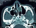
CT scan revealed a well circumscribed expansile mass diplopia, or proptosis [7]. Mucoceles of the middle turbinate
of the middle turbinate with an effect on the lamina pa- have been associated with nasal obstruction, nasal discharge,
pyracea (Fig 1). This mass was isodense with a com- diplopia, exophthalmos and chronic sinusitis symptoms owing
press of the internal rectus muscle. to the blockage of ethmoidal infundibulum [8].
In our case, the patient complained about visual acuity
reduction without any rhinological symptoms.
Physical examination presents with ophtalmic abnormality
like visual disturbance or impaired ocular mobility. Nasal
endoscopy reveal an hypertrophy of the middle turbinate.
CT scan is the most helpfull investigation of mucoceles. They
appear as a homogeneous, well-defined, non-enhancing,
expansile cystic lesion in the middle turbinate [3,5].
In some cases, CT scan inform about causes of mucoceles
Figure 1: Axial computed tomography imaging showing a as adhesions, concha bullosa, septum deviation, paradoxical
mucocele developing from a concha bullosa of the left middle middle turbinate. This exam allows also defining anatomic
turbinate with erosion of the lamina papyracea. variants prior to surgery [3,9]
MRI was indicated because of the orbital extension. MRI allows to differentiate between mucocele or other
The mucocele appeared as homogeneous mass in tumors and to analyze orbital and intracranial extension in
low signal on T1-weighted images, high signal on T2- complement of the CT scan.
weighted images (Fig 2) and with no enhancement In our study, MRI was necessary to evaluate orbital extension.
after gadolinium administration. Orbital content was The compression of the internal rectus muscle and the occular
repulsed and optic nerve was untouched. globe was found in MRI but with no diplopia in ophtalmological
examination [3,10,11].
Mucocele may have different signals on MRI depending on
its composition [12]. A low intense T1 and a high intense T2 is
usually found [3].
The treatment is based on an endoscopic surgery as soon as
possible for symptomatic mucocele [13-15].
This procedure had a low recurrence rate [16]. A middle
turbinoplasty can be also performed.
A special care must be taken when removing mucocele walls
adjacent to the orbit or skull base.
Figure 2: (A): Coronal MRI imaging: Hyperintense nasal
mass on T2- weighted image with orbital extension; (B): Axial
MRI imaging: hypointense nasal mass on T1-weighted image CONCLUSION
with no enhancement after gadolinium administration. The clinical features of middle turbinate mucocele
The patient had an endoscopic treatment. A surgical are diverse. It represents a diagnostic challenge
marsupialization of mucocele and turbinoplasty were to surgeons both in terms of symptoms and risk of
performed. The evolution was favorable with no complications. Its peculiarity lies more in the diagnosis
reccurence during 2 years. Visual acuity in the left than in the management as most of these can be
eye was improved significantly up to 5/10. completely resected or marsupialized endoscopically.
Compliance with ethical standards
DISCUSSION:
Conflict of interest: The authors stated that there is
A mucocele is a cystic mass filled with mucus and lined by
no conflict of interest.
respiratory epithelium. It can cause an erosion of the bone
Funding Statement: The authors received no specific
funding for this work.
76 J. TUN ORL - No 47 MARS 2022D. CHIBOUB, et al MIDDLE TURBINATE MUCOCELE: AN UNUSUAL LOCATION
REFERENCES:
1. E rdogan BA, Unlu N, Aydin S, Avci H. Frontal Mucocele 10. Lee TJ, Li SP, Fu C-H. Extensive paranasal sinus
Extended Orbita and Endoscopic Marsupialization mucoceles: A 15-year review of 82 cases. Am J
Technique. J Craniofac Surg. juin 2018;29(4):e408‑9. Otolaryngol. 2009;30: 234-238.
2. Lee YW, Kim YM. Mucocoele of the inferior turbinate: a 11. Kim YS, Kim K, Lee JG, Yoon JH, Kim CH. Paranasal
case report. Br J Oral Maxillofac Surg. 2016;54(10):1121‑2. sinus mucoceles with ophthalmologic manifestations: A
3. Marrakchi J, Nefzaoui S, Chiboub D. Imaging of paranasal 17-year review of 96 cases. Am J Rhinol Allergy. 2011;
sinus mucoceles. Otolaryngol Open J. 2016;2(3):94–100. 25: 272-275.
4. Pinto JA, Cintra PP, De Marqui ACS, Perfeito DJP, Ferreira 12. Haloi AK, Ditchfield M, Maixner W. Mucocele of the
RDP, Da Silva RH. Middle turbinate mucopyocele: a case sphenoid sinus. Pediatr Radiol. 2006; 36: 987-990.
report. Rev Bras Otorrinolaringol. 2005;71(3):378-81. 13. Ketenci I, İlhan Şahin M, Vural A. Mucopyocele of the
5. Cinar U, Ozgur Y, Uslu B, Alkan S. Pyocele of the middle Concha Bullosa: A Report of Two Cases. Erciyes Med J
turbinate: a case report. Kulak Burun Bogaz Ihtis Derg. 2013; 35(3):157-60.
2004;12:35-8. 14. Lee. Concha bullosa mucocele with orbital invasion and
6. Bahadir O, Imamoglu M, Bektas D. Massive concha secondary frontal sinusitis: a case report. BMC Research
bullosa pyocele with orbital extention. Auris Nasus Larynx. Notes 2013 6:501.
2006;33:195-8. 15. Devars du Mayne M, Moya-Plana A, Malinvaud D,
7. Kim JS, Kim EJ, Kwon SH. An ethmoid mucocele Laccourreye O, Bonfils P. Sinus mucocele: Natural
causing diplopia: A case report. Medicine (Baltimore). history and long-term recurrence rate. European Annals
2017;96(50):e9353. of Otorhinolaryngology, Head and Neck Diseases.
8. Gupta N, Kalia M, Dass A, Singhal SK.Mucocele of the 2012;129:125-30.
middle turbinate: A rare entity. Romanian Journal of 16. Mora-Horna ER, Lopez VG, Anaya-Alaminos R, et al. Optic
Rhinology. 2020;10(38):59-62. neuropathy secondary to a sphenoid-ethmoidal mucocele:
9. Xu Y, Tao Z, Zhan H, Zhou T. Massive concha bullosa case report. Arch Soc Eps Oftalmol 2015;90:582–4.
pyocele with orbital extension--a case report and review
of the literature. J Clin Otorhinolaryngol Head Neck Surg.
2007;21(23):1085‑6.
J. TUN ORL - No 47 MARS 2022 77You can also read


