Multilocular Lipoma in the Middle and Index Fingers: A Case Report - Cureus
←
→
Page content transcription
If your browser does not render page correctly, please read the page content below
Open Access Case
                               Report                                                 DOI: 10.7759/cureus.22172

                                                Multilocular Lipoma in the Middle and Index
                                                Fingers: A Case Report
Review began 01/29/2022
                                                Tuqa A. Alsinan 1 , Tareg M. Alhablany 1 , Hesham R. Alokaili 1 , Abdulla S. Altamimi 1 , Mohammed Ehsan
Review ended 02/05/2022                         Rashidi 1
Published 02/13/2022

© Copyright 2022                                1. Department of Plastic Surgery and Burn Unit, King Saud Medical City, Riyadh, SAU
Alsinan et al. This is an open access article
distributed under the terms of the Creative
                                                Corresponding author: Tuqa A. Alsinan, tuqasinan.15@gmail.com
Commons Attribution License CC-BY 4.0.,
which permits unrestricted use, distribution,
and reproduction in any medium, provided
the original author and source are credited.
                                                Abstract
                                                Lipomas are the most common type of soft tissue tumor, and 95% of them are benign. While lipomas can
                                                present anywhere on the body, 1% of them are found in the fingers. The ultimate goal of management is
                                                surgical excision of the mass with preservation of the neurovascular surroundings. Here, we present the case
                                                of a 24-year-old, morbidly obese Saudi female patient complaining of large non-tender lumps in the index
                                                and middle fingers involving the palmar and dorsal surfaces of the left non-dominant hand. The lumps were
                                                associated with paresthesia and tingling sensations. The article aims to report and highlight the satisfactory
                                                outcomes after total excision of such lipomas and restoring the function as well as the cosmetic results of
                                                the hand.

                                                Categories: Plastic Surgery
                                                Keywords: hand surgery, lipoma of middle digit, lipoma of index digit, hand lipoma, multilocular lipoma

                                                Introduction
                                                Lipomas are benign fat cell tumors that are primarily found in the head and neck, as well as the shoulder and
                                                back [1,2]. Lipomas are considered to be the most common type of soft tissue tumor, and 95% of them are
                                                benign [1,3]. While lipomas can present anywhere on the body, 1% of them are found in the fingers [3].
                                                Lipoma of the hand and fingers is considered to be rare; it usually affects the hypothenar and thenar regions
                                                [2]. The clinical presentation varies depending on the exact location, but it usually presents as a painless and
                                                soft mass felt under the skin [3]. In most cases, lipomas are not treated; they are only observed clinically
                                                unless the patient is symptomatic or aiming for cosmetic results [3]. The ultimate goal of management is
                                                surgical excision of the mass with preservation of the neurovascular surroundings [1-3].

                                                Case Presentation
                                                A 24-year-old, morbidly obese Saudi female patient presented with large non-tender lumps in the index and
                                                middle fingers involving the palmar and dorsal surfaces of the left non-dominant hand. The patient reported
                                                a history of the initial appearance of the lumps at the age of eight and surgical excision four years before
                                                presentation, as well as the history of debulking procedure three years before the surgical excision. The
                                                lumps were asymptomatic until they started to be associated with paresthesia and tingling sensations of the
                                                involved digits, which affected her daily activities. She does not smoke and is not exposed to second-hand
                                                smoke. The rest of her history was unremarkable. The physical examination results were all within the
                                                normal limits, except she had a limited range of motion due to the mass in the affected digits. Soft-tissue
                                                hemangiomas, vascular malformation lipoma, and lipomas were all considered as differential diagnoses
                                                based on the history given.

                                                For further investigations, she underwent Magnetic Resonance Imaging (MRI), which showed an
                                                encapsulated multilocular mass in the proximal and distal sections of the index and the middle digits,
                                                extending to the palmar and dorsal aspects of the left hand until the carpal bone dorsally and from the first
                                                web space to the fourth web space with a volar extension of approximately 3 cm to proximal palmar crease
                                                [Figures 1A, 1B].

                               How to cite this article
                               Alsinan T A, Alhablany T M, Alokaili H R, et al. (February 13, 2022) Multilocular Lipoma in the Middle and Index Fingers: A Case Report. Cureus
                               14(2): e22172. DOI 10.7759/cureus.22172
FIGURE 1: A and B are MRI-T1 images of the left hand showing the
                                                    lipoma of the index and middle digits.

                                                  The patient was booked and taken to the operating theatre under general anesthesia and full aseptic
                                                  technique. Soft, mobile, raised, and fluctuant masses were felt over the second and third digits of the left
                                                  hand with no overlying skin changes [Figures 2A, 2B].

                                                    FIGURE 2: Preoperative photograph of the lipoma over the volar aspect
                                                    of the left hand in image A. Dorsal view of the hand in image B.

2022 Alsinan et al. Cureus 14(2): e22172. DOI 10.7759/cureus.22172                                                                                               2 of 8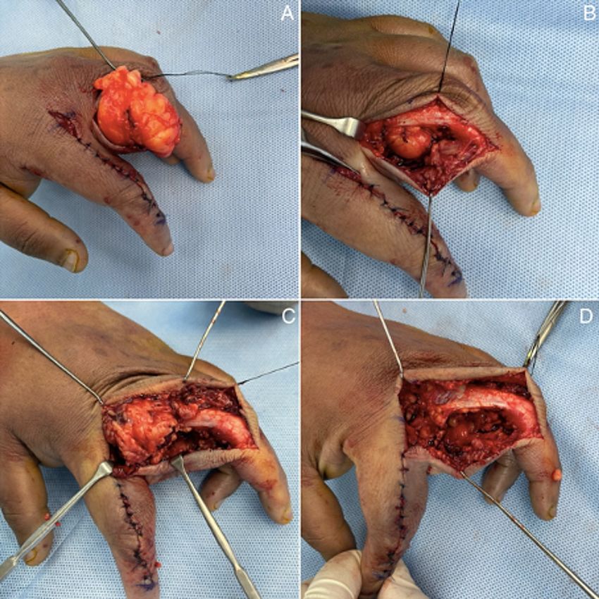
Two lipomatous tumors were felt within the ulnar border of the index finger and the radial border of the
                                                  middle finger. The incision spanned from the superior border of the dorsal proximal interphalangeal (PIP)
                                                  creases distally to the second web space’s dorsal center proximal to the index digit. The incision was kept
                                                  dorsal to the glabrous line [Figures 3A-3D].

                                                    FIGURE 3: (3A, 3B, 3C, 3D). Segmental resection of the lipoma in the
                                                    index digit. Incision of the dorsal PIP creases distally to the second web
                                                    space’s dorsal center proximally.

                                                  The lipoma was found to encompass the zone of incision and was adherent to the extensor mechanism; it
                                                  was also engulfing the neurovascular bundle. Thus, segmental excision rather than en-bloc resection was
                                                  preferred due to the engulfment of the structures. Similarly, another incision was made over the middle digit
                                                  in the radial border, and extensor tendons were repaired, respectively [Figures 4A-4D].

2022 Alsinan et al. Cureus 14(2): e22172. DOI 10.7759/cureus.22172                                                                                                3 of 8
FIGURE 4: (4A, 4B, 4C, 4D). Segmental resection of the lipoma in the
                                                    middle digit.

                                                  The tumor on the middle digit had similar proportions, distribution, and attachments as the tumor on the
                                                  index digit [Figures 5A, 5B].

2022 Alsinan et al. Cureus 14(2): e22172. DOI 10.7759/cureus.22172                                                                                           4 of 8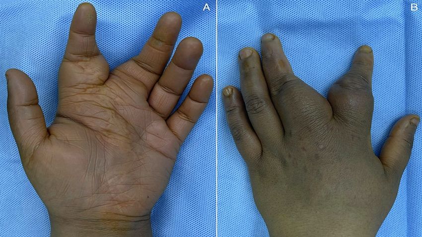
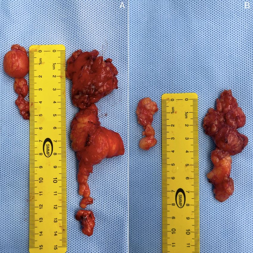
FIGURE 5: Lipoma excised from the index digit in image A. Lipoma
                                                    excised from the middle digit in image B.

                                                  Intra-operative findings were summarized as two lipomatous tumors excised from the index and middle
                                                  digits over the ulnar and radial borders, respectively. Histopathology revealed that the specimens consisted
                                                  of several fragments of fibro-fatty tissue measuring 7.5 x 5.5 x 1 cm, and three ovoid pieces of yellow-tan
                                                  tissue measuring 5.5 x 4 x 3 cm, and 2.5 x 2 x 1 cm, respectively, for both the left index and left middle digits.
                                                  After the procedure, the patient was placed in a compressive dressing and volar splint. The patient tolerated
                                                  the procedure very well and reported satisfactory outcomes with no complications at the one-week follow-
                                                  up appointment at an outpatient clinic [Figures 6A, 6B].

2022 Alsinan et al. Cureus 14(2): e22172. DOI 10.7759/cureus.22172                                                                                                     5 of 8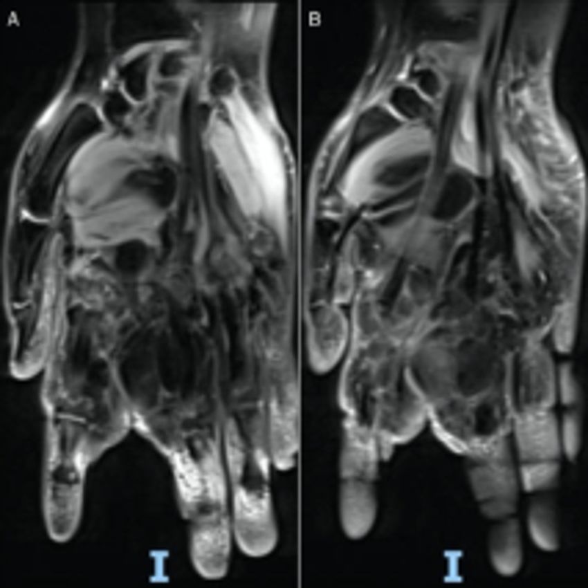
FIGURE 6: One week follow-up post operative. Volar aspect in image A.
                                                    Dorsal aspect in image B.

                                                  Discussion
                                                  Lipomas are the most common form of benign soft tissue tumors with a rare incidence rate in the fingers [1-
                                                  3]. They can be observed or treated with surgical excision, depending on the patient’s symptoms and the
                                                  cosmetic goal [3]. Several articles in the literature have reported on the rarity of lipomas in the hand and
                                                  digits. Recently, two cases were published reporting on a multilocular lipoma of the left thumb and a giant
                                                  lipoma of the index extending to the dorsum of the right hand that was excised by performing a modified
                                                  Bruner incision technique [1-3]. The two cases resulted in a successful surgical outcome with good cosmetic
                                                  results [1-3]. Lipomas in the middle finger are rare, and excision is the best way to restore the normal
                                                  function of the digit. Considering lipoma of the finger as a differential diagnosis and the preoperative use of
                                                  ultrasound and MRI aid in good surgical outcomes, which is highlighted in the literature [2-5]. Furthermore,
                                                  a rare case of a giant lipoma of the hand extending from the thenar to the deep palmar space was reported; it
                                                  was completely excised, resulting in a good surgical outcome [6]. In terms of rarity, a parosteal lipoma of the
                                                  proximal phalanx of the hand accounts for less than 0.1% of all primary bone tumors. In one study, a patient
                                                  with this type of lipoma was treated and managed surgically with no recurrence at the two-year follow-up
                                                  [7]. The review of different articles shows that surgical excision of such lipomas is considered the preferred
                                                  method that results in satisfactory outcomes; in most cases, it also restores the function of the digit. Table 1
                                                  summarizes previous studies.

2022 Alsinan et al. Cureus 14(2): e22172. DOI 10.7759/cureus.22172                                                                                                   6 of 8
Year Authors Title Aim
This case reports a rare location of lipoma of the third finger and
Ramirez-Montaño L, Giant lipoma of the third finger of repercussion in the decision-making process when other more
2013
Lopez RP, Ortiz NS. the hand common similar pathologies with varying prognoses are conceived
[2].
Lipoma of the middle finger: A The case reports a lipoma of the middle finger, and an ultrasound
Hu Z, Yue Z, Tang Y,
2017 case report and review of and MRI should be used to diagnose the finger, lipoma and
Zhu Y.
literature excision was considered the main treatment option [4].
Giant Lipoma Of The Hand To
This case reports a rare giant lipoma of the hand extending from
Cemboluk Ö, Daldal İ, Extending From Thenar Region
2017 thenar to deep palmar space. Should be surgically removed due to
Topçu HN. To Deep Palmar Space: A Case
the potential increased risk of malignancy [6].
Report
Santacoloma K, de Sá
Barreto GM, Loda G, Giant hand lipoma: a surgical This case reports a giant hand lipoma successfully treated with a
2021
Miller MD, de Janeiro R, challenge modified Bruner incision approach [1].
David R.
Wafiq Wafa A, Wani S, This case reports a progressive left thumb lipoma that was treated
Multilocular lipoma of the left
2021 A. Alsinan T, Alkhonizy successfully with total excision and documents satisfactory
thumb of the hand: a case report
S. outcomes [3].
Giant Spindle Cell Lipoma of
Sheeja RT, Bestin T, This case reports an unusual location of giant spindle cell lipoma of
2021 Middle Finger: Case Report and
Aabha DS. the middle finger [5].
Review of Literature
This case reports a parosteal lipoma in a 45-year-old man
Yadav AK, Pawar ED,
Parosteal lipoma of the proximal involving the proximal phalanx of the right middle finger. The tumor
2021 Wadia F, Gs PK, Mane
phalanx of hand. was successfully excised at margins with the osseous attachment
A, Harsoor A.
[7].
TABLE 1: Attached is a table summarizing similar previous studies.
Conclusions
While lipomas are common benign tumors, ones affecting the hand and digits are rare. This case
presentation aims to contribute to the literature on lipomas, specifically articles addressing lipomas of the
index and middle fingers, even if those types of lipomas are rare, to emphasize the importance of
considering it as a differential diagnosis. Total excision of such lipomas is considered to be the surgical
method that is preferred by most plastic and hand surgeons. This article aims to report and highlight the
satisfactory outcomes after total excision of such lipomas and restoring the function as well as the cosmetic
results of the hand.
Additional Information
Disclosures
Human subjects: Consent was obtained or waived by all participants in this study. Conflicts of interest: In
compliance with the ICMJE uniform disclosure form, all authors declare the following: Payment/services
info: All authors have declared that no financial support was received from any organization for the
submitted work. Financial relationships: All authors have declared that they have no financial
relationships at present or within the previous three years with any organizations that might have an
interest in the submitted work. Other relationships: All authors have declared that there are no other
relationships or activities that could appear to have influenced the submitted work.
References
1. Santacoloma K, de Sá Barreto GM, Loda G, Miller MD, de Janeiro R, David R: Giant hand lipoma: a surgical
challenge. Brazil. 2021, 13:
2. Ramirez-Montaño L, Lopez RP, Ortiz NS: Giant lipoma of the third finger of the hand . SpringerPlus. 2013,
2:164. 10.1186/2193-1801-2-164
3. Wafiq Wafa A, Wani S, A Alsinan T, Alkhonizy S: Multilocular lipoma of the left thumb of the hand: a case
report. Case Reports Plast Surg Hand Surg. 2021, 8:130-3. 10.1080/23320885.2021.1968302
4. Hu Z, Yue Z, Tang Y, Zhu Y: Lipoma of the middle finger: a case report and review of literature . Medicine
2022 Alsinan et al. Cureus 14(2): e22172. DOI 10.7759/cureus.22172 7 of 8(Baltimore). 2017, 96:e8309. 10.1097/MD.0000000000008309
5. Sheeja RT, Bestin T, Aabha DS: Giant spindle cell lipoma of middle finger: case report and review of
literature. J Hand Microsurg. 2021, 10.1055/s-0040-1721879
6. Shahbaaz KM, Surendra M, Sanjay K: A rare clinical presentation of lipoma-palmar lipomatosis . J Diagn.
2017, 13:6-12. 10.18488/journal.98.2017.41.6.12
7. Yadav AK, Pawar ED, Wadia F, Kumar Gs P, Mane A, Harsoor A: Parosteal lipoma of the proximal phalanx of
hand. J Hand Surg Am. 2021, 46:933.e1-5. 10.1016/j.jhsa.2020.10.029
2022 Alsinan et al. Cureus 14(2): e22172. DOI 10.7759/cureus.22172 8 of 8You can also read