MCPHERSON EYE RESEARCH INSTITUTE
←
→
Page content transcription
If your browser does not render page correctly, please read the page content below
McPHERSON
                                  2020 2021
                                      CALENDAR
                         ANNUAL
                         REPORT

EYE RESEARCH INSTITUTE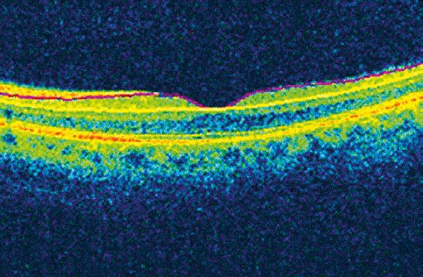
Special
Microscopic image of the mouse UW-Madison’s glaucoma group, a constellation of
retinal ganglion cell layer after AAV2
researchers from the Department of Ophthalmology and
viral vector delivery of the BCL-X
Visual Sciences and collaborating departments—including
Delivery
gene, which produces a protein
that combats ganglion cell death. Drs. Paul Kaufman, Donna Peters, Rob Nickells, Curtis
Cleverly, BCL-X was engineered to Brandt, Colleen McDowell, and Gillian McLellan—is
glow red for ease of visualization.
The nuclei of all retinal cells are
testing various gene delivery strategies to treat glaucoma.
shown in blue. Image courtesy of Viral vectors play a key role in several of these approaches.
the Nickells Lab.
Dr. Rob Nickells’ lab employs viral vectors to short-circuit
FROM THE DIRECTOR the process of ganglion cell death, which is controlled by a
molecular switch involving a pro-death protein called BAX
We have all purchased something online or over the
Viral Transport
and an anti-death protein called BCL-X, which serves to keep
phone this year and then looked forward to a box arriving BAX in check. Using a viral vector called adeno-associated
on our doorstep with the item we needed. Without correct virus type 2 (AAV2), they introduce a lab-created version of
delivery, however, there would be nothing to show for BCL-X into mouse ganglion cells at the onset of glaucomatous
our efforts. Therapeutic development has a similar need Blindness is often caused by mutations in genes, which alter damage. Promising early experiments have shown that
for reliable delivery. Groundbreaking new treatments, proteins and disrupt healthy function. Gene augmentation ganglion cell death is indeed greatly reduced, and the
including gene therapy and stem cell therapy, are in therapy aims to counter these effects by introducing a healthy Nickells Lab is now working to confirm these results and
development for a wide variety of devastating diseases. copy of a gene into cells before they die, which hopefully to establish the safety of their viral vector strategy in further
But in addition to the research being done to create and reverses the tide of the disease. But how does one go about preclinical studies.
refine these novel therapeutic products, McPherson ERI “injecting” a healthy gene into countless microscopic retinal
scientists are busy improving methods to package and cells? By hijacking the most advanced gene microinjector on Dr. Paul Kaufman’s use of viral vector delivery is aimed
place these products safely and reliably in the exact spots earth — the virus. While infamous for their disease-causing at reducing pressures within the eye for those people with
where they are needed in the eye. ability, viruses can be modified so they are unable to cause glaucoma who don’t respond well to current treatments,
disease, yet retain their ability to deliver genetic cargo into or have difficulty with the high frequency and expense of
As you might imagine with an organ so small, delicate, their treatment regimens. To understand Dr. Kaufman’s
and complex, this is no easy task. Many areas within the host cells. These fit-for-purpose biological tools, known
as viral vectors, allow scientists to insert nearly any gene, cutting-edge approach, you first need to understand how
eye that need treatment are in locations that are the postal an eye is “pressurized”. Eye pressure is generated via a
equivalent of a house at the bottom of a lake or on the provided that it fits inside the virus (and not all do). As noted
by Dr. Curtis Brandt, whose lab specializes in the study of balance between fluid production and outflow. One of
side of a steep mountain. In these extreme situations, the the main pathways for fluid to leave the eye is through a
method of treatment delivery often requires as much (if not viruses that affect the eye, “Nature invented gene delivery
way before we took advantage of it.” structure near the front of the eye called the trabecular
more) innovation than the treatment itself. On the next few meshwork. Dr. Kaufman’s lab is pioneering ways to
pages, you’ll learn how McPherson ERI researchers are In McPherson ERI member labs, viral vectors are modulate fluid outflow using a viral vector-based gene
pioneering development of the ocular packages, delivery engineered to deliver genetic material precisely to the retinal therapy strategy that targets the trabecular meshwork and
vans, and distribution routes needed to further our work cells that need them. As a critical first step in this process, lowers outflow resistance. These viral vectors, which are
to end blindness. Some of our researchers are surgeons the genes that the virus normally uses to multiply itself and injected into the front part of the eye, have shown clear
who are involved in advancing the tools and techniques to cause disease are removed. Extensive testing is then potential to improve fluid outflow in preclinical tests (see
of therapeutic delivery, while others are engineers and required to prove that the viral vectors have indeed turned Figures A, B).
biologists who build viral vectors, nanocarriers, and over a new leaf and are safe for use in humans.
microscaffolds that carry cellular and genetic cargo. That’s
what we do best—build and support the most diverse, HOMING
IN ON GLAUCOMA
dedicated, and talented eye research teams in the world,
Glaucoma, the world’s most common cause of irreversible Gene delivery to the trabecular mesh
right here in Wisconsin. work after injection of a viral vector into
vision loss and blindness, is characterized by degeneration
the front of the eye. In panel A, the arrow
of the optic nerve and the death of ganglion cells in the retina. points to the area of injection. Two years
While doctors can treat the high eye pressures associated with after administration of the viral vector,
most forms of glaucoma, scientists are still searching for ways the trabecular meshwork continues
to express the delivered gene, which
RRF Emmett A. Humble Distinguished Director, McPherson ERI to prevent ganglion cell death and optic nerve damage that shows as a thin green line in panel B.
∞ Sandra Lemke Trout Chair in Eye Research often occurs despite treatment. Image courtesy of Dr. Paul Kaufman.
A B
Nanocarrier delivery of CRISPR/Cas

        E
         ngineering
                                                                      machinery results in efficient genome
                                                                      editing in the retinal pigment epithelium
                                                                      (RPE). The area in red shows RPE cells that                                           TARGETING
                                                                                                                                                                     RETINITIS PIGMENTOSA

        Solutions
                                                                      were successfully treated in the mouse
                                                                      retina. Image courtesy of Dr. Pawan Shahi,                                       Viral vectors also can be used to deliver therapeutic
                                                                      Pattnaik Lab.                                                                    genes to the back of the eye to treat inherited retinal
                                                                                                                                                       diseases such as retinitis pigmentosa, or RP. Dr. Bikash
                                                                                                                                                       Pattnaik is using viral vectors in efforts to prevent
    NANOPARTICLE PACKAGING                                                                                                                             blindness caused by an early onset form of RP known as
                                                                        MICROSCAFFOLD INSERTION                                                        Leber congenital amaurosis, or LCA. Dr. Pattnaik has a
Viral vectors might be nature’s gene delivery system, but                                                                                             particular interest in genes that make ion channels, which
they can be costly to make and are limited in what they can         Professor Zhenqiang (Jack) Ma is an exemplar of how sheer                          are molecular gateways that control many cell functions.
carry and what cells they can reach. Therefore, McPherson           engineering know–how can advance biomedical delivery                               Mutations in some ion channel genes lead to LCA and
ERI scientists are also on the forefront of developing non-         systems. Dr. Ma, the Lynn H. Matthias Professor and Vilas                          rapid blindness in infants and young children. Dr. Pattnaik’s
viral delivery systems that may offer superior cargo-carrying       Distinguished Achievement Professor in the Department of                           viral vectors successfully delivered healthy copies of one
capacity, greater precision in cell targeting, broader safety       Electrical and Computer Engineering, has research interests                        type of ion channel gene to the retina of a mouse with LCA,
profiles, and more streamlined manufacturing.                       that span a wide range of engineering disciplines, including                       which resulted in recovery of ion channel function and
Professor Shaoqin “Sarah” Gong and her research group               bioelectronics and biomimetics. His lab recently applied its                       improvement in vision. Efforts are now underway to further
focus on engineering smart, non-viral nanoparticles to deliver      expertise to develop biomedical devices to reconstruct the                         develop this gene therapy for future clinical trials.
drugs, genes, or CRISPR/Cas genome editing machinery to             outer retina in late-stage retinitis pigmentosa (RP) and age-
                                                                    related macular degeneration (AMD).                                                A large team of McPherson ERI researchers, including
cells within the eye and elsewhere in the body. The uses of
                                                                                                                                                       Drs. David Gamm, Krishanu Saha, Bikash Pattnaik, Sarah
these “nanocarriers” are manifold—chemotherapy, gene                Dr. Ma’s lab engineered a solution to a hurdle facing Dr.                          Gong, and Sushmita Roy, is employing a similar viral vector
therapy, immunotherapy, and antimicrobial therapy, among            David Gamm’s efforts to replace photoreceptors and/or                              strategy to fight Best disease, one of the most common
many others.                                                        RPE cells damaged in RP and AMD, as well as many other                             forms of inherited macular degeneration. This same
Collaborating with a group of McPherson ERI investigators           retinal diseases and injuries. The Gamm Lab developed                              group of investigators, as well as Dr. Melissa Skala at UW-
that includes Drs. Pattnaik and Saha, Dr. Gong’s team recently      and patented ways to create photoreceptors and RPE from                            Madison and Dr. Daniel Lipinski at the Medical College
developed several patent-pending nanocarriers that can              human pluripotent stem cells, but they needed a means to                           of Wisconsin, are using viral vectors to deliver genetic
safely and efficiently deliver complex CRISPR/Cas genome            deliver these replacement cells deep within the retina. In                         machinery capable of silencing or fixing mutant genes,
editing machinery to diseased retinal cells without any             collaboration with Dr. Gong, Dr. Gamm, and Dr. Joe Phillips, Dr.                   not just replacing them. This latter technology, known as
apparent toxicity. Importantly, the surface of the nanocarriers     Ma’s lab generated biodegradable microscaffolds suitable for                       CRISPR/Cas genome editing, received the 2020 Nobel
can be decorated with various types of targeting molecules,         safely transporting donor photoreceptors and RPE cells to the                      Prize in Chemistry and has opened the door to treating
thereby allowing them to “home in” on specific retinal cells in a   retina in an organized and highly controlled fashion. Various                      genetic disorders that gene augmentation cannot help.
way similar to (and perhaps better than) viral vectors.             generations of these revolutionary scaffolds have been
                                                                    designed, built, and inserted under the retina in preclinical
With further development and testing, these versatile               models that mimic damage caused by RP or AMD. Results
                                                                                                                                                       A therapeutic gene that produces a protein (tagged with a green fluorescent
                                                                                                                                                       marker) was delivered via a viral vector to stem cell-derived human retinal
nanocarriers may enable therapies for a wide range of               thus far show great promise for these scaffolds to help repair                     pigmented epithelium (RPE) cells created from a patient with LCA (left). The
genetic eye diseases that currently cannot be treated with          the outer retina and restore vision in individuals with RP, AMD,                   number of green cells in the microscopic image on the right reveals that
viral vectors. To achieve this goal, Professor Gong’s team is       and other degenerative disorders.                                                  nearly all of the patient’s RPE cells express the new gene. Images courtesy
broadening their collaborations with scientists and clinicians                                                                                         of the Pattnaik Lab.
within the McPherson ERI and around the world.
                                                                                                              Microscopic image of a “wineglass”
                                                                                                              design photoreceptor scaffold at
                                                                                                              lower (left panel) and higher (middle
                                                                                                              panel) magnifications. The right panel
                                                                                                              shows an image of human pluripotent
                                                                                                              stem cell-derived photoreceptors
                                                                                                              (engineered to glow red) lined up
                                                                                                              neatly within the scaffold. Images
                                                                                                              courtesy of the Ma Lab (left, center)
                                                                                                              and the Gamm Lab (right).
Special Delivery
Optimizing Surgical Techniques
IMPLANTING
PHOTORECEPTORS DISCOVERING
BETTER ROUTES
AND RPE CELLS FOR DRUG DELIVERY
All of the wonderful new therapeutic viral vectors, nano Injections remain the most effective way to deliver
carriers, and microscaffolds must be safely and accurately many types of drugs to the eye, especially those that can’t
placed where they are needed. In many cases, that requires penetrate the surface of the eye as drops. In collaboration
not only skilled surgeons, but innovative ones as well. with pharmaceutical companies, McPherson ERI researchers
at UW-Madison are working hard to develop better means
Vitreoretinal surgeon and newly named Monroe E. Trout
and new routes for drug delivery. Vitreoretinal surgeon and
Chair in Eye Research, Dr. Michael Altaweel has been
researcher Dr. Michael Nork and his colleagues have
working with Drs. Gamm and Phillips to optimize methods
developed and tested many approaches to get a wide range of
to deliver replacement photoreceptors and RPE cells (and
therapeutics to the retina, the tissue primarily affected by most
scaffolds) beneath the retina in a safe and reliable manner that
untreatable blinding disorders. Recently, Dr. Nork’s lab has
can be adopted by other surgeons. Dr. Altaweel has performed
begun testing a method to deliver drugs to the retina through TOP: Image of a rabbit eye that has had two suprachoroidal injections.
retinal surgery on children and adults with complex diseases The areas at the lower left and top show the distribution of a green or
the suprachoroidal space—a potential space between the
and injuries for 20 years, and he has been an investigator for red test dye after injection, respectively, with the area of overlap shown
sclera and the large choroidal blood vessels that supply
many clinical trials that have changed the standard of care for in yellow toward the right side of the image. The image demonstrates
oxygen and nutrients to the outer retina. The procedure, which that two injections are needed to achieve good retinal coverage via the
retinal disease treatment.
involves the use of newly developed microneedles, could suprachoroidal route. Image courtesy of Dr. T. Michael Nork.
To proceed with clinical trials to test stem cell-based revolutionize retinal drug delivery. BOTTOM: A blue dye (arrow in panel A) confirms the accurate injection
therapies for RP and AMD, it is necessary to carefully adapt of material into Schlemm’s canal, which is located next to the trabecular
Dr. Paul Kaufman’s lab is also testing new devices
surgical procedures to support the specialized handling and meshwork and is part of the fluid drainage system of the eye. The arrow
and procedures to deliver novel therapeutics to treat in panel B shows the tip of the microcatheter used to inject material into
delivery requirements of each new therapeutic product. Dr.
glaucoma. Dr. Kaufman’s group has had success using Schlemm’s canal. Images courtesy of Dr. Paul Kaufman.
Altaweel is working with the McPherson ERI team to refine
microcatheters threaded directly through Schlemm’s
and test different surgical strategies, with promising results
canal, a drainage structure that abuts the trabecular
that show proper localization and widespread survival and
meshwork, to inject his viral vectors exactly where they are
integration of donor photoreceptors and RPE cells.
needed to lower eye pressure.
A B
LEFT: Retinal surgeon’s view during a
procedure to implant a photoreceptor
scaffold under the retina. Panel A shows
an intraocular scissors being used
to create an opening in the retina. In
panel B, a custom injector tip is placed
into the opening and the scaffold is
gently inserted under the retina with
high precision. Images courtesy of Dr.
Michael Altaweel and Dr. Kapil Bharti of
the National Eye Institute.
A B
FEATURED RESEARCHERS 2020 Image courtesy
of Andrea Mason.
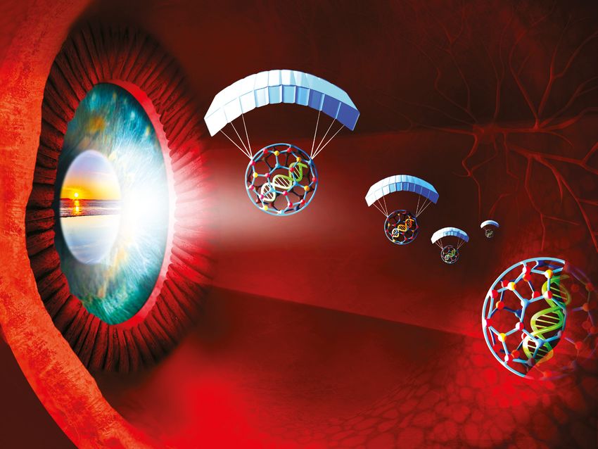
Mid-infrared image of some of the members of the Kats Optical coherence tomography is used to measure the macular ganglion cell-
group, standing behind a Fourier transform infrared (FTIR) inner plexiform layer, outlined by the purple and yellow lines in this image, to
spectrometer. Image courtesy of Mikhail Kats. determine if it is an early marker of neurodegeneration in the brain.
Image courtesy of Adam Paulsen.
ANDREA MASON PhD
Kinesiology, Motor Behavior & Control
∞ School Of Education
Dr. Andrea Mason’s main research interest involves
understanding how people use visual and haptic (touch)
feedback to plan and perform simple and complex
KAREN movements with their hands. She has also studied how
people combine the movements of the upper and lower body
CRUICKSHANKS PhD when grasping objects while walking. Her aim is to determine
MIKHAIL KATS PhD Ophthalmology & Visual Sciences, Population what people need to see and feel, and when that information
Electrical & Computer Engineering Health Sciences ∞ School of Medicine and is needed, to efficiently move and interact with objects in
∞ College of Engineering Public Health their environment. In Mason’s Motor Behavior Laboratory,
research projects have involved children and adults in order
Dr. Mikhail Kats’ research group engages in a broad- As people age, changes in vision and other senses can to determine how the planning and performance of these
ranging experimental and theoretical exploration of make everyday activities more challenging. Recently, coordinated movements change across the lifespan. Mason
topics across optics and photonics, from the visible to there has been growing concern that vision changes also compares performance between natural environments,
the far infrared. Kats—the Jack St. Clair Kilby Associate and age-related eye diseases like cataract and macular where there is an abundance of accurate sensory feedback,
Professor of Electrical and Computer Engineering and degeneration may increase the risk for developing and virtual environments, where synthetic feedback can be
an affiliate faculty member in the departments of Physics cognitive problems and Alzheimer’s disease. crude and suffer from lag.
and Materials Science and Engineering—works on Dr. Karen Cruickshanks’ research group is studying Mason’s recent work, in collaboration with Drs. Kevin
engineering advances that include the development of whether cognitive decline and dementia can be predicted Ponto and Kristen Pickett, explores how people use visual
ultrathin optical components such as flat lenses, tunable by a person’s visual health and function and by the information about objects and their surroundings to
optical components that protect cameras and other thickness of particular layers in the retina (the macular navigate their environment. For example, do features of the
sensors against laser damage, and privacy coatings ganglion cell layer and inner plexiform layer) as measured environment that we are walking in, such as the width of
that can conceal information from infrared cameras. using optical coherence tomography (OCT). This is a hallway, affect the length of the steps we take, the speed
His work often involves new understanding of optical monitored in the Beaver Dam Offspring Study, which at which we walk, or how long we keep both feet on the
properties of emerging materials, including those with began in 2005 and includes thousands of adult children ground? These spatiotemporal measures are known to
complex behaviors such as phase transitions, where of participants in the long-running Beaver Dam Eye Study. be related to fall risk in older adults. Using virtual reality
a material changes to a different physical state. Kats’ Participants are examined every five years. Results have technology, her group can easily manipulate these types
current projects also include hyperspectral light sensors, shown that even in these middle-aged people, small of environmental features and test their effects on walking
light sails, new measurement techniques for optical and changes in visual contrast sensitivity and the thickness performance. Their results have indicated that visual
thermal properties of materials, and, importantly, vision- of the macular ganglion cell–inner plexiform layer were features such as the width of the hallway do, in fact, lead
enhancement technologies. associated with cognitive decline. Currently, the study to subtle changes in walking performance in older adults
For example, Kats and colleagues recently demonstrated aims to determine whether these changes can be used to but not in young adults. In follow-up work, Mason and her
an optical material with a world-record degree of identify people at high risk for dementia decades before collaborators are looking at the effects of performing a
birefringence (a.k.a. double refraction) in the mid-infrared clinical symptoms such as memory loss appear. Early visual dual-task, such as reading information off a computer
spectral range. His team expects that this demonstration identification may help high-risk people get the medical screen mounted to a wall, while walking in a hallway. She
will enable new types of polarization optics for infrared care they need to improve control of blood pressure and hopes to use findings from this work to create an objective
cameras, sensors, and free-space communications. diabetes which are critical for maintaining a healthy brain. measure of fall risk.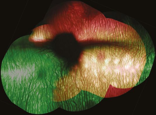
Colormap data visualizations used by the Schloss Lab to study the dark-is-
FEATURED                                                       more bias, from a recent paper in-press at Journal of Vision. Images courtesy
RESEARCHERS                                                    of the Visual Reasoning Lab.

2020
                                                                                     KAREN SCHLOSS PhD
                                                                                     Psychology ∞ College of Letters and Science,
 Blocked iridocorneal angle
                                                                                     Wisconsin Institute for Discovery
 in the eye of an 8-day-old
 PPCD1 mouse. Image
 courtesy of Anna Shen.
                                                               Dr. Karen Schloss is the Principal Investigator of the Schloss
                                                               Visual Reasoning Lab in the Department of Psychology and
                                                               Wisconsin Institute for Discovery at UW-Madison. Her lab
                                                               studies how people interpret meanings from visual features                      Cone photoreceptor distribution in the dog
                                                                                                                                               retina, showing the presence of a macula-like
                                                               like color, shape, and size during visual communication.                        area (red) adjacent to the optic nerve (center
                                                               Visual communication is fundamental to how humans                               blue spot). Image courtesy of Freya Mowat.
                     ANNA SHEN PhD                             share information; they communicate information about
                                                               weather patterns and neural activity through colormap
                     Oncology ∞ School of Medicine and         data visualizations, they communicate information about
                     Public Health
                                                               biological processes and mathematical properties through
                                                               diagrams, and they communicate information about where
                                                                                                                                                                     FREYA MOWAT BVSc, PhD
Dr. Anna Shen’s research, carried out in the laboratory of                                                                                                           Surgical Sciences ∞ School of Veterinary
                                                               to find destinations or discard different types of recyclables
Dr. Chris Bradfield, centers on a mouse model of a human                                                                                                             Medicine · Ophthalmology & Visual Sciences
                                                               through signs. Observers have expectations about how
corneal dystrophy, posterior polymorphous dystrophy                                                                                                                  ∞ School of Medicine and Public Health
                                                               visual features will map onto concepts, such as the “dark-is-
(PPCD). PPCD is characterized by abnormal differentiation
                                                               more” bias to infer that darker colors map to larger quantities
and growth of the corneal endothelium, a cell layer on                                                                                         Dr. Freya Mowat’s research seeks to define the metabolic
                                                               in colormap data visualizations. It is easier to interpret
the inner surface of the cornea. Although many cases                                                                                           pathways important in retinal aging, and to pinpoint ways
                                                               visualizations that match those expectations. Thus, the
are asymptomatic, some affected individuals develop                                                                                            to ameliorate metabolic decline in the outer retina that
                                                               primary goal of the Visual Reasoning Lab is to understand the
corneal clouding, decreased visual acuity, and increased                                                                                       occurs with aging. These pathways determine how well the
                                                               nature of these expectations, including how they are formed,
intraocular pressure and glaucoma, requiring treatment                                                                                         neurons of our retina endure the changes that come with
                                                               and how they can be leveraged in visualization design to
with medication and/or surgery. Overgrowth of abnormal                                                                                         age. They can also predispose us to loss of visual sensitivity
                                                               make visual communication more effective and efficient.
corneal endothelial cells can, in some cases, lead to                                                                                          and blinding diseases such as age-related macular
severe vision loss. Clinical management of this condition      In a secondary line of work, Dr. Schloss’s lab also aims to use                 degeneration (AMD), the most common cause of vision
would be improved with better identification of the factors    information visualizations to facilitate science education                      loss in persons over the age of 65 years.
influencing disease progression and severity.                  in formal and informal learning environments. In the UW
                                                               Virtual Brain Project, they have developed immersive virtual                    A substantial part of Dr. Mowat’s work involves the study
The mouse model of PPCD that Dr. Shen works with                                                                                               of vision in companion (pet) dogs. In addition to cohabiting
                                                               reality (VR) lessons that enable learners to travel through
carries the genetic defect seen in one form of human                                                                                           in their environment and sharing their lifestyles, dogs
                                                               3D diagrams of the human visual system and auditory
PPCD, and it also exhibits the pathological features of                                                                                        share many of the features of their human owners’ retinal
                                                               system. Evidence suggests these VR lessons provide
human PPCD. Shen uses this mouse model to identify the                                                                                         anatomy. Mowat focuses on the unique features of the
                                                               more enjoyable learning experiences than two-dimensional
signaling pathways that lead to aberrant growth of corneal                                                                                     macula of dogs—the central area of the retina that closely
                                                               alternatives, indicating they have exciting potential to
endothelial cells. In addition, the severity of corneal and                                                                                    mirrors the macula of people and is the region most
                                                               motivate interest in science in the classroom and beyond.
retinal disease in this mouse model is dependent on the                                                                                        susceptible to AMD. She is particularly interested in the
genetic background. Using genetic mapping techniques,          Dr. Karen Schloss’s research is funded by a National Science                    function of a protein called PGC1-a, which regulates the
Shen and her colleagues are engaged in an effort to identify   Foundation CAREER award and has been supported by the                           growth of mitochondria, the “energy factories” in cells.
the specific genes that influence the severity of corneal      McPherson ERI Grant Summit program and McPherson                                The study of the age-related vision decline of companion
endothelial cell overgrowth and resulting glaucoma.            ERI Expanding Our Vision program. She was also recently                         dogs will help us to better and more quickly understand
This mouse model will also be used to test therapeutic         awarded the Steven Yantis Early Career Award from the                           the effect of environment and lifestyle on retinal aging and
interventions for PPCD.                                        Psychonomic Society.                                                            susceptibility to disease.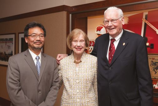
Green fluorescent protein labels the projections from the eye into this Left. A gene regulatory network for photoreceptors. Circles denote genes and
Image courtesy of the
coronal section of zebrafish brain tissue. The projections into this part of the lines denote interactions between genes. The NRL gene is shown as a major
Fundus Photograph
brain, called the optic tectum, mediate prey capture, among other visually hub. Right. Two-dimensional plot of cells of a complex retinal cell population.
Reading Center.
guided behaviors. The isl2b:GFP transgene expression in retinal ganglion Each color represents a different cell population and each dot is a single cell.
cells labels the projection green, while the brain section is counterstained Images courtesy of Sushmita Roy.
white for cell nuclei. Image courtesy of the Veldman Lab.
Bipolar Rods
cells
Cones
Müller
Glia
AMITHA Retinal
Progenitors
DOMALPALLY MD, PhD SUSHMITA ROY PhD
Fundus Photograph Reading Center,
Ophthalmology & Visual Sciences Biostatistics & Medical Informatics
∞ School of Medicine and Public Health ∞ School of Medicine and Public Health
Ophthalmology is fortunate to have cutting-edge imaging Changes in a cell’s genetic activity are controlled by
MATTHEW technologies available to diagnose eye diseases. UW- complex gene regulatory networks that connect regulatory
proteins to target genes in order to control which genes are
VELDMAN PhD Madison’s Fundus Photograph Reading Center (FPRC)
expressed at what time. In effect, these networks convert
is one of the world’s leading diagnostic imaging centers
Cell Biology, Neurobiology, and Anatomy for both patient diagnosis and clinical trials. Dr. Amitha the information encoded in an organism’s genome to
∞ Medical College of Wisconsin
Domalpally, Research Director of the FPRC, investigates actual responses and functions within a cell. Dr. Sushmita
clinical trial imaging endpoints for retinal diseases, Roy’s lab develops computational methods to understand
Optic nerve injury is thought to be the critical step in vision with a focus on employing new imaging techniques to these networks and to examine how they change across
loss due to glaucoma and other optic neuropathies and, understand the natural history and prognostic markers different cell types and tissues or during development and
unfortunately, no treatments are currently available to of diseases such as age-related macular degeneration across evolution.
recover lost vision in these diseases. This is also true in (AMD), diabetic retinopathy, and many others. Together with Dr. David Gamm at the Waisman Center
most animal models of glaucoma such as rats and mice.
Dr. Domalpally’ s research also involves interpretation and Dr. Kris Saha and other collaborators at the Wisconsin
Remarkably however, zebrafish can successfully recover
of the abundant and complex ocular imaging data that Institute for Discovery, Dr. Roy’s lab is developing and
from optic nerve injury and completely recover lost vision.
the FPRC collects. A better understanding of potential applying these computational methods to capture cellular
Dr. Matthew Veldman’s lab at the Medical College of
errors in imaging technologies, for instance, reduces false responses to genetic and environmental perturbations
Wisconsin studies optic nerve injuries and the differences
interpretation of data and improves patient management. in the retina. The group has identified various retinal
in response to these injuries in animals such as mice and
Domalpally has detailed artifacts that are intrinsic to OCT populations from human stem cell-derived retinal
fish, including the zebrafish.
angiography technology that help clinicians quickly flag organoids, from which Roy has inferred gene network
The Veldman Lab uses molecular, genetic, and pharmaco unreliable images that should be discarded. She is also changes that occur in response to genome-based
logical tools to understand the ability of zebrafish to recover involved in developing diagnostic artificial intelligence (AI) therapies. In this way, they can optimize the safety and
lost vision. Thus far, the lab group has identified multiple algorithms for retinal diseases. In collaboration with the efficacy of burgeoning gene therapy technologies. This
signaling molecules and pathways that are activated National Eye Institute, she has developed a deep learning technique was used to assess the safety of genome
in zebrafish during optic nerve regeneration. The lab is algorithm that assists in detecting reticular pseudodrusen, editing for Best macular dystrophy in a study by a team
currently testing their importance in cell survival and a feature associated with AMD that is difficult to identify by of McPherson ERI scientists published earlier this year.
visual recovery. Their goal is to translate these findings in a human observer. In another collaboration, her ongoing Overall, the ability of Roy’s lab to mine and extract hidden
zebrafish to mammalian models of glaucoma to identify work with Dr. Kevin Eliceiri at UW-Madison has resulted in information from large, complex genomic datasets
therapeutic targets for enhancing vision in patients with machine learning models for quantifying retinal lesions. provides an unprecedented opportunity to understand
glaucoma or other optic neuropathies. retinal cells in their normal and abnormal states.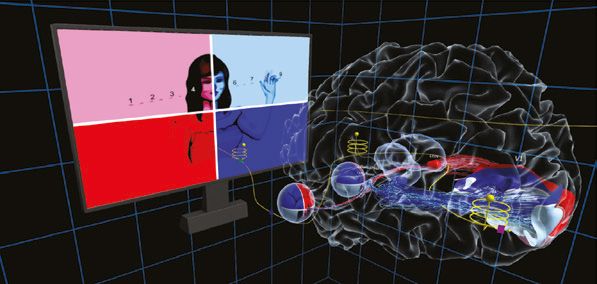
McPHERSON ERI/TROUT FAMILY
ENDOWMENT CHAIRS
In early 2020, Monroe and Sandra Trout endowed their third vision research chair at UW-Madison, the
Monroe E. Trout Chair, and increased the Timothy William Trout Endowment Fund to a Chair level. The
trio of research chairs that the Trouts have endowed will lead the way to better therapies—and, we
hope, a cure—for age-related macular degeneration (AMD), the leading cause of blindness in older
Americans. We are grateful to the Trouts for their visionary—no other word will do!—leadership in
combatting AMD.

                                                                                                                                  AKIHIRO IKEDA DVM, PhD
                 DAVID M. GAMM MD, PHD                                            MICHAEL M.                                      Timothy William Trout Chair in Eye Research
                 Director, McPherson Eye Research Institute                       ALTAWEEL MD
                 ∞ Sandra Lemke Trout Chair in Eye Research                                                                       “Our overall research goal is to identify mechanisms
                                                                                  Monroe E. Trout Chair in Vision Research
                                                                                                                                  underlying aging and age-related diseases in the retina.
                                                                                                                                  Using mouse models, we have successfully discovered
                                                                 “The Monroe E. Trout Chair will allow me to develop
“The Sandra Lemke Trout Chair in Eye Research provides                                                                            several key molecules associated with aging of the retina.
                                                                 and optimize surgical techniques to deliver stem cell-
critical funds to advance our pioneering retinal stem                                                                             One such mouse model showed similar symptoms as
                                                                 derived photoreceptors and RPE cells to the retina in
cell technology toward the clinic to treat patients with                                                                          those observed in patients with age-related macular
                                                                 a safe, effective, and reproducible manner. This will be
devastating degenerative diseases of the retina such as                                                                           degeneration (AMD). We identified a mutation in a
                                                                 accomplished through collaborations with a team of
age-related macular degeneration (AMD). Over the years,                                                                           mitochondrial gene called Tmem135 that is responsible
                                                                 McPherson ERI colleagues, including Dr. Gamm (who
support from this chair has facilitated studies that have                                                                         for accelerated aging and AMD-like phenotypes in the
                                                                 grows the stem cell-derived retinal cells in his lab) and Drs.
improved our ability to generate photoreceptors (rods and                                                                         mouse retina. Most recently, we discovered that over-
                                                                 Gong and Ma (whose labs have developed biodegradable
cones) and retinal pigment epithelium (RPE) in the lab,                                                                           expression of Tmem135 results in abnormal morphology
                                                                 scaffolds to house the photoreceptors). As a vitreoretinal
and to test their capacity to replace these same cell types                                                                       and degeneration of retinal pigment epithelium (RPE)
                                                                 surgeon at UW-Madison with 20 years of experience, I have
in diseased or damaged retinas. As a result, we are now                                                                           cells, suggesting that the proper level of this gene is
                                                                 participated in many clinical trials to advance therapeutics
in a public–private partnership to generate the cells on an                                                                       essential for the health of RPE cells. Interestingly, we
                                                                 for AMD, retinal vascular disorders, inflammatory eye
industrial scale, which is needed to begin clinical trials for                                                                    found that other genes also contribute to the health of
                                                                 disorders, and other indications. For example, I was the
both AMD and retinitis pigmentosa (RP) in the foreseeable                                                                         the RPE in these mice. This finding allows us to search
                                                                 lead UW investigator for a series of clinical trials that led
future. To specifically treat AMD, we have worked                                                                                 for “modifier genes” that interact with Tmem135 to
                                                                 to the FDA approval of the first commercially available
with McPherson ERI researchers Shaoqin Gong and                                                                                   determine how they affect the severity of the RPE
                                                                 anti-VEGF drug that was shown to block abnormal
Zhenqiang Ma to create next-generation biodegradable                                                                              abnormalities. Ongoing studies of Tmem135 will lead to a
                                                                 vascular growth in the eye and improve vision in AMD.
scaffolds that can replace both RPE and photoreceptors.                                                                           better understanding of how RPE cells are maintained in
                                                                 My surgical and research career at UW has led me to this
These scaffolds are now being tested in preclinical studies                                                                       their normal state and how they become diseased. It will
                                                                 new opportunity to restore sight with stem cell therapies
by colleagues at the National Eye Institute and here at                                                                           also help us understand why some people are affected
                                                                 developed entirely here at UW, and I’m enormously
UW by McPherson ERI researcher and retina surgeon                                                                                 by eye diseases like AMD while others are protected.
                                                                 excited to be part of this effort.”
Dr. Michael Altaweel.”                                                                                                            Support from the Timothy William Trout Chair in Eye
                                                                 Further information on Dr. Altaweel's work can be found in       Research is vitally important for us to move our research
                                                                 the Delivery feature, above.                                     effort towards this goal.”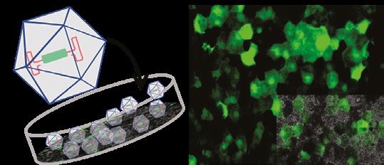
McPHERSON ERI/RETINA BARBARA A. BLODI MD
RESEARCH FOUNDATION CHAIRS Retina Research Foundation Daniel M.
Albert Chair
AND PROFESSORSHIPS “The focus of my vision research as the Daniel M. Albert
Chair has been to help create the Wisconsin Advanced
Imaging of the Visual System (WAIVS) program that
has begun building and augmenting Adaptive Optics
(AO) retinal imaging systems at UW. AO technology
will allow us to visualize the structure and function of
photoreceptors and other retinal cell types in animals
and humans in greater detail than has ever been possible.
DAVID M. GAMM ∞ Retina Research Foundation BIKASH PATTNAIK PhD This year, in order to use our AO systems in clinical
Emmett A. Humble Distinguished Directorship
Retina Research Foundation M. D. Matthews research, our AO team has designed and written a clinical
Professorship trial protocol that will compare AO images and data from
“The RRF Emmett A. Humble Distinguished Directorship 12 human participants across four AO laboratories in the
supports my lab’s efforts to generate human “disease-in- “Our lab research focus is on inherited and acquired Midwest. This trial is being done in order to standardize
a-dish” models of inherited retinal disease (for example, pediatric blindness. We have made some important AO image capture techniques and the formal assessment
retinitis pigmentosa) using induced pluripotent stem cell discoveries regarding the functioning of ion channels— of AO images. In addition, this comparative trial will use
(iPSC) technology that my laboratory patented at UW- proteins that facilitate cellular communication. When the the UW Fundus Photograph Reading Center to grade the
Madison. These models are created from blood samples ion channels are non-functional, they give rise to specific AO images in a masked fashion.”
donated by individuals with blinding disorders, which vision problems in children. We have used patient-derived
we employ as platforms to understand what causes the induced pluripotent stem cells as disease-in-a-dish
diseases and how best to treat them (using drug or gene models to develop a gene therapy treatment for retinal
therapies) in order to preserve or restore vision. Recently, diseases arising from both potassium and chloride JEREMY ROGERS PhD
in collaboration with McPherson ERI researchers Bikash channel mutations. Currently, a gene therapy for one Retina Research Foundation Edwin and
Pattnaik, Krishanu Saha, and Sushmita Roy, we published type of Leber congenital amaurosis (LCA16) is headed Dorothy Gamewell Professorship
a study that used retinal pigment epithelial (RPE) cells toward clinical trials through our close partnership with
generated from patient-derived iPSCs to build a lab- Hubble Therapeutics. In separate studies, we are also “The RRF Edwin and Dorothy Gamewell Professorship
based model of an inherited macular blinding disorder investigating new therapeutic strategies that may be enables my lab to pursue the development of new
called Best disease. We then used these cells to show able to correct a broad class of mutations known as methods to image retinal cells in patients. We are
how two different types of gene therapies (gene “nonsense mutations”. These mutations, which make up investigating how light is scattered and reflected by the
augmentation and CRISPR/Cas genome editing) about 15% of all genetic defects, result in the production retina and using this information in computational models
could potentially be used to treat all forms of this of incomplete and nonfunctional proteins. We are as we optimize and advance cutting-edge imaging
disease. We are now using the same approach using using several approaches, such as small molecules or capabilities, including Adaptive Optics instrumentation
iPSCs from patients with retinitis pigmentosa and engineered tRNA, to overcome the effects of nonsense and Optical Coherence Tomography. Our long-term goal
Usher syndrome.” mutations. We then employ biochemical tests and is to develop functional cellular imaging to aid diagnosis,
advanced electrophysiological techniques that directly optimize therapies, and improve our understanding of the
measure recovery of ion channel function in order to visual system.”
optimize therapeutic dosage and efficacy.”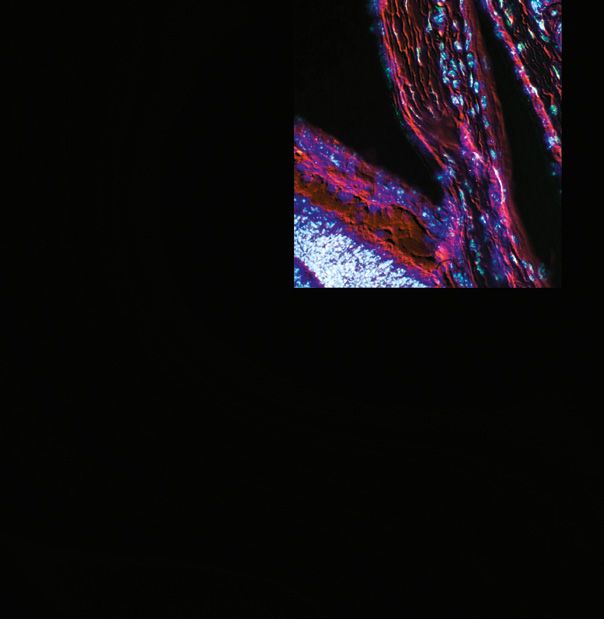
McPHERSON ERI/RETINA
    RESEARCH FOUNDATION CHAIRS                                                                                                                        KRISHANU SAHA PhD
    AND PROFESSORSHIPS                                                                                                                                Retina Research Foundation Kathryn and
                                                                                     MRINALINI HOON PhD                                               Latimer Murfee Chair
                                                                                     Retina Research Foundation Rebecca Meyer
                    KEVIN ELICEIRI PhD                                               Brown Professorship                            “Support from the Retina Research Foundation Kathryn
                                                                                                                                    and Latimer Murfee Chair has enabled me to build further
                    Associate Director, McPherson Eye Research
                                                                    “Our recent research has uncovered a novel role for a cell      momentum for our genome-editing projects in the eye.
                    Institute ∞ Retina Research Foundation Walter
                                                                    adhesion protein called LRRTM4 in the mammalian retina.         With the Chair support, we are developing new methods
                    H. Helmerich Research Chair
                                                                    Using mouse models that lack LRRTM4, we discovered              to understand the safety and efficacy of various genome-
                                                                    that LRRTM4 is necessary for correctly wiring the night-        editing approaches in close collaboration with McPherson
    “The support of the Retina Research Foundation Walter
                                                                    vision (dim-light) circuit of the retina. The retinal neurons   ERI members David Gamm, Bikash Pattnaik, Sarah Gong,
    H. Helmerich Research Chair has allowed me to advance
                                                                    that form this circuit communicate at specialized junctions     Melissa Skala, and others. My lab has now designed
    my current research on computational optics and to
                                                                    called synapses. Without LRRTM4, the formation and              additional viruses and nanoparticles that can be injected
    develop new imaging and computational approaches for
                                                                    function of several types of synapses—‘ribbon’ synapses         into the eye through intravitreal or subretinal injection in
    characterizing changes in the cellular microenvironment.
                                                                    and inhibitory GABAergic synapses surrounding the               order to edit the genome of cells within the retina directly.
    Some highlights of this work include the following:
                                                                    ribbon synapses—are severely perturbed. Importantly,            We have also started to better understand two critical
    • With McPherson ERI members Dr. Amitha Domalpally             these alterations are specific for synapses only along the      concerns with genome-editing strategies in the field:
      and Dr. Barb Blodi, we have enjoyed a successful              night vision pathway. The support of the RRF Rebecca            genomic specificity and potential off-target adverse effects.
      collaboration on automated image analysis at the UW           Meyer Brown Professorship was crucial in enabling us            We are now identifying gene regulatory network changes
      Fundus Photograph Reading Center (FPRC). The Center           to complete this study by combining approaches such             following the administration of a viral genome editor. This
      is using this program for several segmentation tasks in       as 3D electron microscopy, high-resolution confocal             work involves machine learning and bioinformatics, and
      eye images. Recently, we finished implementing a new          microscopy, and single-cell electrophysiology. As a             this pandemic year has been an excellent time to “double-
      platform on machine learning for automated analysis of        follow up to these exciting findings, we are currently          down” on efforts like these, which can be done outside the
      their data.                                                   investigating the mechanistic basis of LRRTM4                   lab. We are also now able to detect off-target modifications
                                                                    function in the retina and extending our findings in
    • With McPherson ERI members Dr. Paul Campagnola               mouse retina to primate retina, as mutations in LRRTM4
                                                                                                                                    to the genome at a higher resolution within retinal pigment
      and Dr. Jeremy Rogers, we have commissioned a novel                                                                           epithelial cells and photoreceptors. This work is helping
                                                                    have been linked to hereditary macular degeneration             us to understand, and ultimately avoid, adverse events in
      multimodal imaging instrument that combines four
                                                                    and macular dystrophy.”                                         patients who are treated with genome-editing therapeutics.”
      tissue-imaging methods ranging from nanometers to
      millimeters in spatial sensitivity. This is being explored
      for multiscale image analysis of sclera organization.

    • My lab continues to develop the open-source image            THE DAVID AND NANCY WALSH FAMILY                                perceptual and anatomical specializations of central vision
      analysis software package ImageJ. We recently received        PROFESSORSHIP IN VISION RESEARCH                                and foveal cone photoreceptors have been known for almost
      an NIH grant to develop and expand this tool. The grant                                                                       a century, our understanding of cone function in the fovea
      includes the addition of new capabilities to our open-
      source platform, including improved approaches for
                                                                                     RAUNAK SINHA PhD                               remains elementary. Using single-cell electrophysiology
                                                                                                                                    in primate cone photoreceptors, our lab is exploring how
      automated analysis and pattern recognition.”                                     “Research in our lab is focused on           cone function in the fovea may be distinct from the rest of
                                                                                       understanding how visual signals are         the primate retina. Our goal is to use this information about
                                                                                       converted in the photoreceptors and how      cone physiology as a baseline for testing cone photoreceptor
                                                                    they are subsequently processed by the downstream neural        function in human stem cell-derived 3D retinal organoids. This
                                                                    circuitry in the vertebrate retina. The David and Nancy Walsh   will help establish a functional assay to probe abnormalities in
David and Nancy
Walsh and family
                                                                    Family Professorship supports our lab's efforts to understand   cone photoreceptor function in retinal organoids derived from
with Dr. Alice                                                      the functional properties of photoreceptors in the fovea—a      human patients with disease mutations, and allow for testing
McPherson                                                           specialized retinal area unique to diurnal primates that        novel pharmaceuticals.”
                                                                    mediates our high-definition central vision. Even though the
RESEARCH
AWARDS & Student Awards
The McPherson ERI sponsors two
GRANTS
student awards, which were given
this year to students who were able to perform
their research remotely (as mandated during the COVID-19
pandemic). The undergraduate Hilldale Award ($4,000) was
given to PJ Derr, a neurobiology and psychology double major
in Raunak Sinha’s lab. The Dan and Ellie Albert Student
Kenzi Valentyn Vision Vision Research Award ($2000), matched through the
Research Awards Expanding NEW IN
2020!
Shapiro Summer Internship Program and funded by Dr. Daniel
Kenzi Valentyn Vision Research Awards, the McPherson Our Vision and Eleanor Albert, supports a summer vision research project
for an SMPH student. Claire Vanden Heuvel, in Dr. Julie
Eye Research Institute’s research grant opportunity for
trainees, were established in 2017. They are named after
Research Grants Mares’ lab, was the recipient for Summer 2020.
Kenzi Valentyn, in honor of her courage and positive attitude The Expanding Our Vision (EOV) grant program was
throughout her long battle with Kearns–Sayre syndrome, started this year to provide awards of up to $10,000 to
a degenerative disease with symptoms including vision researchers performing vision-related research in visual
loss. Kenzi’s many friends and family members, including communication, visual cognition, visual perception/
her parents Nancy and Tim, brothers Brett and Connor, and performance, data visualization, development of novel Grant Summit Program
imaging techniques for the visual system, computer
sister-in-law Mackenzie, have ridden in Cycle for Sight as
“Kenzi’s Team” since 2014. Each award recipient receives a sciences, and/or bioinformatics. Research in these areas is
(GSP), 2019-2020 Awards
grant of $4,000 to support their vision research project. essential for understanding the intricacies of human vision The McPherson ERI initiated the Grant Summit Program
and for enhancing our ability to visualize cells and processes (GSP) in 2018 to provide critical and timely support to bolster
In 2020, three Kenzi Valentyn Vision in the eye, and to analyze large and complex datasets. projects that are under consideration for federal awards but
Research Award recipients were chosen: require additional experiments. Since the program’s inception,
Five projects were awarded EOV Research multiple grants have been resubmitted and successfully
Anjani Chakrala, a graduate student in Neuroscience
mentored by Xin Huang. Project title: Neural Representation
Grants in this initial year: received funding that far exceeded the GSP contribution
Andrea Mason (Kinesiology) and Leigh Ann Mrotek (typically a greater than 20-fold return on investment). Thus,
of Overlapping Motion Surfaces Located at Different Depths
& Robert Scheidt (Biomedical Engineering, Marquette the GSP is a very cost-effective way to support vision research
in Visual Area MT: Effects of Selective Attention on this
University), Impact of visual cue saliency during bimanual within the Institute.
Representation.
visually guided reach-to-grasp In 2019 and 2020, GSP awards were given to
Ralph W. Nelson, a graduate student in Kinesiology
– Motor Control & Behavior, mentored by Andrea Mason. Ari Rosenberg (Neuroscience), Hierarchical cortical three McPherson ERI researchers:
Project title: The Assessment of Dual-Task Interference processing of three-dimensional visual motion
Colleen McDowell, Ophthalmology and Visual Sciences,
Using Walking and Carrying Tasks in Children With and Sushmita Roy (Biostatistics & Medical Informatics), received $10,000 for her grant resubmission, titled Toll-like
Without Autism Spectrum. Computational approaches for characterizing cell receptor 4 signaling in the glaucomatous optic nerve head.
Abhilash Sawant, a graduate student in Neuroscience, type dynamics in human retinal tissue
Ismail Zaitoun, Ophthalmology and Visual Sciences, received
mentored by Raunak Sinha. Project title: Understanding Karen Schloss (Psychology), Understanding dimensional $10,000 for his grant resubmission, titled Gender impact and
the Mechanism and Function of Postsynaptic Inhibition in structure underlying color-concept associations to advance retinal damage in hypoxic-ischemic encephalopathy.
the Mouse ON-Alpha Retinal Ganglion Cell. visual communication
Karen Schloss, Psychology, received $9,686 to facilitate
We are grateful to the Valentyn family for continued support Andreas Velten (Biostatistics & Medical Informatics), resubmission of an NSF CAREER award proposal, titled
for these awards, which go to the next generation of young Measuring ocular retroreflectance for remote ocular Understanding visual reasoning for scientific communication.
vision researchers! diagnostics Dr. Schloss’s proposal was recently approved for $558,702.Image Credit: Visualization by Tananun Songdechakraiwut, Moo K. Chung; Processing by Zhan Luo, Ian C. Carroll; Acquisition by
JANUARY Andrew L. Alexander, H. Hill Goldsmith. | In the above image, the reconstructed white matter fibers of the human brain from diffusion
tensor imaging (DTI) reveal the structural connectivity between brain regions. The fibers are colored based on the strength of functional
connectivity obtained from resting-state functional magnetic resonance imaging (rsfMRI).
TUE WED THU FRI SAT SUN MON TUE WED THU FRI SAT SUN MON TUE WED THU FRI SAT SUN MON TUE WED THU FRI SAT SUN MON TUE WED THU
DEC
1 2 3 4 5 6 7 8 9 10 11 12 13 14 15 16 17 18 19 20 21 22 23 24 25 26 27 28 29 30 31
SUNDAY MONDAY TUESDAY WEDNESDAY THURSDAY FRIDAY SATURDAY
27 28 29 30 31 1 2
NEW YEAR’S EVE NEW YEAR’S DAY
3 4 5 6 7 8 9
10 11 12 13 14 15 16
17 18 19 20 21 22 23
MARTIN LUTHER KING, JR. DAY
24 25 26 27 28 29 30
31
MON TUE WED THU FRI SAT SUN MON TUE WED THU FRI SAT SUN MON TUE WED THU FRI SAT SUN MON TUE WED THU FRI SAT SUN
FEB
1 2 3 4 5 6 7 8 9 10 11 12 13 14 15 16 17 18 19 20 21 22 23 24 25 26 27 28Image courtesy of Elizabeth Capowski, PhD | This is a confocal microscope image of the inner and outer nuclear layers of a self-
organizing, human stem cell-derived retinal organoid. Cones (in red) form a single line at the top of the outer nuclear layer while rods
FEBRUARY (blue nuclei and yellow outer segments) form 4-6 layers under the cones, as seen in human retinas. The green nuclei in the inner layer
are bipolar cells which form the second link in the neuronal chain that detects light and projects the signal to the visual cortex. Retinal
organoids such as these, grown in the laboratory of Dr. David Gamm, are used to model human retinal development and to test novel
therapeutics for blinding disorders.
FRI SAT SUN MON TUE WED THU FRI SAT SUN MON TUE WED THU FRI SAT SUN MON TUE WED THU FRI SAT SUN MON TUE WED THU FRI SAT SUN
JAN
1 2 3 4 5 6 7 8 9 10 11 12 13 14 15 16 17 18 19 20 21 22 23 24 25 26 27 28 29 30 31
SUNDAY MONDAY TUESDAY WEDNESDAY THURSDAY FRIDAY SATURDAY
31 1 2 3 4 5 6
GROUNDHOG DAY
7 8 9 10 11 12 13
CHINESE NEW YEAR
14 15 16 17 18 19 20
VALENTINE’S DAY PRESIDENTS’ DAY MARDI GRAS ASH WEDNESDAY
21 22 23 24 25 26 27
28 1 2 3 4 5 6
MON TUE WED THU FRI SAT SUN MON TUE WED THU FRI SAT SUN MON TUE WED THU FRI SAT SUN MON TUE WED THU FRI SAT SUN MON TUE WED
MAR
1 2 3 4 5 6 7 8 9 10 11 12 13 14 15 16 17 18 19 20 21 22 23 24 25 26 27 28 29 30 31Image courtesy of Briana Ebbinghaus, Hoon Lab | Dr. Mrinalini Hoon’s lab studies the development of connectivity in the neural retina
MARCH
and how this connectivity is affected in disease states. Shown here is a retinal ganglion cell, the type of output neuron for this circuit. This
is a maximum intensity projection of a 3D image taken with a confocal microscope. Ganglion cells transmit visual information to the brain,
so a problem with this cell type will have a noticeable effect on vision. Glaucoma and diabetic retinopathy are two of the known diseases
which involve changes in the retinal ganglion cells.
MON TUE WED THU FRI SAT SUN MON TUE WED THU FRI SAT SUN MON TUE WED THU FRI SAT SUN MON TUE WED THU FRI SAT SUN
FEB
1 2 3 4 5 6 7 8 9 10 11 12 13 14 15 16 17 18 19 20 21 22 23 24 25 26 27 28
SUNDAY MONDAY TUESDAY WEDNESDAY THURSDAY FRIDAY SATURDAY
28 1 2 3 4 5 6
7 8 9 10 11 12 13
14 15 16 17 18 19 20
DAYLIGHT SAVING
TIME BEGINS ST. PATRICK’S DAY FIRST DAY OF SPRING
21 22 23 24 25 26 27
PASSOVER BEGINS
28 29 30 31 1 2 3
PALM SUNDAY
THU FRI SAT SUN MON TUE WED THU FRI SAT SUN MON TUE WED THU FRI SAT SUN MON TUE WED THU FRI SAT SUN MON TUE WED THU FRI
APR
1 2 3 4 5 6 7 8 9 10 11 12 13 14 15 16 17 18 19 20 21 22 23 24 25 26 27 28 29 30Image courtesy of Michael Taylor, PhD | This image, generated by laser confocal microscopy, displays the blood vessels in the
APRIL brain of a zebrafish larva. The color is false color—the color spectrum represents depth in the image, with red/yellow being nearest
the surface, green intermediate, and blue/violet the deepest. Dr. Michael Taylor is an Assistant Professor in the School of Pharmacy,
Pharmaceutical Sciences Division.
MON TUE WED THU FRI SAT SUN MON TUE WED THU FRI SAT SUN MON TUE WED THU FRI SAT SUN MON TUE WED THU FRI SAT SUN MON TUE WED
MAR
1 2 3 4 5 6 7 8 9 10 11 12 13 14 15 16 17 18 19 20 21 22 23 24 25 26 27 28 29 30 31
SUNDAY MONDAY TUESDAY WEDNESDAY THURSDAY FRIDAY SATURDAY
28 29 30 31 1 2 3
APRIL FOOLS' DAY GOOD FRIDAY
4 5 6 7 8 9 10
EASTER
11 12 13 14 15 16 17
RAMADAN BEGINS
18 19 20 21 22 23 24
EARTH DAY
25 26 27 28 29 30 1
SAT SUN MON TUE WED THU FRI SAT SUN MON TUE WED THU FRI SAT SUN MON TUE WED THU FRI SAT SUN MON TUE WED THU FRI SAT SUN MON
MAY
1 2 3 4 5 6 7 8 9 10 11 12 13 14 15 16 17 18 19 20 21 22 23 24 25 26 27 28 29 30 31Image courtesy of Pupa Gilbert, PhD | Dr. Pupa Gilbert’s image of human tooth enamel, displayed in the Mandelbaum & Albert
MAY
Family Vision Gallery show Demystify: Seeing the Unseeable, was generated by Polarization-dependent Imaging Contrast (PIC)
mapping. The technique uses polarized X-rays to detect the orientation of individual crystals in a microcrystalline material such as
tooth enamel. Variations in orientation are displayed as variations in color and intensity. Dr. Gilbert is a Professor in the Department
of Physics at UW-Madison.
THU FRI SAT SUN MON TUE WED THU FRI SAT SUN MON TUE WED THU FRI SAT SUN MON TUE WED THU FRI SAT SUN MON TUE WED THU FRI
APR
1 2 3 4 5 6 7 8 9 10 11 12 13 14 15 16 17 18 19 20 21 22 23 24 25 26 27 28 29 30
SUNDAY MONDAY TUESDAY WEDNESDAY THURSDAY FRIDAY SATURDAY
25 26 27 28 29 30 1
2 3 4 5 6 7 8
ORTHODOX EASTER CINCO DE MAYO
9 10 11 12 13 14 15
MOTHER’S DAY EID AL-FITR BEGINS
16 17 18 19 20 21 22
SHAVUOT BEGINS
23 24 25 26 27 28 29
30 MEMORIAL DAY
31
TUE WED THU FRI SAT SUN MON TUE WED THU FRI SAT SUN MON TUE WED THU FRI SAT SUN MON TUE WED THU FRI SAT SUN MON TUE WED
JUN
1 2 3 4 5 6 7 8 9 10 11 12 13 14 15 16 17 18 19 20 21 22 23 24 25 26 27 28 29 30You can also read