Journal Club Antivirale Therapie der idiopathischen Fazialisparese - GUAD Netz
←
→
Page content transcription
If your browser does not render page correctly, please read the page content below
32. Journal Club
Antivirale Therapie der idiopathischen Fazialisparese

Background & Objectives

Corticosteroids are widely used in the treatment of idiopathic facial paralysis (Bell's palsy),
but the effectiveness of additional treatment with an antiviral agent is uncertain. This review
was first published in 2001 and most recently updated in 2015. Since a significant benefit of
corticosteroids for the early management of Bell's palsy has been demonstrated, the main
focus of this update, as in the previous version, was to determine the effect of adding
antivirals to corticosteroid treatment. Objectives: To assess the effects of antiviral treatments
alone or in combination with any other therapy for Bell's palsy.

Selection Criteria & Main results

We considered randomised controlled trials (RCTs) or quasi‐ RCTs of antivirals with and
without corticosteroids versus control therapies for the treatment of Bell's palsy.

Fourteen trials, including 2488 participants, met the inclusion criteria. Most were small, and
most were at high or unclear risk of bias in multiple domains. We included four new studies
at this update.

Incomplete recovery

A combination of antivirals and corticosteroids may have little or no effect on rates of
incomplete recovery in people with Bell's palsy compared to corticosteroids alone (risk ratio
(RR) 0.81, 95% confidence interval (CI) 0.38 to 1.74; 3 trials, N = 766; random‐ effects;
low‐ certainty evidence). We excluded 10 trials that were at high or unclear risk of bias in
several domains from this analysis and limited all analyses to studies at lower risk of bias.
Recovery rates were better in participants receiving corticosteroids alone than antivirals
alone (RR 2.69, 95% CI 0.73 to 10.01; 2 trials, N = 667; random‐ effects), but the result was
imprecise and allowed for the possibility of no effect. The rate of incomplete recovery was
lower with antivirals plus corticosteroids than with placebo or no treatment (RR 0.56, 95% CI
0.42 to 0.76; 2 trials, N = 658; random‐ effects). Antivirals alone had no clear effect on
incomplete recovery rates compared with placebo, but the result was imprecise (RR 1.10,
95% CI 0.87 to 1.40; 2 trials, N = 658; fixed‐ effect). For people with severe Bell's palsy
(House‐ Brackmann score of 5 and 6, or equivalent on other scales), we found that the
combination of antivirals and corticosteroids had no clear effect on incomplete recovery at
month six compared to corticosteroids alone, although the result was again imprecise (RR
0.82, 95% CI 0.57 to 1.17; 2 trials, N = 98; random‐ effects).

Motor synkinesis or crocodile tears

Antivirals plus corticosteroids reduced the proportion of participants who experienced these
long‐ term sequelae from Bell's palsy compared to placebo plus corticosteroids (RR 0.56,
95% CI 0.36 to 0.87; 2 trials, N = 469; fixed‐ effect; moderate‐ certainty evidence). Antivirals
plus corticosteroids reduced long‐ term sequelae compared to placebo but there was no
clear difference in this outcome with antivirals alone compared to placebo.

Authors' conclusions

The combination of antivirals and corticosteroids may have little or no effect on rates of
incomplete recovery in comparison to corticosteroids alone in Bell's palsy of various degrees
of severity, or in people with severe Bell's palsy, but the results were very imprecise.
Corticosteroids alone were probably more effective than antivirals alone and antivirals plus
corticosteroids were more effective than placebo or no treatment. There was no clear benefit
from antivirals alone over placebo.

The combination of antivirals and corticosteroids probably reduced the late sequelae of
Bell's palsy compared with corticosteroids alone. Studies also showed fewer episodes of
long‐ term sequelae in corticosteroid‐ treated participants than antiviral‐ treated participants.

We found no clear difference in adverse events from the use of antivirals compared with
either placebo or corticosteroids, but the evidence is too uncertain for us to draw
conclusions.

Fazit:
Cortison alleine war effektiver als antivirale Medikation. Cortison mit antiviraler Medikation
war auch nicht besser als Cortison alleine.
Die Kombination hat aber wohl etwas besser die Folgekrankheiten lindern können.
Evidence alerts

Association Between the Use of Antidepressants and the Risk of Type 2 Diabetes: A Large,
Population-Based Cohort Study in Japan

Abstract

OBJECTIVE: This study aimed to reveal the associations between the risk of new-onset type
2 diabetes and the duration of antidepressant use and the antidepressant dose, and between
antidepressant use after diabetes onset and clinical outcomes.

RESEARCH DESIGN AND METHODS: In this large-scale retrospective cohort study in
Japan, new users of antidepressants (exposure group) and nonusers (nonexposure group),
aged 20-79 years, were included between 1 April 2006 and 31 May 2015. Patients with a
history of diabetes or receipt of antidiabetes treatment were excluded. Covariates were
adjusted by using propensity score matching; the associations were analyzed between risk of
new-onset type 2 diabetes and the duration of antidepressant use/dose of antidepressant in the
exposure and nonexposure groups by using Cox proportional hazards models. Changes in
glycated hemoglobin (HbA1c) level were examined in groups with continuous use,
discontinuation, or a reduction in the dose of antidepressants.

RESULTS: Of 90,530 subjects, 45,265 were in both the exposure and the nonexposure group
after propensity score matching; 5,225 patients (5.8%) developed diabetes. Antidepressant
use was associated with the risk of diabetes onset in a time- and dose-dependent manner. The
adjusted hazard ratio was 1.27 (95% CI 1.16-1.39) for short-term low-dose and 3.95 (95% CI
3.31-4.72) for long-term high-dose antidepressant use. HbA1c levels were lower in patients
who discontinued or reduced the dose of antidepressants (F[2,49] = 8.17; P < 0.001).

CONCLUSIONS: Long-term antidepressant use increased the risk of type 2 diabetes onset in
a time- and dose-dependent manner. Glucose tolerance improved when antidepressants were
discontinued or the dose was reduced after diabetes onset.

FAZIT:
Diabetes als Laborwert ist ja nur ein Surrogatparameter.
Gibt es möglicherweise Confounder?
Insgesamt spannend zu wissen und das möglicherweise bei der Diabetikerbehandlung mit
zu beachten. Zumindest als Assoziation. Es wäre möglicherweise ein Fingerzeig, vor einem
medikamentösen Versuch noch den Diabetes zu bedenken.
Quarantine alone or in combination with other public health
measures to control COVID‐ 19: a rapid review
Cochrane Systematic Review - Prototype Version published:
08 April 2020

Helfen Quarantänemaßnahmen wirklich dabei, die Ausbreitung des neuen Coronavirus
einzudämmen?

Zum Hintergrund: In vielen Ländern, darunter auch Deutschland, Österreich und der
Schweiz, müssen sich all jene in häusliche Quarantäne begeben, die engen Kontakt mit
einer infizierten Person hatten. Quarantäne kann aber auch auf Ebene ganzer Städte oder
Regionen angewendet werden, so wie es seit Februar in Norditalien der Fall ist. Ziel von
Quarantäne ist es, Infektionsketten zu unterbrechen und dadurch den Ausbruch unter
Kontrolle zu bekommen.
Der aktuelle Rapid Review suchte nun nach Evidenz für die Wirksamkeit solcher
Maßnahmen. Weitere Fragestellungen waren, inwiefern eine solche Quarantäne mit
anderen Maßnahmen wie räumlicher Distanzierung, Schulschließungen oder einer
verbesserten Handhygiene zusammenwirkt und ob Quarantäneregeln auch für Menschen
sinnvoll sind, die von einer Reise aus einem Risikogebiet zurückkommen.

Für ihren Rapid Review zu diesen Fragen sichtete das Autorenteam mehr als 2000 Studien,
von denen sie am Ende 29 als relevant einstuften und für den Review auswerteten. Zehn
dieser Arbeiten sind mathematische Modellierungen, die in den letzten Monaten direkt zu
COVID-19 publiziert wurden. 15 weitere Modeling-Studien und vier Kohortenstudien lieferten
Evidenz zur Quarantäne bei den Lungenkrankheiten SARS und MERS, die von nahe
verwandten Coronaviren ausgelöst werden. Diese Studien sind also nur indirekt relevant.

Die Ergebnisse all dieser Studien sind weitgehend konsistent und legen nahe, dass
Quarantäne eine wichtige Maßnahme zur Eindämmung der Pandemie ist. Allerdings reicht
Quarantäne alleine wahrscheinlich nicht aus, um den Ausbruch von COVID-19 zu
kontrollieren. Sie sollte daher zusammen mit anderen Maßnahmen erfolgen. Denn bei
COVID-19 können Ansteckungen bereits vor Auftreten von Symptomen geschehen. Das
erschwert es, Fälle rechtzeitig zu erkennen und deren Kontaktpersonen zu finden und unter
Quarantäne zu stellen.
Nur vergleichsweise geringe Effekte fand der Review für eine individuelle Quarantäne allein
auf Basis eines vorhergehenden Aufenthalts in einem Risikogebiet – das legen zumindest
Studien aus der Zeit des SARS-Ausbruchs der Jahre 2002 und 2003 nahe.
Die Autoren machen allerdings deutlich, dass die Evidenzbasis für diese Schussfolgerungen
alles andere als optimal ist: Alle zehn Studien, die sich direkt auf COVID-19 beziehen, sind
mathematische Modellierungen des Pandemieverlaufs. In diese Modelle gehen zahlreiche
Annahmen ein, etwa zu Übertragungsraten, Inkubationszeiten oder Krankheitsverläufen.
Diese Annahmen beruhen auf dem aktuell noch sehr lückenhaften Wissen über SARS-CoV-
2, beziehungsweise COVID-19, welches sich aber laufend erweitert – sie sind deshalb mit
einem hohen Maß von Unsicherheit verbunden. Die übrigen 19 Studien (15 davon ebenfalls
Modellierungen) liefern indirekte Evidenz zu SARS und MERS. Es ist zwar plausibel, dass
zwischen diesen von nahe verwandten Viren ausgelösten Krankheiten Parallelen bestehen,
doch genauso gibt es offensichtlich erhebliche Unterschiede, etwa in der Infektiosität oder
der Schwere des Krankheitsverlaufs. Insofern sind solche Rückschlüsse von einer Krankheit
auf eine andere stets mit großer Vorsicht zu behandeln.
Die Aussagekraft dieser Studien, und damit auch die des Rapid Reviews, ist aufgrund dieser
Limitationen begrenzt. Doch der Anspruch evidenzbasierter Medizin ist es, die beste
verfügbare Evidenz zu einer Fragestellung zu berücksichtigen. In einer beispiellosen
Situation wie der momentanen Krise durch COVID-19 bedeutet dies, auch mathematische
Modellierungen zu berücksichtigen, solange keine bessere Evidenz vorhanden ist.

Im Fall des aktuellen Rapid Reviews stufen die Autoren die Vertrauenswürdigkeit der
Evidenz aufgrund der zahlreichen Limitationen als niedrig bis sehr niedrig ein.

FAZIT:
Sehr schwieriges Thema, zu dem wir kein Fazit ziehen konnten.
Review: Postinfarkt-Remodeling und medikamentöse
Sekundärprophylaxe
Inhalt:
1. Theoretische Grundlagen
2. Von aktuellen Leitlinien
3. Fazit
4. Postinfarkt-Algorithmus
--------------------------------------------------------------------------------------------------------------
1. Theoretische Grundlagen:
Pathological Ventricular Remodeling (Circulation 2013, Xie et al.)
https://www.ahajournals.org/doi/10.1161/CIRCULATIONAHA.113.001879
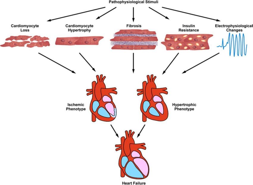
Mechanisms of pathological ventricular remodeling:
In response to pathophysiological stimuli such as ischemia/reperfusion or excessive mechanical load,
multiple molecular and cellular processes contribute to ventricular remodeling.
These include cardiomyocyte loss through cell death pathways such as necrosis, apoptosis, or
possibly excessive autophagy.
Cardiomyocytes become hypertrophic in response to both mechanical and neurohumoral triggers.
Accumulation of excess extracellular matrix leads to fibrosis. Metabolic derangements, insulin
resistance, and lipotoxicity can occur.
Finally, structural changes and alterations in ion transporting processes culminate in a proarrhythmic
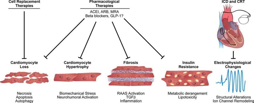
phenotype.https://www.ahajournals.org/doi/10.1161/CIRCULATIONAHA.113.001879 Therapeutic interventions in pathological ventricular remodeling: Clinically proven pharmacological agents reduce morbidity and mortality, including angiotensin- converting enzyme inhibitors (ACEIs), angiotensin receptor blockers (ARBs), β-blockers, and mineralocorticoid receptor antagonists (MRAs), by reducing cell death, hypertrophy, and fibrosis. https://www.ahajournals.org/doi/10.1161/CIRCULATIONAHA.113.001879 Kardiales Remodelling nach Myokardinfarkt (Springer Medizin 2017, Ertl et al.) https://link.springer.com/article/10.1007/s11298-017-5979-0 Medikamentöse Prävention und Therapie des Remodelling: Nachdem zunächst Bemühungen, die Infarktgröße durch medikamentöse Interventionen zu reduzieren, zwar im Tierexperiment erfolgreich [28], im Einsatz am Menschen aber frustran waren, konnten Pfeffer et al. zunächst an der Ratte und dann auch am Menschen zeigen, dass ACE-Hemmer die negativen morphologischen und hämodynamischen Konsequenzen des Myokardinfarkts verhindern oder verzögern [29, 30]. Das Besondere der ACE-Hemmer in diesem Zusammenhang war, dass sie trotz ihrer auch vasodilatatorischen Eigenschaften nicht zu einer refektorischen Aktivierung des sympathischen Nervensystems und zu einer Refextachykardie führten. Schließlich zeigten Pfeffer et al. einen Überlebensvorteil von Ratten und dann in der SAVE-Studie auch von Patienten nach großen Herzinfarkten durch den ACE-Hemmer Captopril [31, 32] . Diese Ergebnisse wurden vollumfänglich in der SOLVD-Studie bestätigt und etablierten die sekundär-präventive Therapie von Patienten nach Myokardinfarkt mit ACE-Hemmern [33, 34]. Angiotensin-1-Rezeptor (AT1)-Blocker waren den ACE-Hemmern nicht überlegen [35]. Da initial die Intoleranz gegenüber ACE-Hemmern als hoch eingeschätzt wurde [36], wurden AT1-Blocker als Alternative evaluiert [37], konnten sich aber als Erstlinientherapie nicht durchsetzen. Auch für Betablocker konnte gezeigt werden, dass sie ähnlich wie ACE-Hemmer das LV- Remodelling bei der KHK aushalten können, weshalb sie inzwischen zur etablierten Therapie bei der Prävention der ischämischen Kardiomyopathie gehören (Abb. 3; [38, 39]).
Mineralokortikoidrezeptorantagonisten (MRA) haben neben diuretischen Effekten eine
antitrombrotische und endothelschützende Wirkung und reduzieren die neurohumorale
Aktivierung [40]. Transgene Mäuse, die keinen myokardialen Mineralokortikoidrezeptor
haben, sind gegen das ungünstige Remodelling geschützt [41, 42]. Die frühzeitige
intravenöse Gabe des MRA Canrenoat , gefolgt von Spironolacton, verhinderte das
Remodelling und supprimierte bei Patienten nach einem ersten Vorderwandinfarkt einen
Marker für die im Remodelling induzierte Kollagensynthese über 4 Wochen (Abb. 4; [43]).
Die Ausgangs-LVEF lag in dieser Studie knapp über 45 %, alle Patienten waren mit einem
ACE-Hemmer, jedoch nur etwa ein Drittel mit einem Betablocker behandelt. Diese
Ergebnisse konnten allerdings bis zu einem Jahr nach Infarkt in einem Kollektiv bestätigt
werden, das eine Ausgangs-LVEF um 50 % hatte und dessen Patienten fast alle sowohl
einen ACE-Hemmer oder AT1-Blocker als auch einen Betablocker erhielten [44].
Medikamentöse Interventionen an neurohumoralen Systemen waren also erfolgreich darin,
ein Remodelling zu verhindern oder zu verzögern.

Zusammenfassung

Die Herzinsuffizienz ist eine führende Todesursache und häufigster Anlass für
Hospitalisierungen in Deutschland, obwohl die therapeutischen Möglichkeiten v. a. bei
reduzierter Pumpfunktion in den letzten Jahren erheblich zugenommen haben. Meist wird,
bevor sich eine Herzinsuffizienz auch klinisch manifestiert, eine Phase des kardialen
Remodellings durchlaufen. Remodelling wurde ursprünglich morphologisch und mechanisch
definiert, vollzieht sich allerdings auch auf zellulärer und molekularer Ebene. So sind
Heilungsvorgänge nach einem akuten Myokardinfarkt geprägt von Inflammation, zellulärer
Migration und Narbenbildung. Das kardiale Remodelling wird zudem von systemischen
Anpassungsvorgängen begleitet.

Ein primäres Ziel der Therapie sollte daher sein, das Remodelling zu verhindern. Wichtig
sind hierfür die möglichst schnelle Diagnostik und Therapieeinleitung. Frühzeitige
Reperfusionstherapie limitiert die Infarktgröße und trägt dazu bei, die linksventrikuläre
Pumpfunktion zu erhalten.

Als medikamentöse Standardtherapie sind nach Myokardinfarkt Angiotensinkonversions-
hemmer, Angiotensin-1-Rezeptor-Blocker und Betablocker etabliert. Auch Mineralokortikoid-
rezeptorantagonisten zeigen günstige Wirkungen. Spezifische Medikamente, die darauf
abzielen, schon in die Heilung des Infarkts einzugreifen, sind in Entwicklung.

   2. Von aktuellen Leitlinien

           ●   Up-to-date
           ●   Deximed
           ●   NVL
           ●   ESC
           ●   ACC/AHA
           ●   NICE

Up-to-date (2020):

Betablocker

https://www.uptodate.com/contents/acute-myocardial-infarction-role-of-beta-blocker-therapy
While all patients with acute myocardial infarction (MI) should receive long-term beta blocker therapy, the optimal duration, dose, and agent are not known. We recommend the use of a long-acting, once daily beta blocker to improve treatment adherence. We believe the evidence supports the use of beta blockers in patients with MI for as long as three years. The evidence supporting a longer duration, or indefinite therapy, is limited. However, many patients with prior MI have an indication for continued beta blocker therapy such as heart failure with reduced ejection fraction, asymptomatic left ventricular systolic dysfunction, hypertension, or angina. We suggest doing so in patients with high-risk features at presentation such as cardiogenic shock, heart failure, or chronic kidney disease. For patients without these high-risk features, we suggest that practitioners discuss the potential benefits and risks of continued therapy with patients and have them participate in decision making. (See 'Contraindications' below.) Although the adjusted incidence rate of hospitalization for acute MI has declined by 4 to 5 percent per year since 1987, approximately 200,000 recurrent episodes of acute MI occur annually [39]. Whereas ischemic heart disease has become the leading contributor to the burden of disease as assessed on the basis of disability-adjusted life-years [40], and survival from acute MI is associated with the increasing incidence of heart failure in the United States, there exists a sound argument for prolonged, indefinite beta-blocker therapy post-MI. We believe it is reasonable to discontinue beta blocker therapy, using a tapering protocol carried out over a few weeks, in patients with unacceptable side effects, for whom the financial burden is unacceptable, or in those for whom the use of multiple medications is problematic (polypharmacy). There are no known life-threatening side effects (such as proarrhythmia or malignancy) of long-term beta blocker therapy [41]. Many patients have been continued on this therapy indefinitely based on a 1999 meta-analysis of over 50,000 patients that showed a 23 percent reduction in death at a mean follow-up of 1.4 years [23]. In addition to the relatively short duration of follow-up, application of the conclusions of this meta- analysis is limited as reperfusion and medical (aspirin and statin) therapies were underutilized routinely. The findings in this meta-analysis were supported by a large observational study published in 1998 [21]. A more contemporary evaluation of the potential benefit from long-term beta blocker use was made in a 2012 observational study of over 14,000 patients with known prior MI enrolled in the international REACH registry [42]. Patients were enrolled in 2003 and 2004 and followed prospectively for up to four years. The primary outcome was a composite of cardiovascular death, nonfatal MI, or nonfatal stroke. Propensity score matching identified 3599 pairs of patients with and without beta blocker use. Aspirin and statin use (each) was approximately 75 percent. After a median follow-up of 44 months, there was a trend toward a lower incidence of the primary outcome with beta blocker therapy (16.9 versus 18.6 percent, respectively; hazard ratio 0.90, 95% CI 0.79-1.03). However, little difference was seen in the event rates in the beta blocker and no beta blocker groups as early as two years. A 2013 observational study evaluated outcomes in 5628 patients with ST-elevation MI (STEMI) treated with primary percutaneous coronary intervention. During a median follow-up of nearly four years, mortality rates did not differ between patients with and without beta blocker therapy (5.2 versus 6.2 percent) [25]. The absence of a significant difference persisted after multivariate and propensity score matching. However, subgroup analyses revealed that beta blocker treatment was associated with a significantly lower mortality for only high-risk patients, such as those with heart failure. ACE-Hemmer https://www.uptodate.com/contents/angiotensin-converting-enzyme-inhibitors-in-acute-myocardial- infarction A meta-analysis concluded that administration of an ACE inhibitor within 3 to 16 days of infarction can slow the progression of cardiovascular disease and improve the survival rate (figure 1) [1]. The mechanisms by which ACE inhibitors improve survival after MI will be reviewed here, beginning with a discussion of infarct expansion. The clinical data supporting the use of ACE inhibitors in this setting are presented elsewhere. (See "Angiotensin converting enzyme inhibitors and receptor blockers in acute myocardial infarction: Clinical trials".)
Attenuation of remodeling is thought to play an important role in the survival benefit associated with
angiotensin converting enzyme (ACE) inhibitors after myocardial infarction. These drugs also may
have favorable effects on ischemic preconditioning, recurrent myocardial infarction and ischemia,
sudden death, and fibrinolysis (reduction in plasma plasminogen activator inhibitor-1). (See
'Reduction in recurrent myocardial infarction and ischemia' below.)

The effect of ACE inhibitors on infarct expansion reflects a chronic benefit. There is also experimental
evidence that ACE inhibitors have more acute benefits, limiting the following changes that can occur
during acute ischemia: myocardial injury, degradation of high-energy phosphate stores, and
impairment of endothelium-dependent arteriolar responses [19,20]. These observations constitute part
of the rationale for the early administration of ACE inhibitors (within the first 24 hours) after the
diagnosis of acute MI has been made [21]. (See "Angiotensin converting enzyme inhibitors and
receptor blockers in acute myocardial infarction: Recommendations for use".)

ACE inhibitors appear to reduce the incidence of recurrent myocardial infarction. As an example, the
Survival and Ventricular Enlargement (SAVE) Trial of 2231 patients found that captopril was
associated with a significant reduction in recurrent MI by 25 percent. This benefit was noted in all
patient groups, including those who received other adjunctive therapies such as fibrinolysis, aspirin,
and/or beta blockers [23].

ACE inhibitors reduce overall cardiovascular and sudden-death mortality [37]. There are several
mechanisms that might contribute to the reduction in sudden death (see "Pathophysiology and
etiology of sudden cardiac arrest")

The addition of angiotensin converting enzyme (ACE) inhibitor or receptor blocker to standard medical
therapy (including aspirin, thienopyridine, beta blocker, and statin) in patients with recent myocardial
infarction improves cardiovascular outcomes.

Attenuation of remodeling is thought to play a critically important role in the survival benefit associated
with ACE inhibitors after myocardial infarction. (See 'Attenuation of remodeling' above.)

Mineralocorticoid receptor antagonists

https://www.uptodate.com/contents/overview-of-the-non-acute-management-of-st-elevation-
myocardial-infarction

For patients with STEMI who are receiving an ACE inhibitor and a beta blocker, and who have a left
ventricular ejection fraction less than or equal to 40 percent and either HF or diabetes, we prescribe a
mineralocorticoid receptor antagonist, also referred to as an aldosterone blocker or antagonist [3,4].
Therapy should be begun before discharge, since a mortality benefit is seen within 30 days [40]. The
serum potassium should be monitored closely during treatment. Although uncommon, life-threatening
hyperkalemia can occur due to the combination of aldosterone inhibition, reduced aldosterone
secretion associated with ACE inhibitor therapy, and a progressive decline in renal perfusion induced
due to HF. Elderly patients with renal insufficiency are at greatest risk. The use of aldosterone
antagonists is discussed in detail elsewhere. (See "Secondary pharmacologic therapy in heart failure
with reduced ejection fraction (HFrEF) in adults", section on 'Evidence'.)

Although a 2018 meta-analysis found benefit to the addition of aldosterone antagonist in patients with
STEMI without heart failure and with an ejection of more than 40 percent, we believe significant
limitations to this study prevent us from recommending this therapy for these patients [41,42].

https://www.uptodate.com/contents/overview-of-the-non-acute-management-of-unstable-angina-and-
non-st-elevation-myocardial-infarction

We recommend an aldosterone antagonist, also referred to as an aldosterone receptor antagonist or
a mineralocorticoid receptor antagonist, to all NSTEMI patients who are receiving a therapeutic dose of
an ACE inhibitor, have an LVEF ≤40 percent, have heart failure or diabetes mellitus, and are free of
significant renal dysfunction or hyperkalemia [1].

https://www.uptodate.com/contents/secondary-pharmacologic-therapy-in-heart-failure-with-reduced-
ejection-fraction-hfref-in-adults
The following are two partially overlapping indications for mineralocorticoid receptor antagonist (MRA)
therapy. MRA use is limited to patients whose serum potassium and renal function can be carefully
monitored and who have baseline serum potassium
■   neuere Analysen mit uneinheitlichen Ergebnissen zum Nutzen von
                      Betablockern in der Reperfusionsära44,59
          ○   Betablocker sind auf jeden Fall indiziert bei einer LVEF ≤ 40 % und/oder Zeichen
              der Herzinsuffizienz.31,35

   ●   ACE-Hemmer
          ○ ACE-Hemmer sind indiziert bei LV-Dysfunktion und/oder Zeichen der
              Herzinsuffizienz oder bei Hypertonie.31,35
          ○ Ein Myokardinfarkt ist nicht per se eine Indikation für ACE-Hemmer, wenn der
              Blutdruck nicht erhöht und die kardiale Kontraktilität im Echo nicht eingeschränkt
              sind.
   ●   Aldosteronantagonisten
          ○   Aldosteronantagonisten sind indiziert bei einer LVEF ≤ 40 % und/oder Zeichen der
              Herzinsuffizienz.

https://deximed.de/home/b/herz-gefaesse-kreislauf/krankheiten/koronare-
herzkrankheit/koronarerkrankungen-sekundaerpraevention/#medikament-se-pr-vention

Betablocker

   ●   Patienten nach Myokardinfarkt sollte für 1 Jahr ein Betarezeptorenblocker empfohlen
       werden und dann die weitere Gabe bzw. das Absetzen reevaluiert werden.2
   ●   Betablocker reduzieren die Herzfrequenz, senken den Blutdruck und verbessern so die
       Leistungsfähigkeit des Herzens.
   ●   Die Häufigkeiten von Re-Infarkten werden im 1. Jahr nach Infarkt so vermindert.19 Im
       weiteren Verlauf besitzen Betablocker keinen Vorteil vor anderen Antihypertensiva.
   ●   Darüber hinaus verringern sie das Auftreten von Arrythmien.

ACE-Hemmer, AT1-Rezeptorantagonisten und Aldosteronantagonisten

   ●   Diese Medikamente sind nicht primär hilfreich in der Sekundärprävention bei KHK,
       kommen aber oft zum Einsatz im Rahmen eines gleichzeitig bestehenden
       Hypertonus oder einer Herzinsuffizienz.

NVL (2019):
https://www.leitlinien.de/nvl/html/nvl-chronische-khk/5-auflage/kapitel-7

Betarezeptorenblocker
Die strukturierte Suche nach Übersichtsarbeiten ergab einen systematischen Review der NICE- Guideline zur Sekundärprävention nach Myokardinfarkt. [263] Der Surveillance-Report der Leitlinie [292] identifizierte eine Metaanalyse von Bangalore et al. [293] als relevante neue Evidenz, die eine Aktualisierung des NICE-Reviews erforderlich mache. In unsere qualitative Analyse wird sowohl der NICE-Review als auch die Metaanalyse von Bangalore einbezogen. Die Analyse von NICE schloss nur Studien ein, bei denen maximal 30% der Patienten an Herzinsuffizienz litten. Bei Initiierung der Betablockertherapie innerhalb von drei Tagen nach Symptombeginn (48 RCTs mit 77 719 Patienten, GRADE moderat) zeigte sich ein Trend zu einer reduzierten Gesamtmortalität (8,3% vs. 8,7%, RR 0,95 (95% KI 0,91; 1,0)). Ein knapp signifikantes Ergebnis bestand für den Behandlungszeitraum von bis zu 12 Monaten nach Myokardinfarkt (8 RCTs mit 19 406 Patienten, GRADE moderat, 11,2% vs. 12,2%, RR 0,91 (95% KI 0,84; 0,99)). Studien mit einem späteren Therapiebeginn (14 RCTs, 17 642 Patienten) zeigten einen deutlicheren Effekt auf die Gesamtmortalität (RR 0,76 (95% KI 0,69; 0,83)), ihre methodische Qualität wurde jedoch als sehr niedrig bewertet. [263] Die Re-Infarktrate war bei initialer Betablocker-Therapie (13 RCTs mit 65 846 Patienten, GRADE low) um ca. 20% reduziert (2,1% vs. 2,5%, RR 0,81 (95% KI 0,73; 0,89)), entsprechend 5 verhinderten Myokardinfarkten bei 1 000 Patienten (3 weniger bis 7 weniger). Ein ähnliches Ergebnis zeigte die Analyse von Studien mit verzögertem Therapiebeginn (13 RCTs mit 17 089 Patienten, GRADE very low, RR 0,79 (95% KI 0,69; 0,91)). [263] Die Wirksamkeit der verschiedenen Vertreter dieser Stoffgruppe ist nicht vergleichend untersucht worden. Nach Einschätzung der Autoren der NVL liegen bei Hypertonie und Z.n. Myokardinfarkt die besten Daten für Metoprolol vor, bei Herzinsuffizienz zusätzlich auch für Bisoprolol und Carvedilol. Vorteilhaft bei Patienten mit Diabetes mellitus oder COPD sind Beta-1-selektive Rezeptorenblocker ohne partielle antagonistische bzw. intrinsische sympathomimetische Aktivität (z. B. Bisoprolol, Metoprolol). Ein Jahr nach Myokardinfarkt besteht aus Sicht der Leitliniengruppe keine eigenständige Indikation für einen Betarezeptorenblocker und die Gabe bzw. das Absetzen sollte daher re-evaluiert werden. Ihrer Einschätzung nach ist der Nutzen von Betarezeptorenblocker nicht ausreichend belegt für Patienten mit KHK, bei denen keine anderweitige Indikation zur Therapie besteht, z. B. auf Grund eines Hypertonus, einer Herzinsuffizienz oder spezifischen arrythmischen Indikationen. Bezüglich der Behandlung von Patienten mit KHK und linksventrikulärer Dysfunktion bzw. Herzinsuffizienz wird auf die NVL Chronische Herzinsuffizienz [53] verwiesen. Die abgeschwächte Empfehlung spiegelt die Unsicherheit bzgl. des Zeitraums von einem Jahr wieder. ACE-Hemmer Die strukturierte Suche nach Übersichtsarbeiten ergab zwei systematische Reviews von NICE zur ACE-Hemmer-Therapie bei Patienten mit KHK und erhaltener systolischer Pumpfunktion (LVEF > 40%). NICE identifizierte einen RCT (n = 1 252, Follow-up 1 Jahr), der ACE-Hemmer bei Patienten nach Myokardinfarkt mit erhaltener LV-EF untersuchte. Dieser RCT zeigte keinen signifikanten Effekt auf die Gesamtmortalität und die Häufigkeit von Re-Infarkten, wobei seine methodische Qualität u. a. auf Grund einer hohen Abbruchrate als sehr gering eingeschätzt wurde. [263] Bezüglich der Behandlung von Patienten mit stabiler KHK wurden drei RCTs identifiziert. Die Gesamtmortalität war unter ACE-Hemmern um 15% reduziert (2 RCTs, n = 11 047, Follow-up 3-5 Jahre, GRADE moderat, 9,2% vs. 10,8%, RR 0,85 (95% KI 0,76; 0,96)). Das Risiko für einen Re- Infarkt war ebenfalls signifikant geringer (3 RCTs, n = 19 337, Follow-up 3-5 Jahre, GRADE moderat, 7,4% vs. 8,6%, RR 0,86 (95% KI 0,78; 0,95)). [216] Auf Ebene der Einzelstudien fand nur ein RCT einen signfikanten Effekt auf die Gesamtmortalität und Infarktrate [294]. Nach Einschätzung von NICE beruhen diese Effekte am ehesten auf abweichenden Baseline-Patientencharakteristika: Im Vergleich zu anderen RCTs litten in dieser Population mehr Patienten an Diabetes mellitus, der durchschnittliche Ausgangsblutdruck war höher und weniger Patienten erhielten Lipidsenker. [216] Die Leitliniengruppe betrachtet eine KHK nicht als eigenständige Indikation für einen ACE- Hemmer: Ihrer Einschätzung nach ist der Nutzen von ACE-Hemmern nicht ausreichend belegt für Patienten mit KHK, bei denen keine anderweitige Indikation zur ACE-Hemmer Therapie besteht, z. B. auf Grund eines Hypertonus oder einer Herzinsuffizienz. Bezüglich der Behandlung von Patienten mit KHK und linksventrikulärer Dysfunktion bzw. Herzinsuffizienz wird auf die NVL Chronische Herzinsuffizienz [53] verwiesen.
AT1-Rezeptorantagonisten
Ein NICE-Review identifizierte einen RCT, der AT1-Rezeptorantagonisten mit ACE-Hemmern bei
Patienten mit normaler linksventrikulärer Funktion nach Myokardinfarkt verglich (n = 429, Follow-
up 60 Tage). Die Mortalität und die Myokardinfarktrate unterschieden sich in beiden Gruppen nicht
signifikant [263]. Analog zu den ACE-Hemmern betrachtet die Leitliniengruppe eine KHK nicht als
eigenständige Indikation für eine Behandlung mit AT1-Rezeptorantagonisten. Bezüglich der
Behandlung von Patienten mit KHK und linksventrikulärer Dysfunktion bzw. Herzinsuffizienz wird
auf die NVL Chronische Herzinsuffizienz [53] verwiesen.

Aldosteronantagonisten
Eine selektiv recherchierte Metaanalyse (10 RCTs, n = 4 147) fand eine signifikante Reduktion der
Mortalität durch Aldosteronantagonisten bei Patienten nach STEMI (2,4% vs. 3,9%; OR 0,62 (95%
KI 0,42; 0,91); p = 0,01). Kein Unterschied zeigte sich bei der Häufigkeit von Myokardinfarkten,
den Neudiagnosen von Herzinsuffizienz und dem Auftreten ventrikulärer Arrhythmien. Ein
Einschlusskriterium der Metaanalyse war eine erhaltene LV-Funktion (LVEF > 40%) [295].
Allerdings erfolgte in einer der eingeschlossenen Primärstudien keine Überprüfung der LVEF [296]
und in sieben weiteren lag die durchschnittliche bzw. mediane LVEF zwar über 40%, eine
linksventrikuläre Dysfunktion war jedoch kein Ausschlusskriterium. Ein unbekannter Anteil der
Patienten hatte somit bei Studienbeginn eine linksventrikuläre Dysfunktion (durchschnittliche bzw.
mediane LVEF in den eingeschlossenen RCTs 40%-52%, SD bis 12%) [297], [298], [299], [300],
[301], [302], [303].

In der ALBATROSS-Studie, die das Gesamtergebnis der Metaanalyse wesentlich bestimmte, wurde
außerdem keine LVEF-Bestimmung bei Studienbeginn durchgeführt, sondern erst zu einem
unbestimmten Zeitpunkt bis sechs Monate nach Randomisierung. Zur Subgruppe der Patienten mit
LVEF > 40% gehörten deshalb in der ALBATROSS-Studie auch Patienten mit initialer
linksventrikulärer Dysfunktion, die sich im Studienverlauf unter optimaler Behandlung besserte.
[299]

Nach Einschätzung der Leitliniengruppe können die Ergebnisse der Metaanalyse nicht sicher auf
Patienten mit STEMI und erhaltener linksventrikulärer Funktion bezogen werden, da nur zwei der
zehn Primärstudien ausschließlich Patienten mit LVEF > 40% einschlossen. Analog zu den ACE-
Hemmern und AT1-Rezeptorantagonisten betrachtet die Leitliniengruppe eine KHK nicht als
eigenständige Indikation für eine Behandlung mit Aldosteronantagonisten. Bezüglich der
Behandlung von Patienten mit KHK und linksventrikulärer Dysfunktion bzw. Herzinsuffizienz wird
auf die NVL Chronische Herzinsuffizienz [53] verwiesen.

https://www.kbv.de/media/sp/nvl_herzinsuffizienz_lang.pdf

ACE-Hemmer
In RCTs [153–156] und Metaanalysen [157,158] wurde nachgewiesen, dass ACE-Hemmer bei
Patienten mit leichter, mäßiger und schwerer HFrEF (NYHA II-IV) die Gesamtsterblichkeit senken, die
Progression der Pumpfunktionsstörung verzögern, die Hospitalisierungsrate senken sowie die
Symptomatik und Belastungstoleranz verbessern. Bei herzinsuffizienten Patienten nach
Myokardinfarkt senken ACE-Hemmer darüber hinaus die Re-Infarktrate [153–155].

Aldosteronantagonisten
Die Empfehlung zu Mineralokortikoidrezeptorantagonisten (MRA, auch: Aldosteronantagonisten)
beruht auf inter-nationalen Leitlinien [13,17]. Nach Prüfung der dort zitierten Evidenz durch die
Leitliniengruppe wurden Inhalt und Empfehlungsgrad übernommen.
Der Nutzen von MRA bei chronischer Herzinsuffizienz wurde in mehreren randomisierten Studien
belegt:
• Spironolacton 12,5-50 mg/Tag (RALES) [202]: NYHA III/IV, LVEF ≤ 35%, n = 1 663; Follow-up 24
Monate; Gesamtsterblichkeit signifikant reduziert (ARR 11%, NNT = 9); Rate der
Krankenhauseinweisungen aufgrund der Herzinsuffizienz signifikant reduziert (ARR 29%, NNT = 4);
• Eplerenon 25-50 mg/Tag (EPHESUS) [203]: Patienten 3-14 Tage nach akutem Myokardinfarkt, LVEF ≤
40%, mit Herzinsuffizienzsymptomen oder Diabetes mellitus, n = 6 632; Gesamtmortalität signifikant
gesenkt (ARR 2,3%, NNT = 43); Komposit-Endpunkt Risiko kardiovaskuläre Sterblichkeit und
Hospitalisierung aufgrund kar-diovaskulärer Ereignisse signifikant reduziert (ARR 3%, NNT = 34);
• Spironolacton [204]: NYHA I/II, LVEF ≤ 40%, Follow-up 6 Monate, n = 168; LVEF signifikant erhöht (p
< 0,001), positive Effekte auf Remodeling und diastolische Funktion;
• Eplerenon (EMPHASIS-HF) [205]: NYHA II, EF ≤ 30% (≤ 35% bei QRS > 130ms), Hospitalisierung aus
kardiovaskulären Gründen < 6 Monate oder erhöhte BNP-Werte, Follow-up 21 Monate, n = 2 737;
Komposit-Endpunkt Risiko kardiovaskuläre Mortalität und herzinsuffizienzbedingte Hospitalisierung
signifikant reduziert (ARR 7,7%, NNT = 13), Gesamtmortalität reduziert (ARR 3%, NNT = 33);
• Metaanalyse NYHA I//II, n = 3 929 [206]: Gesamtmortalität reduziert (RR 0,79 (95% KI 0,66; 0,95)),
Rehospitalisierungen aus kardialen Gründen reduziert (RR 0,62 (95% KI 0,52; 0,74)).

ESC (2017):
https://academic.oup.com/eurheartj/article/39/2/119/4095042 (STEMI)

Beta-blockers (Mid- and long-term beta-blocker treatment)

The benefit of long-term treatment with oral beta-blockers after STEMI is well established, although
most of the supporting data come from trials performed in the pre-reperfusion era.353 A recent
multicentre registry enrolling 7057 consecutive patients with AMI showed a benefit in terms of
mortality reduction at a median follow-up of 2.1 years associated with beta-blocker prescription at
discharge, although no relationship between dose and outcomes could be identified.354 Using
registry data, the impact of newly introduced beta-blocker treatment on cardiovascular events in 19
843 patients with either ACS or undergoing PCI was studied.355 At an average of 3.7 years of follow-
up, the use of betablockers was associated with a significant mortality reduction (adjusted HR 0.90,
95% CI 0.84–0.96). The association between beta-blockers and outcomes differed significantly
between patients with and without a recent MI (HR for death 0.85 vs. 1.02; Pint = 0.007).
Opposing these results, in a longitudinal observational propensity-matched study including 6758
patients with previous MI, beta-blocker use was not associated with a lower risk of cardiovascular
events or mortality.356
Based on the current evidence, routine administration of beta-blockers in all post-STEMI patients
should be considered as discussed in detail in the heart failure guidelines;6 beta-blockers are
recommended in patients with reduced systolic LV function (LVEF
receptor antagonist, has been shown to reduce morbidity and mortality in these patients. The
Eplerenone Post-AMI Heart failure Efficacy and SUrvival Study (EPHESUS) randomized 6642 post-MI
patients with LV dysfunction (LVEF 177mmol/L (2.0mg/dL) in women] and routine monitoring of
serum potassium is warranted.
https://academic.oup.com/eurheartj/article/37/3/267/2466099 (N-STEMI)

ACE inhibition
ACE inhibitors are recommended in patients with systolic LV dysfunction or heart failure, hypertension
or diabetes (agents and doses of proven efficacy should be employed). ARBs are indicated in patients
who are intolerant of ACE inhibitors.478 – 480,530,531

Beta-blockers
Beta-blockers are recommended, in the absence of contraindications, in patients with reduced systolic
LV function (LVEF ≤40%). Agents and doses of proven efficacy should be administered.482 – 486
Betablocker therapy has not been investigated in contemporary RCTs in patients after NSTE-ACS
and no reduced LV function or heart failure. In a large-scale observational propensity-matched study
in patients
with known prior MI, beta-blocker use was not associated with a lower risk of CV events or
mortality.532

Mineralocorticoid receptor antagonist therapy
Aldosterone antagonist therapy is recommended in patients with LV dysfunction (LVEF ≤40%) and
heart failure or diabetes after NSTE-ACS. Eplerenone therapy has been shown to reduce morbidity and
mortality in these patients after ACS.487,488,525

ACC/AHA (2013/2014):
https://www.ahajournals.org/doi/pdf/10.1161/circ.110.9.e82 (STEMI)

Beta-Blockers

1. All patients after STEMI except those at low risk (normal or near-normal ventricular function,
successful reperfusion, absence of significant ventricular arrhythmias) and those with
contraindications should receive beta-blocker therapy. Treatment should begin within a few days of
the event, if not initiated acutely, and continue indefinitely. (Level of Evidence: A)

2. Patients with moderate or severe LV failure should receive beta-blocker therapy with a gradual
titration scheme. (Level of Evidence: B)
It is reasonable to prescribe beta-blockers to low-risk patients after STEMI who have no
contraindications to that class of medications. (Level of Evidence: A)

Inhibition of the Renin-Angiotensin-Aldosterone System

1. An ACE inhibitor should be administered orally during convalescence from STEMI in patients who
tolerate this class of medication, and it should be continued over the long term. (Level of Evidence: A)

2. An ARB should be administered to STEMI patients who are intolerant of ACE inhibitors and have
either clinical or radiological signs of heart failure or LVEF less than 0.40. Valsartan and candesartan
have demonstrated efficacy for this recommendation. (Level of Evidence: B)

3. Long-term aldosterone blockade should be prescribed for post-STEMI patients without significant
renal dysfunction (creatinine should be less than or equal to 2.5 mg/dL in men and less than or equal
to 2.0 mg/dL in women) or hyperkalemia (potassium should be less than or equal to 5.0 mEq/L) who
are already receiving therapeutic doses of an ACE inhibitor, have an LVEF of less than or equal to
0.40, and have either symptomatic heart failure or diabetes. (Level of Evidence: A)

https://www.ahajournals.org/doi/epub/10.1161/CIR.0000000000000134 (N-STEMI)

Beta-Adrenergic Blockers

1. Oral beta-blocker therapy should be initiated within the first 24 hours in patients who do not have
any of the following: 1) signs of HF, 2) evidence of low-output state, 3) increased risk for cardiogenic
shock, or 4) other contraindications to beta blockade (eg, PR interval >0.24 second, second- or
thirddegree heart block without a cardiac pacemaker, active asthma, or reactive airway disease).240–
242
(Level of Evidence: A)

2. In patients with concomitant NSTE-ACS, stabilized HF, and reduced systolic function, it is
recommended to continue beta-blocker therapy with 1 of the 3 drugs proven to reduce mortality in
patients with HF: sustained-release metoprolol succinate, carvedilol, or bisoprolol.
(Level of Evidence: C)

3. Patients with documented contraindications to beta blockers in the first 24 hours of NSTE-ACS
should be re-evaluated to determine their subsequent eligibility. (Level of Evidence: C)
It is reasonable to continue beta-blocker therapy in patients with normal LV function with NSTE-ACS.
241,243 (Level of Evidence: C)

Inhibitors of the Renin-Angiotensin-Aldosterone System

1. ACE inhibitors should be started and continued indefinitely in all patients with LVEF less than 0.40
and
in those with hypertension, diabetes mellitus, or stable CKD (Section 7.6), unless
contraindicated.275,276
(Level of Evidence: A)

2. ARBs are recommended in patients with HF or MI with LVEF less than 0.40 who are ACE inhibitor
intolerant. 277,278 (Level of Evidence: A)

3. Aldosterone blockade is recommended in patients post–MI without significant renal dysfunction
(creatinine >2.5 mg/dL in men or >2.0 mg/dL in women) or hyperkalemia (K+ >5.0 mEq/L) who are
receiving therapeutic doses of ACE inhibitor and beta blocker and have a LVEF 0.40 or less, diabetes
mellitus, or HF.279 (Level of Evidence: A)

NICE (2013):

https://www.nice.org.uk/guidance/CG172/chapter/1-Recommendations#drug-therapy-2

Offer all people who have had an acute MI treatment with the following drugs:

          ●   ACE (angiotensin-converting enzyme) inhibitor
          ●   dual antiplatelet therapy (aspirin plus a second antiplatelet agent)
          ●   beta-blocker
          ●   statin. [2007, amended 2013]

ACE inhibitors

Offer people who present acutely with an MI an ACE inhibitor as soon as they are haemodynamically
stable. Continue the ACE inhibitor indefinitely. [new 2013]

Titrate the ACE inhibitor dose upwards at short intervals (for example, every 12–24 hours) before the
person leaves hospital until the maximum tolerated or target dose is reached. If it is not possible to
complete the titration during this time, it should be completed within 4–6 weeks of hospital discharge.
[new 2013]

Do not offer combined treatment with an ACE inhibitor and an angiotensin II receptor blocker (ARB) to
people after an MI, unless there are other reasons to use this combination. [new 2013]

Offer people after an MI who are intolerant to ACE inhibitors an ARB instead of an ACE inhibitor.
[new 2013]

Renal function, serum electrolytes and blood pressure should be measured before starting an ACE
inhibitor or ARB and again within 1 or 2 weeks of starting treatment. Patients should be monitored as
appropriate as the dose is titrated upwards, until the maximum tolerated or target dose is reached,
and then at least annually. More frequent monitoring may be needed in patients who are at increased
risk of deterioration in renal function. Patients with chronic heart failure should be monitored in line
with Chronic heart failure (NICE clinical guideline 108). [2007]

Offer an ACE inhibitor to people who have had an MI more than 12 months ago. Titrate to the
maximum tolerated or target dose (over a 4–6-week period) and continue indefinitely. [new 2013]
Offer people who have had an MI more than 12 months ago and who are intolerant to ACE inhibitors
an ARB instead of an ACE inhibitor. [new 2013]
Beta-blockers
Offer people a beta-blocker as soon as possible after an MI, when the person is haemodynamically
stable. [new 2013]
Communicate plans for titrating beta-blockers up to the maximum tolerated or target dose – for
example, in the discharge summary. [new 2013]
Continue a beta-blocker for at least 12 months after an MI in people without left ventricular systolic
dysfunction or heart failure. [new 2013]
Continue a beta-blocker indefinitely in people with left ventricular systolic dysfunction. [new 2013]
Offer all people who have had an MI more than 12 months ago, who have left ventricular systolic
dysfunction, a beta-blocker whether or not they have symptoms. For people with heart failure plus left
ventricular dysfunction, manage the condition in line with Chronic heart failure (NICE clinical guideline
108). [new 2013]
Do not offer people without left ventricular systolic dysfunction or heart failure, who have had an MI
more than 12 months ago, treatment with a beta-blocker unless there is an additional clinical
indication for a beta-blocker. [new 2013]
Aldosterone antagonists
For patients who have had an acute MI and who have symptoms and/or signs of heart failure and left
ventricular systolic dysfunction, initiate treatment with an aldosterone antagonist licensed for post-MI
treatment within 3–14 days of the MI, preferably after ACE inhibitor therapy. [2007]
Patients who have recently had an acute MI and have clinical heart failure and left ventricular systolic
dysfunction, but who are already being treated with an aldosterone antagonist for a concomitant
condition (for example, chronic heart failure), should continue with the aldosterone antagonist or an
alternative, licensed for early post-MI treatment. [2007]
For patients who have had a proven MI in the past and heart failure due to left ventricular systolic
dysfunction, treatment with an aldosterone antagonist should be in line with Chronic heart failure
(NICE clinical guideline 108). [2007]
Monitor renal function and serum potassium before and during treatment with an aldosterone
antagonist. If hyperkalaemia is a problem, halve the dose of the aldosterone antagonist or stop the
drug. [2007]
3. Fazit
● Remodeling ist ein sehr komplexer und nicht ganz abgeklärter
pathophysiologischer Prozess
● Durch kardiale Ischämie (MI) oder Hypertrophie (HOCM, AS,
Hypertonus etc.) entsteht Bild einer Herzinsuffizienz (meist kombiniert
systolisch + diastolisch) mit möglichem Übergang in die
Kardiomyopathie bzw. kardiogenen Schock
● Es gibt präklinische und klinische Hinweise, dass die RAAS-Blockade
die Remodeling-Prozesse hemmt
● RAAS-Blockade involviert Betablocker, ACEI (+ARB) und MRA● Aktuelle Datenlage von verschiedenen Autoritäten anders interpretiert,
industrienahe Kardio-Gesellschaften natürlich weniger zurückhaltend
(v.a. ESC)
● Betablocker
○ Schon ein etablierter Remodeling-Hemmer (v.a. bei STEMI und
HFrEF)
○ In allen relevanten LL postischämisch empfohlen, fraglich bleibt
die Therapiedauer
■ Up-to-date: 3 Jahre,
■ NVL+Deximed: 1 Jahr, dann Reevaluation nach
Komorbiditäten
■ ESC 1-3 Jahre,
■ ACC/AHA: unbegrenzt
■ NICE: 1 Jahr, dann Reevaluation nach Komorbiditäten
○ Relativ gut belegte Reduktion der KV- und Gesamtmortalität
nach dem MI (v.a. bei STEMI und HFrEF)
● ACEI
○ Positive Rolle bei STEMI und postischämischer HFrEF gut
belegt, sogar ein Grundstein der HFrEF-Therapie (schon ab
NYHA I)
○ Bei MI+HFpEF ziemlich widersprüchliche Datenlage
○ ARB schlechtere Ergebnisse, nur bei ACEI-Unverträglichkeit
○ Von LL verschiedene Empfehlungen
■ NVL+Deximed: nur bei Komorbiditäten (HFrEF,
Hypertonus, DM)
■ ESC: bei STEMI (Ia bei HFrEF, IIa bei HFpEF),
Therapiedauer nicht begrenzt
■ ACC/AHA: bei STEMI eine Dauertherapie
■ NICE: min. 1 Jahr
● MRA
○ Noch wenig untersucht bei Z.n. MI (ESC IIb), obwohl schon
positive Ergebnisse bei MI+HFrEF (EPHESUS, REMINDER,
ALBATROSS)
○ Bei HFpEF nur eine Metaanalyse (2018 Dahal et al.) mit dem
Benefit aber kontroverse Methodik
○ Spironolakton als MRA der Wahl (Eplerenon nur bei
Gynäkomastie)
arznei-telegramm.de/html/2013_10/1310093_01.html
○ Indikation:
■ Up-to-date: HFrEF oder NYHA III oder DM
■ NVL+Deximed: HFrEF oder NYHA III
■ ESC: HFrEF oder NYHA III oder DM
■ ACC/AHA: HFrEF oder NYHA III oder DM
■ NICE: HFrEF oder NYHA III oder DM
4. Postinfarkt-Algorithmus
● → Betablocker (z.B. Biso) 1 Jahr, dann Reevaluation nach
Komorbiditäten● STEMI?, HFrEF?, NYHA I oder mehr?, Hypertonus?, DM? → ACEI (z.B.
Rami), so lange bis die Indikation gilt
● HFrEF?, NYHA III-IV?, DM? → MRA (Spiro), so lange bis die Indikation
gilt
Fazit:
Betablocker sind eigentlich nach jedem Infarkt indiziert. ACE-Hemmer je nach Co-
Morbiditäten, Aldosteronantagonisten wie ACE-Hemmer.
Wichtig ist, ein Jahr nach dem Infarkt durch einen klugen (!) Kardiologen die Indikation der
einzelnen Medikamente kritisch überprüfen zu lassen.Über Evidenz der Craniosacrale Therapie bei Chronischen Schmerzen Haller H, Lauche R, Sundberg T, et al. Craniosacral therapy for chronic pain: a systematic review and meta-analysis of randomized controlled trials. BMC Musculoskelet Disord. 2019 Dec 31;21(1):1. doi: 10.1186/s12891-019-3017-y. (Systematic review) Abstract OBJECTIVES: To systematically assess the evidence of Craniosacral Therapy (CST) for the treatment of chronic pain. METHODS: PubMed, Central, Scopus, PsycInfo and Cinahl were searched up to August 2018. Randomized controlled trials (RCTs) assessing the effects of CST in chronic pain patients were eligible. Standardized mean differences (SMD) and 95% confidence intervals (CI) were calculated for pain intensity and functional disability (primary outcomes) using Hedges' correction for small samples. Secondary outcomes included physical/mental quality of life, global improvement, and safety. Risk of bias was assessed using the Cochrane tool. RESULTS: Ten RCTs of 681 patients with neck and back pain, migraine, headache, fibromyalgia, epicondylitis, and pelvic girdle pain were included. CST showed greater post intervention effects on: pain intensity (SMD = -0.32, 95%CI = [- 0.61,-0.02]) and disability (SMD = -0.58, 95%CI = [- 0.92,-0.24]) compared to treatment as usual; on pain intensity (SMD = -0.63, 95%CI = [- 0.90,-0.37]) and disability (SMD = -0.54, 95%CI = [- 0.81,-0.28]) compared to manual/non-manual sham; and on pain intensity (SMD = -0.53, 95%CI = [- 0.89,-0.16]) and disability (SMD = -0.58, 95%CI = [- 0.95,-0.21]) compared to active manual treatments. At six months, CST showed greater effects on pain intensity (SMD = -0.59, 95%CI = [- 0.99,-0.19]) and disability (SMD = -0.53, 95%CI = [- 0.87,-0.19]) versus sham. Secondary outcomes were all significantly more improved in CST patients than in other groups, except for six-month mental quality of life versus sham. Sensitivity analyses revealed robust effects of CST against most risk of bias domains. Five of the 10 RCTs reported safety data. No serious adverse events occurred. Minor adverse events were equally distributed between the groups. DISCUSSION: In patients with chronic pain, this meta-analysis suggests significant and robust effects of CST on pain and function lasting up to six months. More RCTs strictly following CONSORT are needed to further corroborate the effects and safety of CST on chronic pain. Fazit: Für chronische Schmerzpatienten ist Berührung wichtig. Vielleicht ist es auch das, was den Effekt auslöst. Vielleicht müssen wir die Patienten mit chronischen Schmerzen besser vorab einschätzen, welche Therapie denn dann bei ihnen hilft.
Bonanca MP, Bauersachs RM, Anand SS. Rivaroxaban in peripheral artery disease

after revascularization [published online March 28, 2020]. N Engl J Med. 2020; DOI:

10.1056/NEJMoa2000052

Hintergrund: pAVK-Patienten haben ein hohes Risiko für akute Durchblutungsstörungen und
kardiovaskuläre Ereignisse nach Revaskularisierung der unteren Extremitäten. Die
Effektivität und Sicherheit von Rivaroxaban (Xarelto) bei solchen Patienten ist noch nicht
gesichert.

Methode: In einer doppelblinden Studie wurden pAVK Patienten nach Revaskularisation in
zwei Gruppen geteilt: die erste erhielt Rivaroxaban (2x2,5 mg) plus ASS, die andere Plazebo
plus ASS. Der primäre kombinierte Endpunkt bestand aus akuten Durchblutungsstörungen
der Beine, Amputation, Herzinfarkten, Schlaganfällen und kardiovaskulärem Tod. Der
primäre und sekundäre Sicherheitsendpunkt beinhaltete Majorblutungen (primär: TIMI
Klassifizierung, sekundär: ISTH Klassifizierung)

Ergebnisse: 6564 Patienten wurden randomisiert. 3286 waren in der Rivaroxaban-, 3278 in
der Plazebogruppe. Der primäre Endpunkt trat 508mal in der ersten, 584mal in der zweiten
Gruppe. Laut Kaplan-Meier Schätzung würde das ein Auftreten von 17,3% bzw. 19,9 %
bedeuten (Hazard ratio, 0.85, 95% confidence interval [CI], 0.76 to 0.96; P = 0.009). TIMI
Majorblutung trat bei 62 Patienten in der ersten und bei 44 Patienten in der zweiten Gruppe.
(2.65% and 1.87%; hazard ratio, 1.43; 95% CI, 0.97 to 2.10; P = 0.07) Es kam zu 140 bzw.
zu 100 ISTH Majorblutung in der Rivaroxaban bzw. in der Plazebogruppe. (5.94% and
4.06%; hazard ratio, 1.42; 95% CI, 1.10 to 1.84; P = 0.007)

Fazit: Bei pAVK Patienten nach Revaskularisierung der unteren Extremitäten kam es zu
weniger ischämischen Ereignissen unter der postinterventionellen Therapie mit Rivaroxaban
+ ASS. Die Inzidenz von TIMI Majorblutung war nicht signifikant unterschiedlich. ISTH
Majorblutungen traten in der Rivaroxabangruppe signifikant vermehrt auf.

(Die Studie wurde durch Bayer and Janssen Pharmaceuticals finanziert; VOYAGER PAD
ClinicalTrials.gov number, NCT02504216)

Fazit:
Für ein Jahr gibt es einen minimalen Vorteil, nach einem Jahr nicht mehr. Die
Vergleichsgruppe war nicht leitliniengerecht ASS+Clopidrogel. Das ist einfach nicht die
richtige Fragestellung. Seeding-Trial?
You can also read