Graphene-Based Antimicrobial Biomedical Surfaces
←
→
Page content transcription
If your browser does not render page correctly, please read the page content below
Graphene-Based Antimicrobial Biomedical Surfaces

Downloaded from: https://research.chalmers.se, 2023-08-16 17:28 UTC

     Citation for the original published paper (version of record):
     Pandit, S., Gaska, K., Kádár, R. et al (2021). Graphene-Based Antimicrobial Biomedical Surfaces.
     ChemPhysChem, 22(3): 250-263. http://dx.doi.org/10.1002/cphc.202000769

N.B. When citing this work, cite the original published paper.

research.chalmers.se offers the possibility of retrieving research publications produced at Chalmers University of Technology.
It covers all kind of research output: articles, dissertations, conference papers, reports etc. since 2004.
research.chalmers.se is administrated and maintained by Chalmers Library

                                           (article starts on next page)
Minireviews
     ChemPhysChem                                         doi.org/10.1002/cphc.202000769

 1
 2
 3     Graphene-Based Antimicrobial Biomedical Surfaces
 4
 5     Santosh Pandit,[a] Karolina Gaska,[b, c] Roland Kádár,*[b] and Ivan Mijakovic*[a, d]
 6
 7
 8     Biomedical application of graphene derivatives have been                            sensors and drug delivery. This review focuses on the recent
 9     intensively studied in last decade. With the exceptional                            advancement in the research of biomedical devices with the
10     structural, thermal, electrical, and mechanical properties, these                   coatings or highly structured polymer nanocomposite surfaces
11     materials have attracted immense attention of biomedical                            of graphene derivatives for antimicrobial activity and sterile
12     scientists to utilize graphene derivatives in biomedical devices                    surfaces comprising an entirely new class of antibacterial
13     to improve their performance or to achieve desired functions.                       materials. Overall, we aim to highlight on the potential of these
14     Surfaces of graphene derivatives including graphite, graphene,                      materials, current understanding and knowledge gap in the
15     graphene oxide and reduce graphene oxide have been                                  antimicrobial behavior and biocompatibility to be utilized of
16     demonstrated to pave an excellent platform for antimicrobial                        their coatings to prevent the cross infections.
17     behavior, enhanced biocompatibility, tissue engineering, bio-
18
19
       1. Introduction                                                                     colonization and later cause infections, which is commonly
20
                                                                                           known as device associated infections. The use of topical or
21
       Nosocomial infection, which refers to healthcare associated                         systemic antibiotics is major treatment strategy for such
22
       infections (HAI) stands as a one of the major global healthcare                     infections. To address this issue many preventive measures has
23
       concerns since it comprises ~ 10 % of all admitted patients with                    been explored by scientists using biologically inspired agents
24
       a ~ 1 % mortality rate and a ~ 3 % mortality contribution to                        such as antimicrobial peptides, bacteriophages, immunomodu-
25
       other diseases, amounting to millions of unnecessary deaths                         lators, quorum sensing inhibitors, and even predatory
26
       worldwide.[1] The Center for Disease Control (USA) reported                         microorganisms.[4] Like traditional antibiotics, each explored
27
       nearly 2 million of HAI cases and out of that 50–70 % can be                        bio-inspired agent prevents microbial growth, colonization and
28
       attributed to indwelling medical devices.[2] Attributable mortal-                   damages the microbial cells by targeting the biological and
29
       ity is highly device-dependent but can range from < 5 % for                         metabolic factor, which again generates the concern of resistant
30
       dental implants and Foley catheters and up to > 25 % for                            development towards such agents. In parallel, various nano-
31
       mechanical heart valves.[3] The use of biomedical devices such                      technological approaches are emerging to prevent or combat
32
       as heart valves, endovascular stents, catheters, joint prostheses,                  such cross infections by employing metallic nanoparticles,
33
       implantable meshes, artificial lenses or cochlear implants,                         nanocarbons, nanogels and nanocomposites. The proposed
34
       ventricular assist devices, artificial hearts, and deep brain                       methodologies for such nanocoating’s are relatively simple,
35
       stimulators are being widely used to improve the body                               affordable and tunable compared to other approaches and
36
       functions in case of certain abnormalities. Such devices or                         have shown great potential to be used as antimicrobial coatings
37
       materials in human body are always in the risk of microbial                         / surfaces in biomedical devices. Despite having great potential
38
                                                                                           and possibility, the exploitation of antimicrobial nanomedicine
39
       [a] Dr. S. Pandit, Prof. I. Mijakovic                                               in actual clinical practice is currently very limited. Only silver
40
           Department of Biology and Biological Engineering                                nanoparticles have been used in several clinical trials and
41         Chalmers University of Technology                                               shown clinical success.[5,6] In spite of the partial clinical success
42         Kemivägen 10, 412 96 Göteborg, Sweden
           E-mail: ivan.mijakovic@chalmers.se                                              of nano-silver, it is realized that these metallic nanoparticles are
43
       [b] Dr. K. Gaska, Prof. R. Kádár                                                    not perfect for the substitution of traditional antibiotics. Many
44         Department of Industrial and Materials science                                  evidences suggest the accumulation of these nanomaterials in
45         Chalmers University of Technology
           412 96 Göteborg, Sweden                                                         tissues, local and systemic adverse effect due to the release
46
           E-mail: roland.kadar@chalmers.se                                                silver ions, allergic reaction and overall cellular toxicity.[7,8]
47     [c] Dr. K. Gaska                                                                    Furthermore, the antimicrobial phenomenon of silver nano-
48         Department of Aerospace Engineering,
           University of Bristol, BS8 1TR Bristol, UK                                      particles is primarily based on the ionic interaction with
49
       [d] Prof. I. Mijakovic                                                              intracellular components and disruption of intracellular bio-
50         The Novo Nordisk Foundation Center for Biosustainability,                       chemical targets like most antibiotics, which again leaves the
51         Technical University of Denmark,
           2800 Kgs. Lyngby, Denmark                                                       possibility of resistant development by target microorganisms.
52
           © 2020 The Authors. ChemPhysChem published by Wiley-VCH GmbH.                   On the other hand, graphene derivatives offer excellent
53
           This is an open access article under the terms of the Creative Commons          antimicrobial behavior which is attained through damaging the
54         Attribution Non-Commercial NoDerivs License, which permits use and              cellular envelopes via the combinatorial effect of physiochem-
55         distribution in any medium, provided the original work is properly cited,
           the use is non-commercial and no modifications or adaptations are               ical interaction. This unique underlying phenomenon is sug-
56
           made.
57

       ChemPhysChem 2020, 21, 1 – 15                                                   1              © 2020 The Authors. ChemPhysChem published by Wiley-VCH GmbH

                    These are not the final page numbers! ��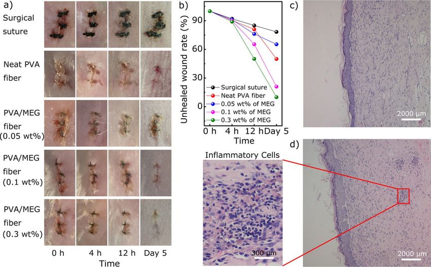
Minireviews
     ChemPhysChem                                 doi.org/10.1002/cphc.202000769

       gested to overcome or delaying the resistant development                  surfaces, nanocomposites and membranes. Herein, this review
 1
       ability of microbial cells.                                               article is an effort towards: (i) antimicrobial potential of
 2
            Graphene and its derivatives offer unique characteristics            graphene derivatives and understanding on mechanistic in-
 3
       such as, large surface area, high stability in physiological              sight, (ii) current advancement in translation of antimicrobial
 4
       environment, antimicrobial properties, good biocompatibility,             potential of graphene derivatives on surfaces, nanocomposite
 5
       easy-modification, and multifunctional behavior. It is obvious            surfaces and (iii) approach for understanding of biocompatibil-
 6
       that with these key multifunctional properties, graphene                  ity and biosafety. This review also discusses the strength and
 7
       derivatives have shown their potential and became favorite                weakness of graphene to be used or apply in biomedical
 8
       materials for many biomedical scientists, to design new                   devices to prevent the possibility of cross-infection and current
 9
       materials or hybrid materials or coatings of these nanomaterials          knowledge gap on mechanistic behavior of these materials on
10
       to existing biomedical devices to overcome with the possibility           antimicrobial activity and biocompatibility. Overall, numbers of
11
       of device associated infections. Since the first observation of           current challenge on the utilization of these materials in
12
       antimicrobial behavior of graphene oxide (GO) and reduced                 biomedical fields are highlighted. We hope that the roadmap
13
       graphene oxide (rGO) at 2010,[9] interest towards the antimicro-          provided here will inspire researchers to move towards the
14
       bial coatings of these materials or design of antimicrobial               development of graphene based antimicrobial surfaces or
15
       composites in biomedical field is rapidly emerging. As an initial         biomedical devices and towards an in-depth analysis of
16
       aspect, large number of studies focused into antimicrobial                antimicrobial assessment and their biocompatibility for their
17
       assessment of these materials. Most of the obtained results               future use in biomedical fields.
18
       clearly demonstrated the concentration dependent bacterio-
19
       static and bactericidal activity of these graphene
20
       derivatives.[10,11] Later physiochemical interaction of graphene
21                                                                               2. Antimicrobial Potential of Graphene
       derivatives with the microbial cells was demonstrated as
22
       fundamental mechanistic insight for the overall antimicrobial             Derivatives: Overview
23
       behavior.
24
            Unlike antimicrobial behavior of graphene derivatives in             Since the beginning of antimicrobial demonstration of gra-
25
       solution, there is controversy regarding the antimicrobial                phene derivatives, large scale of investigations has been carried
26
       efficiency of these materials on surfaces.[12,13] A number of             out to figure out their potential to be utilized in biomedical
27
       studies demonstrated the antimicrobial activity graphene                  fields. All the graphene derivatives including nanographite
28
       derivative coating, composites, membrane and paper like                   (GnP), monolayer graphene, GO and rGO, are well explored in
29
       structure.[9,14–16] However other studies did not observe signifi-        their different form, concentrations, purity and thickness of
30
       cant antimicrobial properties of graphene coated surfaces.[13,17]         materials. Among them, studies conducted on antimicrobial
31
       There are number of reports suggesting the variation in                   behavior of GO and rGO are significantly more advanced in
32
       antimicrobial ability with different graphene derivatives. Major-         compared to other derivatives. The first study showed strong
33
       ity of studies find GO as most effective by damaging the                  antimicrobial behavior of both GO and rGO by deactivating the
34
       microbial cells with mechanical interaction of sharp edges as             98.5 % and 90 % of Escherichia coli (E. coli) DH5α cells.[9]
35
       well as by the generating reactive oxygen species (ROS).[10,18]           Furthermore, restricted growth of E. coli was observed on
36
       Others found rGO in the form of nano walls as more effective              freestanding paper fabricated by using GO and rGO. Later Liu
37
       due to the higher hydrophobicity, sharp edges, charge transfer            et al.[10] compared the time and concentration dependent
38
       and its interaction with hydrophobic layer of lipid in microbial          antimicrobial activity of graphite, graphite oxide, GO and rGO
39
       cell surface.[19] Since the physiochemical interaction of graphene        dispersion against E. coli. The highest antibacterial activity was
40
       with microbial cells is key to exhibit antimicrobial activity,            observed with GO, which inactivated 69.3 � 6.1 % of bacterial
41
       graphene derivatives in solution apparently have higher affinity          cells in comparison to graphite, graphite oxide, and rGO where
42
       or dynamics to interact with free floating microbial cells thus           26.1 � 4.8 %, 15.0 � 3.7 % and 45.9 � 4.8 % of killing efficiency
43
       provide strong antimicrobial behavior. When it comes to                   were observed, respectively. Most of the bactericidal effect was
44
       coatings or development of antimicrobial surfaces or microbial            observed in the first four hour of interaction with these
45
       resistant biomedical devices, instant and continuous reaction of          graphene derivatives.[10] The generation of oxidative stress was
46
       surfaces with microbial cells is expected to either prevent the           suggested to play a major role on the antimicrobial activity of
47
       bacterial attachment, inhibit the microbial growth or damage              these materials. It was speculated that graphene materials,
48
       the microbial cells which come in contact to such surfaces.               which contain a higher density of functional groups, and are
49
       Hence it is crucial to maintain the material exposure, enough             smaller in size, have more chances to interact with bacterial
50
       roughness (height), density and distribution of graphene                  cells, resulting in cell disruption. The antimicrobial activity of
51
       derivatives to fully reflect or translate the similar antimicrobial       graphene nanosheets was observed even more effective than
52
       behavior to biomedical surfaces.                                          standard antibiotic kanamycin.[20] However, antimicrobial effi-
53
            In recent years, many reviews have been published mainly             ciency was varying with the different bacterial species.
54
       covering the antimicrobial properties of graphene materials and           Graphene nanosheets synthesized by hydrothermal method in
55
       fundamental underlying mechanisms. However, there are few                 alkaline condition exhibited excellent antibacterial activity
56
       reviews covering antimicrobial activity of these materials on             against E. coli, Salmonella typhimuirum (S. typhimuirum), Bacillus
57

       ChemPhysChem 2020, 21, 1 – 15    www.chemphyschem.org                 2              © 2020 The Authors. ChemPhysChem published by Wiley-VCH GmbH

                    These are not the final page numbers! ��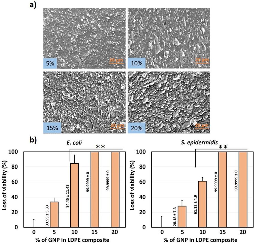
Minireviews
     ChemPhysChem                                  doi.org/10.1002/cphc.202000769

       subtilis (B. subtilis), and Enterococcus faecalis (E. faecalis). The       various mechanism including membrane stress, generation of
 1
       minimum inhibitory concentration (MIC) values of graphene                  oxidative stress, trapping behavior, basal plane and photo-
 2
       nanosheets against these bacteria are very low compared with               thermal activity. Fine and sharp edges of graphene derivatives
 3
       the MIC values of the standard antibiotic, suggesting that                 are proven to tear the bacterial cell membrane to inactivate the
 4
       graphene can be effectively used as an antibacterial agent.                bacterial cells by storming out of intracellular materials.[16,28] The
 5
       Toxicity of GO and rGO were also tested against the                        graphene derivatives mediated oxidative stress is generated via
 6
       Pseudomonas aeruginosa (P. aeruginosa) and detected the                    production of ROS, which is known to damage DNA, causes
 7
       strong inhibitory activity on growth and viability of cells due to         mitochondrial dysfunction followed by the deactivation of
 8
       the generation of reactive oxygen species, leading to cell death           bacterial cells. Moreover, when microbial cells get trapped in
 9
       through resulting nuclear fragmentation.[21] Later the toxicity of         graphene sheets, get totally isolated from the external environ-
10
       both GO and rGO were compared against E. coli and realized                 ment due to the blockage of gas/ion/nutrient exchange.[10,29]
11
       that GO produce more superoxide anions compared to rGO                     This trapping prevents the proliferation of microbial cells and
12
       leading more DNA fragmentation resulting higher toxicity.[22]              later loss the viability due to the lack of nutrient and respira-
13
       Afterwards rGO was tested against the few fungal species and               tional activity. With these proven excellent phenomena, recent
14
       found the strong inhibitory activity suggesting the antimicrobial          research activity is focusing more towards the development of
15
       activity of graphene derivatives regardless of type and species            composites by incorporating graphene materials with other
16
       of microorganisms.[23] A year later Tu et al, shed light on the            antimicrobial materials, polymers, functionalization with bio-
17
       underlying mechanism behind the physical disruption of                     active molecules not only to improve the antimicrobial activity
18
       bacterial cells after interaction with graphene sheets.[24] With           but also to stabilize the nanosheets which could be used later
19
       the computer simulation analysis, the interaction of graphene              to modify or develop antimicrobial biomedical surfaces. There
20
       sheet with lipid membrane of microbial cells and extraction of             are also efforts going on the method development and
21
       phospholipid was observed. These simulation results were                   optimization for coatings of graphene on the surfaces with
22
       further validated by using transmission electron microscopy                enhanced antimicrobial behavior.
23
       (TEM), where degradation of E. coli cell membrane can be seen
24
       clearly. Following the trend interaction of GO and rGO were
25
       evaluated against the copper resistant plant pathogens and                 3. Graphene-Based Antimicrobial Surfaces
26
       reveled the significant inhibition in growth and viability of
27
       bacterial by disruption of cell membrane and release of                    Due to the multidisciplinary nature of research on graphene
28
       cytoplasm.[25] Subsequently, the broad-spectrum antimicrobial              based antimicrobial surfaces we must make a few notes about
29
       activity of GO was demonstrated by evaluating the antimicro-               terminology and classification. In this section, by the term
30
       bial activity against the phytopathogen and fungal species.[26]            ‘nanocomposite’ and'composite’ we refer to materials consisting
31
       SEM imaging revealed the disruption of bacterial cells and                 of at least two phases, whereby one phase, i. e. nanoparticles or
32
       fungal spores suggesting the damage of membrane integrity of               particles, are included in bulk phases such as polymers, i. e.
33
       these cells by thin GO sheets via perturbation and trapping. The           matrix.[30] However, the term ‘nanocomposite’ is also used in
34
       broad-spectrum antimicrobial activity of GO was further dem-               several publications with an emphasis on the hybrid nature of
35
       onstrated by testing its bactericidal effect against dental                the nanoparticles, especially GO-Ag structures, rather than on
36
       pathogens including Streptococcus mutans (S. mutans), Fusobac-             their inclusion in a bulk phase.[31,32]
37
       terium nucleatum (F. nucleatum) and Porphyromonas gingivalis                    The tailoring of graphene based antimicrobial materials
38
       (P. gingivalis) causative agents of tooth decay and                        relies on the use of suitable procedures in order to obtain
39
       periodontitis.[27]                                                         structures that allow for the exploitation of graphene and its’
40
           In addition to physical perturbation, charge transfer is               derivatives’ antibacterial potential. Such antibacterial surfaces
41
       considered as other dynamics for the generation of oxidative               can be tailored as scaffolds and membranes, surface coatings or
42
       stress to bacterial cells by graphene materials. This was                  from bulk nanocomposites. Antibacterial surfaces can be
43
       demonstrated by Li et al., where monolayer graphene films                  obtained as e. g. paper, scaffolds and membranes from
44
       fabricated on conductive, semi conductive and non-conductive               dispersions using for example drying,[15,33] filtration,[34,35]
45
       substrates and their antimicrobial activity was examined against           lyophilization[36] or interfacial self-assembly.[37] Graphene and
46
       E. coli and Staphylococcus aureus (S. aureus).[12] Their results           graphene derivatives can be attached to other surfaces as
47
       showed that antibacterial activity of pristine graphene could              coatings for example by electrical and chemical deposition
48
       only be obtained when conducting substrate is beneath. The                 methods,[14–16,38–40] layer-by-layer assembly[41,42] or various surface
49
       electron transfer from bacterial cells membrane to graphene                spreading       methods.[43–48]       Finally,    gel          spinning,[49]
50                                                                                                  [50,51]                             [41,52,53]
       was proposed to damage bacterial cells rather than ROS                     electrospinning,          layer-by-layer assembly                or melt
51
       mediated cell disruption. This phenomenon remains controver-               extrusion[54] have been used to create graphene based anti-
52
       sial since a year later Dellieu et al., demonstrated that the              bacterial nanocomposites. The method of obtaining the anti-
53
       conductive character of the substrate has no influence on the              bacterial surfaces is essential as it dictates the extent to which
54
       viability of S. aureus and E. coli bacteria in contact with chemical       such surfaces can be tuned in terms of orientation and
55
       vapor deposition (CVD) graphene films.[13] Until now the                   distribution of the graphene and their upscaling potential.
56
       antimicrobial effect of graphene derivatives attributed via
57

       ChemPhysChem 2020, 21, 1 – 15    www.chemphyschem.org                  3                © 2020 The Authors. ChemPhysChem published by Wiley-VCH GmbH

                    These are not the final page numbers! ��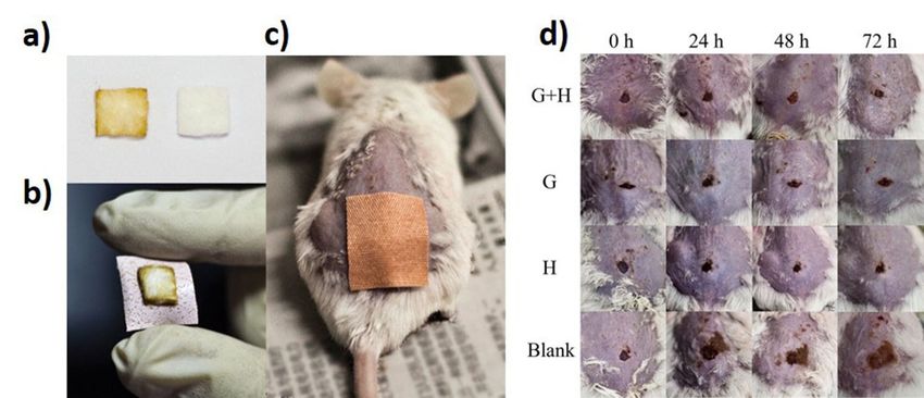
Minireviews
     ChemPhysChem                                 doi.org/10.1002/cphc.202000769

           In addition to the direct use of graphene derivatives, hybrid         sized with defined exposure, in terms of grafted polymer
 1
       nanomaterials are also getting attention in scientific community          coverage and functionality, and isoelectric points.[54] The
 2
       to obtain additive or synergistic antimicrobial activity. Graphene        synthesized materials had strong bactericidal activity via
 3
       based hybrid nanomaterials are mainly developed by using                  trapping of bacterial cells as well as piercing of cell membrane
 4
       nano silver, zinc, ferrous and other 2d materials, which are              due to nano knives effect suggesting that improvement in the
 5
       demonstrated to enhance the antimicrobial potential of                    solidity of agglomeration and orientation of nanomaterials
 6
       graphene based antimicrobial surfaces.                                    could enhance the antimicrobial activity.[57] Another study
 7
                                                                                 designed the strategy for the synthesis of a polycationic
 8
                                                                                 peptide functionalized graphene–silver hybrid nanoparticle
 9
       3.1. Graphene-Based Assembled Structures                                  structures. The nanocomposite was demonstrated to facilitate
10
                                                                                 enhanced biofilm inhibition and disruption properties to
11
       The assembly of graphene and graphene derivates into self-                eliminate the biofilm development of gram-negative bacteria.[32]
12
       sustaining structures in the form of scaffolds, paper and                 In another approach, GO sheets and functionalized rGO nano-
13
       membranes is a rather facile method for screening their                   sheets were self-assembled spontaneously onto solid titanium
14
       antibacterial properties. This is particularly important for              (Ti), using an evaporation-assisted electrostatic assembly proc-
15
       graphene derivatives where other particles are attached to e. g.          ess and a mussel-inspired one-pot assembly process. These
16
       GO. A graphene-based paper with antimicrobial behavior was                assemblies strongly mitigated bacterial adhesion and showed
17
       developed by Hu et al., suggesting the possibility of fabrication         considerable antibiofilm activities.[33] Zou et al developed the
18
       or modification of surfaces having antimicrobial potential by             wrinkled GO films by vacuum filtration of a GO suspension
19
       using graphene derivatives.[9] Following that, Pham et al.                through a prestrained filter. Where highly wrinkled GO surface
20
       produced graphene nanofilms with different edge lengths and               observed to bear bactericidal activity with contact-based
21
       different angles of orientation of the graphene sheets to                 mechanism.[34]
22
       compare the antimicrobial behavior with regards to morphol-
23
       ogy and orientation of nanosheets. These graphene nanofilms
24
       exhibited strong but variable bactericidal behavior against P.            3.2. Graphene-Based Surface Coatings
25
       aeruginosa and S. aureus. The density of the exposed edges of
26
       graphene was as a major parameter contributing to the                     The application of graphene-based coatings to arbitrary
27
       antibacterial behavior of graphene nanosheets. This suggested             surfaces has a significant potential for applications to biomed-
28
       that the bactericidal activity arises from the formation of pores         ical devices. The coating procedure varies depending on the
29
       in the bacterial cell wall, causing a subsequent osmotic                  substrate material, e. g. metals, polymers, and controlling the
30
       imbalance.[55] Furthermore, GO nanowalls reduced by hydrazine             morphology of the coatings is strongly dependent on the
31
       were more toxic to the bacteria than the unreduced GO                     application method.
32
       nanowalls due to the better charge transfer between the                        Polyethyleneimine grafted GO nanosheets incorporated Ni-
33
       bacteria and the more sharpened edges of the reduced                      P coatings on copper plates depicted to offer surface free
34
       nanowalls during contact. Another study provided the facile               energies to have significant influence on bacterial adhesion
35
       strategy to decorate AgNPs onto rGO by the simultaneous                   there by possibility of its use as a preventive measures of
36
       reduction of silver ions and graphene oxide nanosheets within             bacterial biofilm formation on the surface.[15] In a recent study,
37
       one system, and further to fabricate a dimension-adjustable               antimicrobial activity of GO- and rGO coated aluminum plates
38
       rGO/AgNP multi-layered film by a thermal-driven self-assembly             was tested against E. coli and revealed excellent antibacterial
39
       process.[37] Previously, titanate nanosheets were incorporated            efficacy of the rGO-coated surface. The intracellular ROS
40
       into rGO films to develop free standing hybrid films. This rGO            production was observed to play significant role in oxidative
41
       layered titanate films showed enhanced mechanical strength,               damage of the bacterial cells.[58] Graphene in the form of
42
       high surface roughness, chemical stability and hydrophobicity.            graphene nanowalls coated on stainless steel by electrophoretic
43
       The prepared composite films of rGO and metal oxide nano-                 deposition exhibited strong antimicrobial activity against both
44
       sheets exhibited excellent antimicrobial behavior in compared             gram positive and negative bacterial cells. The cell membrane
45
       to pure rGO film resulting in the complete sterilization of E. coli       damage of bacterial cells due to the direct contact with sharp
46
       O157:H7 ( � 100 %) in the very short time, i. e. 15 min.[56] The          edges of the nanowalls was clearly demonstrated as mechanism
47
       antimicrobial activity was observed mainly due to the irrever-            in the bacterial inactivation.[19]
48
       sible destruction of E. coli cells by sharp edges nanosheets.                  By understanding the interaction of graphene derivatives to
49
       Since rGO and copper both antimicrobial behaviors, rGO-copper             microbial cells, recent studies have been more focused towards
50
       free-standing films were synthesized to boost the biocidal                the practical applications of materials for antimicrobial coating
51
       activity. The nanocomposites were further co-deposited with               of surfaces and the development of self-cleaning membranes
52
       polydopamine (PDA) onto an ultrafiltration support and tested             and films. With the advanced designs, methods of fabrication
53
       for antimicrobial potential. The membrane exhibited a strong              and coatings have been proven for the development of devices
54
       antibacterial performance with a 97.9 % reduction in viability of         with superior antimicrobial efficiency. Previously GO with differ-
55
       E. coli.[35] In another study, different graphene derivatives
                                                                                                                                      *
                                                                                 ent oxidation, hydroxyl, and carbon radical ( C) levels were
56
       including zwitterionic graphene nanomaterials were synthe-                tested to figure out the role of functional group on antimicro-
57

       ChemPhysChem 2020, 21, 1 – 15    www.chemphyschem.org                 4              © 2020 The Authors. ChemPhysChem published by Wiley-VCH GmbH

                    These are not the final page numbers! ��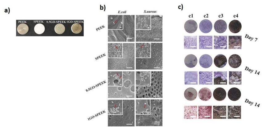
Minireviews
ChemPhysChem doi.org/10.1002/cphc.202000769
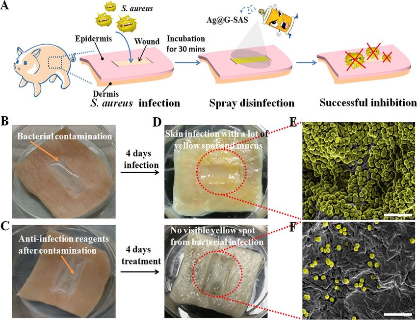
bial potential of this material and found that density of carbon catheter showed 2.2-fold of log reduction in the antimicrobial
1
radicals on hydrated GO (hGO) as a major factor to have strong resistant E. coli in compared to non-coated catheter, suggesting
2
antimicrobial effect with enhanced membrane binding and that, such coatings on biomedical devices could potentially be
3
induction of lipid peroxidation.[43] The hGO coated silicon used to deactivate drug resistant bacteria (Figure 1).[43] Yadhav
4
5
6
7
8
9
10
11
12
13
14
15
16
17
18
19
20
21
22
23
24
25
26
27
28
29
30
31
32
33
34
35
36
37
38
39
40
41
42
43
44
45
46
47
48
49
50
51
52
Figure 1. Inhibition of antibiotic resistant (AR) bacteria growth by noncovalently coated hydrated GO (hGO-2) films on a glass substrate. (A) AFM imaging of
53 hGO-2 coated substrates. A series of substrates (S-1, S-2, S-3, and S-4) with different GO coverage and thickness were characterized by AFM. (B) SEM imaging
54 of changes in the bacterial morphology after incubation with the hGO-2-coated substrate. (C) Visualization and (D) quantification of bacterial death on hGO-2
55 films by confocal microscopy. Following 6 h incubation of AR E. coli with substrates S-1 to S-4, the cells were stained with PI, fixed, and washed with 70 %
ethanol to determine the percentages of dead cells on substrates. The morphological changes of bacteria were visualized by SEM; *p < 0.05 compared to
56 control. Adapted from reference [43] with permission. Copyright 2018 American Chemical Society.
57
ChemPhysChem 2020, 21, 1 – 15 www.chemphyschem.org 5 © 2020 The Authors. ChemPhysChem published by Wiley-VCH GmbH
These are not the final page numbers! ��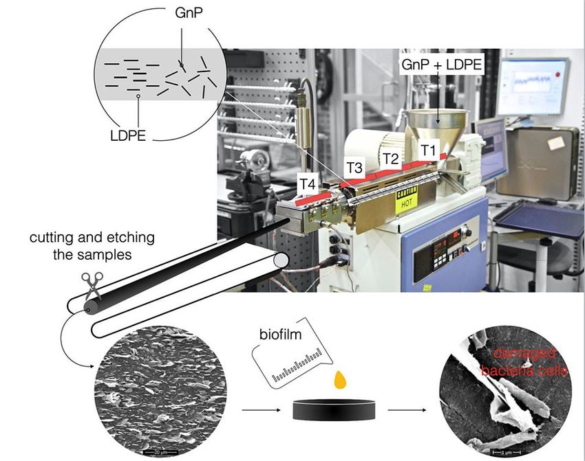
Minireviews
     ChemPhysChem                                 doi.org/10.1002/cphc.202000769

       et al., coated synthesized GO by in improved Hummers method               bling oppositely charged polyelectrolyte-stabilized rGO sheets
 1
       and coated to polymeric well plate and examined the                       (PEL-rGO) on a quartz substrate with the layer-by-layer (LBL)
 2
       antimicrobial potential. The coated well plates exhibited toxicity        technique.[62] The PEL-rGO surface rapidly generated localized
 3
       towards both gram positive and negative bacteria due to the               heating upon solar irradiation and damages > 90 % airborne
 4
       mechanical interaction as well as the generation of ROS.[44] GO           bacteria, including antibiotic-tolerant cells on contact. The
 5
       coatings on titanium surface was achieved with three kinds of             bactericidal effect is induced likely by permeabilizing their
 6
       combination types including drop with gravitational effects,              cellular membranes, suggesting the potential use of multilayer
 7
       electrostatic interaction and electrophoretic deposition. Ob-             PEL-rGO surface to be utilized on biomedical implant coatings
 8
       tained results showed that the combination of such coatings               to prevent the microbial colonization.[62] Northan et al, exam-
 9
       provides significant numbers of wrinkled areas with sharp                 ined the antimicrobial efficiency of rGO coatings on collagen
10
       edges in compared to single types of coating methods. With                scaffolds. The rGO coatings not only improved the mechanical
11
       the higher exposed sharp edges GO coatings effectively                    properties of collagen scaffold but also showed significant
12
       deactivated S. aureus and prevented the gathering of bacterial            inhibitory impact against growth and adhesion of E. coli, S.
13
       cells around the coated surface physical disruption as well as by         aureus and Streptococcus pyogenes (S. pyogenes). Going further,
14
       production of ROS.[38] Panda et al, demonstrated the antimicro-           scaffolds coated with rGO (400 μg/ml) were shown to enhance
15
       bial activity of natural shellac-derived GO coatings on metallic          the growth and proliferation of cardiomyocytes suggesting
16
       films, such as Zn, Ni, Sn, and steel via electron transfer                their potential use as a cardiac patch.[46] Choudhary et al.,
17
       mechanism and consequent ROS mediated oxidative stress to                 developed a rGO-protein nanoframework by using rGO synthe-
18
       the bacteria. It was proposed that a synchronous activity of GO           sized from a biosynthetic route. The developed coatings on
19
       acting as an electron pump and subsequent charge transfer                 glass substrates exhibited excellent antimicrobial activity by
20
       from cell membrane to functional oxygen groups on the surface             damaging 94 % of E. coli cells and prevented bacterial
21
       of GO induces ROS production. It was further speculated that              colonization. The bactericidal mechanism of rGO involved again
22
       the loss of cell membrane integrity is due to the electron                mechanical disruption of the cell membrane and later altering
23
       transfer at an initial stage and later compromises bacterial              transmembrane potential of the cells leading to leakage of
24
       metabolism and membrane structure eventually causing cell                 cytoplasmic materials.[47] rGO was also shown to induce
25
       death.[59] Coatings of silver/hydroxyapatite/graphene hybrid              oxidative stress through intracellular ROS production and
26
       particles on titanium surface exhibited strong antibacterial              inactivates respiratory chain dehydrogenases causing metabolic
27
       activity against S. aureus and E. coli after only 3 h of exposure,        imbalance in the cells. Arun et al, synthesized magnetic GO
28
       suggesting the potential of suppressing harmful biofilm for-              paint by incorporating cobalt ferrite and GO along with paint
29
       mation with no toxic effect on poly nuclear blood cells.[60] In           materials via high energy ball milling and deposited the mixture
30
       another study, a large CVD grown monolayer graphene covered               on a galvanized iron substrate, which was then subsequently
31
       silver nanowires thin film coating was developed, showing                 peeled off. The resulting hybrid coating film significantly
32
       broad spectrum antimicrobial activity by deactivating bacterial           inhibited growth and deactivated the E. coli and S. typhimurium
33
       and fungal cells. The strong antimicrobial behavior was                   cells. The fundamental antimicrobial activity was described by
34
       attributed to the sustained release of Ag + from the silver               corelating with generation of ROS followed by the mechanical
35
       nanowires due to graphene coverage.[39]                                   stress to bacterial cell membrane.[48] Zhao et al., prepared novel
36
            He et al., fabricated hemo-compatible and antibacterial              fabric materials by combining GO sheets onto cotton fabrics
37
       dual-layered polymeric membrane by coating a top layer of                 firmly by adsorption or by radiation-induced crosslinking using
38
       graphene oxide and sulfonated polyanion co-doped hydrogel                 triallyl isocyanurate (TAIC) as a crosslinker. The GO containing
39
       thin film on a bottom membrane substrate. The dual layered                fabrics showed significant antibacterial properties by inactivat-
40
       membrane exhibited significant antimicrobial activity against E.          ing 98 % of bacterial cells. This combined fabric materials were
41
       coli and S. aureus after in situ loading of silver-nanoparticles by       able to kill > 90 % bacteria even after being washed for 100
42
       maintaining the sustained release of silver ions.[45] Uniformly           times demonstrates the sustainable antimicrobial behavior of
43
       distributed silver nanoparticles GO nanosheets through in-situ            materials suggesting its potential to be used to coatings or
44
       reduction of Ag + subsequently wrapped with a thin layer of               production of self-sterilizing fabric based medical products and
45
       type I collagen observed to have synergistic antimicrobial                could potentially reduce the use of antibiotics on such
46
       activity on implant surface by rapidly deactivating the 96.3 %            products.[63] In another study, bioinspired polydopamine
47
       and 99.4 % E. coli and S. aureus respectively via physical                chemistry was used to fabricate GO-functionalized membranes
48
       interaction as well as light inspired photodynamic action.[61]            via coating and blending. GO coated membranes, where
49
       Another study demonstrated the enhanced antimicrobial                     nanosheets are externally exposed, exhibited enhanced biofoul-
50
       activity of rGO-hybridized zinc oxide electrochemically depos-            ing resistance with strong bactericidal effect compared to that
51
       ited thin film indium-doped tin oxide (ITO) glass substrate               of the GO-blended membrane. This emphasized the importance
52
       against the S. aureus with photoinactivation.[40] The rGO hybrid-         of GO exposure and interaction with microbial cells for
53
       ization with ZnO increased amount of oxygen vacancies cross-              antimicrobial behavior.[64] With the help of coated graphene
54
       ponding to higher concentration of active sites for ROS                   quantum dots (GQD) with low concentration of H2O2, wound
55
       formation was postulated as mechanism for antimicrobial                   healing band aids were prepared. The developed materials
56
       behavior. Hui et al developed antimicrobial surface by assem-             showed excellent antibacterial properties against both E. coli
57

       ChemPhysChem 2020, 21, 1 – 15    www.chemphyschem.org                 6              © 2020 The Authors. ChemPhysChem published by Wiley-VCH GmbH

                    These are not the final page numbers! ��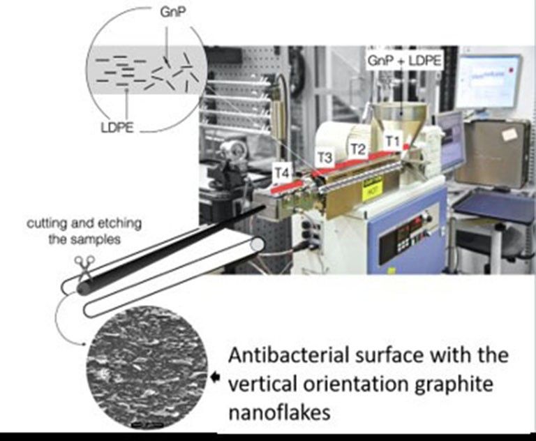
Minireviews
     ChemPhysChem                                        doi.org/10.1002/cphc.202000769

       and S. aureus in vitro and vivo with the assistance of low                        accelerate the proliferation and osteogenic differentiation of
 1
       concentration of H2O2 (Figure 2). The ability of GQD was                          osteoblast-like MG-63 cells, suggesting the potential use of GO
 2
       speculated to catalyze H2O2 into 3 OH to exhibit enhanced                         coatings on orthopedic implants to prevent the associated
 3
       bactericidal activity.[65] Recently, sulfonated polyetheretherke-                 infection as well as to enhance the bone integration (Fig-
 4
       tone (SPEEK) was coated with GO by using simple dip coating                       ure 3).[66]
 5
       method and found a close deposition of GO on the surface due                          In this framework, we initially tested the antimicrobial
 6
       to π-π stacking interaction between polyetheretherketone                          activity of GO coatings on the titanium surface, which was
 7
       (PEEK) and GO. The GO coatings were demonstrated to inhibit                       observed to deactivate E. coli.[14] Later we developed the
 8
       biofilm formation and excellent bactericidal efficiency against of                horizontal and vertical arrays of graphene coatings on metallic
 9
       E. coli and S. aureus. In addition, GO coatings were observed to                  and nonmetallic surfaces by using plasma enhanced chemical
10
11
12
13
14
15
16
17
18
19
20
21
22
23
24
25
26
27
28
29
       Figure 2. a) The cotton fabric absorbed with (left) and without (right) GQDs. (b) The obtained GQD-Band-Aid. c) The mouse treated with GQD-Band-Aid. d)
30
       Photographs of wound on the mice from the four groups at different times during the therapeutic process. G + H:H2O2 + GQD-Band-Aid group; G: GQD-Band-
31     Aid group; H: H2O2 + Blank-Band-Aid group; Blank: Saline + Blank-Band- Aid. Adapted from reference [65] with permission. Copyright 2014 American
32     Chemical Society.
33
34
35
36
37
38
39
40
41
42
43
44
45
46
47
48
49
50
51
52
53
54     Figure 3. a) The digital pictures of polyetheretherketone (PEEK), sulfonated PEEK (SPEEK), and GO-SPEEK samples. b) SEM images of bacteria on the substrates.
55     The red arrow shows the morphology of E. coli and S. aureus in high magnification. C) ALP staining and Alizarin Red S staining of different samples. c1) PEEK,
       c2) SPEEK, c3) 0.5GO-SPEEK, and c4) 1 GO-SPEEK. Adapted from reference [66] with permission. Copyright 2018 WILEY-VCH Verlag GmbH & Co. KGaA,
56     Weinheim.
57

       ChemPhysChem 2020, 21, 1 – 15         www.chemphyschem.org                    7               © 2020 The Authors. ChemPhysChem published by Wiley-VCH GmbH

                    These are not the final page numbers! ��
Minireviews
     ChemPhysChem                                       doi.org/10.1002/cphc.202000769

       vapor deposition (PE-CVD) method. The coatings of pristine                       Another nano-agent with strong antimicrobial activity was
 1
       CVD graphene observed to be neutral with bacteria similar to                     developed by using dopamine-conjugated polysaccharide
 2
       results obtained by Dellieu et al, neither deactivated the                       sulfate-anchored and -protected Ag-graphene. The synthesized
 3
       bacterial cells nor prevented the attachment.[13,16] By contrast,                nanocomposite exhibited robust antibacterial activity against
 4
       vertical arrays of graphene flakes showed pronounced killing                     both E. coli and S. aureus in vitro and demonstrated to inhibit S.
 5
       effect and effectively prevented both E. coli and Staphylococcus                 aureus infection on wounded pig skin without or with NIR laser
 6
       epidermidis (S. epidermidis) suggesting that the geometry of                     (Figure 4).[42]
 7
       graphene derivatives on the surface is key parameter deactivate
 8
       the microbial cells.[16] The vertical array of graphene was
 9
       observed to damage bacterial cells mainly with physical                          3.3. Graphene-Based Nanocomposites
10
       interaction and no ROS generation was detected. Similarly, Liu
11
       et al., explored the antimicrobial activity of GO based on the                   Polymer based graphene nanocomposites have substantial
12
       orientation, where vertically oriented GO flakes on composite                    potential applications due to the outstanding properties of
13
       surface showed enhanced antimicrobial activity in compared to                    graphene and versatility of the polymers.[30] We note here that
14
       non-oriented and randomly oriented GO flakes.[28] Both physical                  in the context of nanocomposites, monolayer graphene is rarely
15
       interaction with cells and ROS generation was realized as an                     used due to the difficulties in obtaining it in sufficient
16
       underlying mechanism for deactivation of bacterial cells. With                   quantities. Thus, graphene derivatives such as graphene nano-
17
       excellent antimicrobial behavior and enhancing osteogenic                        platelets, graphite nanoplatelets, graphene oxide and reduced
18
       differentiation, such coatings to biomedical implants could                      graphene oxide are most commonly used. Broadly speaking,
19
       provide essence benefit by preventing the possibility of cross                   the most common applications of graphene nanocomposites
20
       infections as well as by enhancing the osseointegration.                         are mainly directed towards mechanical reinforcement, elec-
21
22
23
24
25
26
27
28
29
30
31
32
33
34
35
36
37
38
39
40
41
42
43
44
45
46
47
48
49
50
51
52
53     Figure 4. (A) Procedures of S. aureus infection on pig skin and wound disinfection by spraying Ag@G-SAS (Ag @ graphene- sodium alginate sulfate). (B)
54     Photographs of the bacterial contamination procedure for sterilizing pig skin. (C) Photographs of the disinfection-reagent-treated wound after bacterial
55     contamination. (D) Photographs of morphologies of the infected wound and uninfected wound, respectively. (E, F) Typical scanning electron microscopy
       (SEM) images of the infected wound and uninfected wound, respectively. Scale bar: 10 μm. Adapted from reference [42] with permission. Copyright 2017
56     American Chemical Society.
57

       ChemPhysChem 2020, 21, 1 – 15        www.chemphyschem.org                    8               © 2020 The Authors. ChemPhysChem published by Wiley-VCH GmbH

                    These are not the final page numbers! ��
Minireviews
ChemPhysChem doi.org/10.1002/cphc.202000769
trical and thermal conductivity[30,67–69] and gas barrier potential to transform polymeric surfaces into antibacterial
1
properties.[70] For most of such applications, some the main surfaces, including on biomedical devices. The tailored sand-
2
challenges revolve around achieving maximal e. g. mechanical, wich composite showed great bactericidal activity and potential
3
electrical and thermal, improvements with a minimal amount of for biofilms inhibition.[74] In addition, GO-poly(methyl methacry-
4
filler and controlling the morphology, e. g. orientation for gas late) (PMMA) nanofibers prepared by pressurized gyration also
5
barrier properties. demonstrated to reduce the viability of E. coli in compared to
6
Yan et al developed the spray mediated assembly of a bio- PMMA nanofibers.[75] Nanocomposites produced by dispersing
7
inspired Ag at reduced graphene-sodium alginate nanocompo- mechanically exfoliated graphene into a poly vinyl alcohol
8
site film for effective wound healing. The composite film matrix and then formed into fibers via gel spinning exhibited
9
effectively inactivated the P. aeruginosa, E. coli and Candida antimicrobial behavior and were found to be biocompatible.
10
albicans (C. albicans) demonstrating the ability of protecting Furthermore, the wounds treated with this type of fibers with
11
wound from pathogenic microbial infections. This was shown to 0.3 wt % graphene showed improved wound healing after the
12
promote the recovery of wound sites in rats, suggesting the 5 days of surgery (Figure 5).[49] Composites containing graphene
13
potential use of such composite films for wound healing oxide and quaternary ammonium salt also exhibited antimicro-
14
application.[52] The GO/benzylpenicillin/Mg–Al layered double- bial activity while being biocompatible to mammalian cells in
15
hydroxide hybrid composite film system prepared via solvent both in vitro and in vivo analysis (Figure 5). The composites
16
evaporation, was shown to maintain the sustained release of demonstrated the ability to deactivate bacterial cells synergisti-
17
antibiotics to achieve enhanced antimicrobial activity suggest- cally with mechanical stress, membrane perforation and gen-
18
ing the possible use of graphene derivative composites for erating oxidative stress. The composite was also shown to
19
efficient delivery of antibiotics/antimicrobial agents.[53] In anoth- eradicate multidrug-resistant bacteria more effectively than
20
er work, poly-lactic acid (PLA) was loaded with cellulose conventional antibiotics and rapid wound healing in vivo.[76]
21
nanocrystal and rGO as reinforcing nanofillers and a membrane Recently, polyurethane and cellulose acetate and GO/Ag
22
was produced through solution casting. The nanocomposite electrospun nanocomposite nanofibrous scaffold mats were
23
membrane shown to have antibacterial potential against S. shown to gain strong antimicrobial activity. The hybrid
24
aureus and E. coli while being non-toxic to NIH-3T3 cells.[71] composite mats were able to deactivate bacterial cells by
25
Recently, multilayer nanofilms were fabricated using a layer by- contact killing. Additionally, the addition of curcumin was found
26
layer technique (LBL) with alternative deposition of GO and to enhance antimicrobial activity by inactivating 95 % and
27
lysozyme (Lys) as a novel coating strategy. These nanofilms 100 % of gram positive and gram-negative bacteria respectively.
28
with Lys as the outmost layer exhibited stronger antibacterial The hybrid composite mats also demonstrated the ability to
29
ability against S. aureus and E. coli with enhancing osteogenic significantly promote the wound healing process and regener-
30
differentiation of dental pulp stem cells, through combing the ation of the epidermis layer supporting the wound healing
31
strong bacterial property of Lys and osteogenic profile of GO.[41] properties of graphene derivatives.[51]
32
In order to develop wound healing patches Lu et al., used Recently, we developed the method to prepare antibacterial
33
electrospinning to prepare chitosan–PVA nanofibers containing surfaces from highly structured highly filled polymer nano-
34
graphene. Graphene here was used mainly to obtain the composites based on graphite nanoplatelets[54] which showed
35
antimicrobial behavior in composite. Indeed, the composite strong bactericidal activity by eliminating 99.99 % of bacteria.
36
patches were found effective to inhibit E. coli, Agrobacterium, One should highlight the antimicrobial activity achieved by the
37
yeast and enhanced the wound healing rate.[50] In another study graphite nanoplatelets (GNP) and low-density polyethylene
38
nanocomposite was developed by using polyethyleneimine (LDPE) composite is several orders of magnitude stronger than
39
(PEI)-modified and AgNP-decorated GO. The nanocomposite any previously reported using pristine graphene, graphene
40
acquired excellent stability in physiological solutions and oxide, and reduced graphene oxide, used either as a coating or
41
electropositivity, showing substantially higher antimicrobial in solution.[14,19,55] As previously mentioned, the processing stage
42
efficacy. The sustainability in antimicrobial efficiency of compo- is crucial for obtaining a microstructure with desired properties
43
site was established by preserving > 99 % efficiency against in polymer nanocomposites. In the previous research[28] it has
44
Gram-negative bacteria, and > 95 % efficiency against Gram- been observed that vertical arrays of GO flakes showed
45
positive bacteria and fungi even after 1-week storage.[36] pronounced killing effect. Therefore, we have selected extrusion
46
Furthermore, graphene-polyindole nanocomposite were dem- as a processing method from which one can benefit in
47
onstrated to enhance the antimicrobial potential even against achieving alignment of the GNP flakes along the polymer flow
48
methicillin resistant S. aureus with minimal toxicity towards direction as well as deagglomeration, and efficient dispersion of
49
mammalian cells. The composite demonstrated to heal S. aureus particles.[77,78] It’s worth to highlight that extrusion is one of the
50
associated skin infection in mice.[72] In another study, GO most common processing techniques used in the plastic
51
nanosheets were immobilized to the surface of a polyamide industry. Moreover, selected filler in comparison to lab-scale
52
thin film composite forward osmosis membranes. The coatings produced CVD graphene is easy to manufacture in an
53
of these membrane significantly mitigated biofouling with economically beneficial and scalable process based on prelimi-
54
synergistic antibacterial properties (99.9 %) in compared to nary orientation studies.[77,78] Previous studies by authors
55
pristine membrane.[73] A recent study developed laser induced revealed also other interesting physical properties that these
56
graphene composite as multifunctional surfaces, with great nanocomposites with aligned GNPs exhibit as: gas barrier
57
ChemPhysChem 2020, 21, 1 – 15 www.chemphyschem.org 9 © 2020 The Authors. ChemPhysChem published by Wiley-VCH GmbH
These are not the final page numbers! ��Minireviews
ChemPhysChem doi.org/10.1002/cphc.202000769
1
2
3
4
5
6
7
8
9
10
11
12
13
14
15
16
17
18
19
20
21
22
23
24
25 Figure 5. Wound healing evaluation of mice model. a) Representative wounds at different times after surgery, b) unhealed wound rate in five groups during 5
26 postoperative days. c) Histological examination of nanocomposite fiber with 0.3 wt % of MEG-treated group and (d) common surgical suture-treated group.
Adapted from reference [49] with permission. Copyright 2018 American Chemical Society.
27
28
29
30
properties,[79,80] enhanced mechanical performance,[81] or aniso- matic conditions (i. e., apparent shear rate see) were applied.[54]
31
tropic electrical and thermal conductivity.[78,81] A schematic of The polymer matrix with the higher viscosity exhibited more
32
the manufacturing process is shown on Figure 6, with more consistent deagglomeration and filler orientation and for that
33
details found in our previous study.[54] Moreover, the exper- reason was chosen for further study. the density of the exposed
34
imental trials have included two types of LDPE, differing in edges of graphene of is the most important parameters
35
viscosity by around one order of magnitude. The same kine- contributing to the antibacterial behavior of graphene
36
nanosheets.[55] The antimicrobial activity of the nanocomposite
37
surfaces was found to be strongly dependent on the density
38
and orientation of sharp edges of graphite nanoplatelets
39
(Figure 7).[54] Overall, the results have the potential as low-cost
40
and straightforward mass production method of GNP–polymer
41
nanocomposites with outstanding antibacterial properties. We
42
note that due to the unique the manufacturing strategy, the
43
low material costs and overall results, the developed nano-
44
composites could constitute an entirely new class of high-
45
performance antibacterial surfaces.
46
Based on the structural analogy with graphene-like materi-
47
als, boron nitride (BN) and its derivatives were investigated for
48
their potential antibacterial applications.[82] Boron nitride (BN)
49
exhibits a honeycomb structure similar to graphene, with
50
alternating boron and nitrogen atoms. The results have shown
51
that BN nanoflakes on the extruded BN-LDPE composite interact
52
with the bacterial cell membrane as anticipated, to cell damage
53
and rupture. Significant loss in viability of E. coli, S. aureus and S.
54
Figure 6. Schematic diagram for the method of extrusion process of graphite epidermidis was observed compared to the pure LDPE control
55
nanoplatelets (GNP) -LDPE composite with the vertically oriented graphite surface. However, in comparison to GNP-LDPE nanocomposites
56 nanoflakes on the surface. the antibacterial effect of BN-LDPE nanocomposites is weaker.
57
ChemPhysChem 2020, 21, 1 – 15 www.chemphyschem.org 10 © 2020 The Authors. ChemPhysChem published by Wiley-VCH GmbH
These are not the final page numbers! ��Minireviews
ChemPhysChem doi.org/10.1002/cphc.202000769
1
2
3
4
5
6
7
8
9
10
11
12
13
14
15
16
17
18
19
20
21
22
23
24
25
26
27
28
29
30
31
32
33
34
35
36
37
Figure 7. a) SEM images demonstrating the surface morphology of extruded GNP–LDPE polymer composite, b) Loss of viability of E. coli and S. epidermidis by
38 the longitudinal section of GNP–LDPE composite materials compared to control (LDPE). Adapted from reference [54] with permission. Copyright 2020 WILEY-
39 VCH Verlag GmbH & Co. KGaA, Weinheim.
40
41
42
As we noticed BN has higher tendency to agglomerate during as large surface area, mechanical stability and conductivity is
43
processing and as an effect poor orientation of this filler is believed to endorse the possibility of enhanced extracellular
44
achieved. protein adhesion facilitating cell growth and proliferation.[14,89,90]
45
Graphene derivatives are demonstrated as biocompatible
46
materials by majority of studies and even suggested the
47
potential use of materials in tissue engineering.[91–93] Results
48 4. Current Understanding on Biocompatibility obtained from in vitro and in vivo suggested enhanced growth
49
and Biosafety and proliferation of mammalian cells resulting the acceleration
50
of bone integration, wound healing and tissue
51
The materials must be biocompatible to be used in biomedical development.[49,76,94] In contrast, other studies found adverse
52
devices not only being neutral to the human cells but also be effect of these materials such as cytotoxicity, inflammatory cell
53
able to cope with in vivo environment and enhanced functional recruitment and tissue fibrosis suggesting that detail inves-
54
integration to host tissue.[83,84] As like antimicrobial behavior, tigation of biocompatibility should be revealed before using
55
many studies have evaluated the biocompatibility of graphene such materials in biomedicine either as device coatings or drug
56
derivatives.[85–88] The characteristics of graphene materials such delivery.[95,96] The biocompatibility of nanomaterials is generally
57
ChemPhysChem 2020, 21, 1 – 15 www.chemphyschem.org 11 © 2020 The Authors. ChemPhysChem published by Wiley-VCH GmbH
These are not the final page numbers! ��Minireviews
ChemPhysChem doi.org/10.1002/cphc.202000769
dependent with the type of materials, size, shape, chemical biomedical devices. Furthermore, the release of graphene
1
content, thickness and charge particles. Being 2D material particles from such surfaces could be to toxic to other cells in
2
biocompatibility of graphene derivatives have shown to differ surrounding microenvironment or even show systemic toxicity.
3
with the types (pristine graphene, GO and rGO), number of The possibility of particle release should be taken seriously
4
layers, dynamics of orientation and carbon-to oxygen atomic while developing graphene based antimicrobial surfaces.
5
ratio. Moreover, any modification and functionalization of these Despite having advancement in development of graphene
6
materials for any macroscale applications might have different based antimicrobial surfaces, still their knowledge gap on the
7
physiochemical properties in compared to original state and how graphene derivatives show antimicrobial behavior and
8
therefore offer distinct biocompatible behavior.[96] Indeed, biocompatibility in molecular level. Which is due to the lack of
9
experimental evidence suggests the distinct interaction with studies covering detail investigation of changes in molecular
10
mammalian cells have been observed with different graphene dynamics in the bacterial as well as mammalian cells up on
11
derivatives, morphology and orientation of materials and size of exposure to graphene-based surfaces. Yet there are not enough
12
the particles. To address this each type and modification of reports for translation of in vitro antimicrobial activity of
13
materials should be tested for the biocompatibility in order to graphene-based surfaces to in vivo environment. Hence future
14
confirm the no adverse effect towards exposed mammalian studies needed to be focused on the in vivo utilization of such
15
cells, tissues and organs. Most of the studies regarding the graphene based antimicrobial surfaces to revel the actual
16
biocompatibility of graphene derivatives mainly evaluated by in vivo antimicrobial performance and biocompatibility.
17
viability of cells after the few hours to few days of interactions
18
with the materials, which hardly response to question of long-
19
term toxicity. Currently there is knowledge gap on the changes Acknowledgements
20
in molecular dynamics of host cells upon the exposure to
21
graphene derivatives to define or predict the biocompatibility This work was supported by grants from SIO-Grafen, a joint
22
or toxicity in the case of long-term exposure. There are very few investment of VINNOVA, Formas, and Energimyndigheten, Formas
23
reports on the biocompatibility of these materials with respect and ÅForsk to RK and IM. Novo Nordisk Foundation grant
24
to comparison in types of materials, size, and orientation NNF10CC1016517 and Independent Research Fund Denmark –
25
dynamics. Hence still there is no conclusive remark regarding FTP to IM.
26
the biocompatibility of graphene derivatives.
27
Graphene based antimicrobial surfaces are either developed
28
by coatings, impregnation and composites matrices. The Conflict of Interest
29
biocompatibility of such surfaces is mostly being examined by
30
growing mammalian cells, which possibly be as non-reactive or The authors declare no conflict of interest.
31
even enhance the proliferation of cells. However, possibility of
32
nanomaterials release from the such surfaces cannot be Keywords: antimicrobial · biocompatibility · biofilms ·
33
neglected and is the major raising concern. Even though, biomedical devices · graphene derivatives
34
developed antimicrobial surfaces might show biocompatibility
35
in local area, once the nanomaterials release from the surfaces
36
might show systemic toxicity to various organ and functional
37
systems. Hence the possibility of graphene-based materials
38
release should be examined carefully while developing anti- [1] World Health Organization. Health care associated infections Fact sheet.
39 World Health Organization 2015, 4.
microbial surfaces for biomedical applications.
40 [2] J. S. VanEpps, J. G. Younger, Shock 2016, 46, 597–608.
41 [3] R. A. Weinstein, R.O Darouiche, Clin. Infect. Dis. 2001, 33, 1567–1572.
[4] H. E. Karahan, C. Wiraja, C. Xu, J. Wei, Y. Wang, L. Wang, F. Liu, Y. Chen,
42
5. Future Perspectives Adv. Healthcare Mater. 2018, 7, e1701406.
43 [5] J. Fong, F. Wood, Int. J. Nanomed. 2006, 1, 441.
44 [6] A. M. Allahverdiyev, K. V. Kon, E. S. Abamor, M. Bagirova, M. Rafailovich,
The orientation dynamics and exposure of graphene derivatives Expert Rev. Anti-Infect. Ther. 2011, 9, 1035.
45 [7] D. E. Marx, D. J. Barillo, Burns 2014, 40, S9.
on the surfaces are key parameter to obtain the enhanced
46 [8] B. H. Mao, Z. Y. Chen, Y. J. Wang, S. J. Yan, Sci. Rep. 2018, 8, 2445.
antimicrobial and anti-biofouling efficiency. However, a major [9] W. Hu, C. Peng, W. Luo, M. Lv, X. Li, D. Li, Q. Huang, C. Fan, ACS Nano
47
challenge is to obtain the desired density of oriented graphene 2010, 4, 4317.
48 [10] S. Liu, T. H. Zeng, M. Hofmann, E. Burcombe, J. Wei, R. Jiang, J. Kong, Y.
flakes on the surface to efficiently inhibit the biofilm formation.
49 Chen, ACS Nano 2011, 5, 6971–6980.
Although few methods have been applied to achieve the right [11] W. Sun, F. G. Wu, Chem. Asian J. 2018, 13, 3378–3410.
50
orientation and density of exposed graphene flakes, these [12] J. Li, G. Wang, H. Zhu, M. Zhang, X. Zheng, Z. Di, X. Liu, X. Wang, Sci.
51 Rep. 2014, 4, 4359.
existing methods have several drawbacks and cannot be
52 [13] L. Dellieu, E. Lawarée, N. Reckinger, C. Didembourg, J.-J. Letesson, M.
applied to develop coatings or surfaces of arbitrary shapes that Sarrazin, O. Deparis, J.-Y. Matroule, J.-F. Colomer, Carbon 2015, 84, 310–
53
could be readily applied to all biomedical devices. Hence, there 316.
54 [14] C. Zhao, S. Pandit, Y. Fu, I. Mijakovic, A. Jesorka, J. Liu, RSC Adv. 2016, 6,
is a need for the development of simple and scalable methods
55 38124.
which could be used to create arbitrary surfaces containing [15] W. Shao, J. Wu, H. Liu, G. Dong, S. Wang, H. Min, M. Huang, RSC Adv.
56
vertically aligned graphene flakes on different types of 2016, 6, 46270–46277.
57
ChemPhysChem 2020, 21, 1 – 15 www.chemphyschem.org 12 © 2020 The Authors. ChemPhysChem published by Wiley-VCH GmbH
These are not the final page numbers! ��You can also read