FNAC IN DIAGNOSIS OF BREAST LESION (FOR PUBLICATION )
←
→
Page content transcription
If your browser does not render page correctly, please read the page content below
FNAC IN DIAGNOSIS OF BREAST LESION (FOR PUBLICATION )
 DR. BIJALI CHAKRABARTI , Assistant Professor of Pathology ,DR.HIMAXI RATHOD, Junior
Lecturer of Pathology, DR. J. M. SHAH , Prof & HOD Of Pathology Department.
: DR.HIMAXI RATHOD email id himaxi27rathod@gmail.com
PATHOLOGY DEPARTMENT, AMC MET MEDICAL COLLEGE, L. G. HOSPITAL
MANINAGAR, AHMEDABAD

ABSTRACT:

INTRODUCTION: Breast carcinoma is the leading most common malignant tumor and leading
cause of carcinoma death with more than 1000000 cases occurring world wide annually 1. FNAC
(Fine Needle Aspiration Cytology) has become widely accepted as a reliable diagnostic tool for
diagnosis of breast masses3.The aim of the study was to classify Breast lesions & correlate with
histopathology Report.

MATERIALS & METHOD: This was a retrospective observational study done over a period
of one year (Jan. 17- Dec. 17) in Cytology Division of Department of Pathology of AMC MET
Medical College at L.G.Hospital Campus ,a tertiary Health Care located in Maninagar
Ahmedabad .Of total 1104 FNAC were done in the department among these 215 were Breast
Lesions. Breast lesions were categorized into Inflammatory, Benign with No Risk, Benign with
moderate Risk, Suspicious & Malignant.

RESULTS: The Maximum number of cases was in the age group of 18-30 in Benign Breast
Lesion. Malignant Breast Lesion was found in the age group of above 41.

CONCLUSION: FNAC is minimally invasive,produces speedy results and is inexpensive.

KEY WORDS: Breast Lesions, Fine Needle Aispiration, Cytology, Fibroadenoma.

INTRODUCTION :
Breast carcinoma is the leading most common malignant tumor and leading cause of carcinoma
death with more than 1000000 cases occurring world wide annually 1.It is the second most
common cancer in women after cervical cancer. The most important risk factor is a history of
breast cancer in a close relative 2. The risk of breast cancer is minimally increased in women
who take hormonal contraceptives but risk is no longer present 10 years after cessation of
medication2. By 2020 breast cancer is set to overtake cervical cancer as most common type of
cancer among all women in india3. FNAC (Fine Needle Aspiration Cytology) has become widely
accepted as a reliable diagnostic tool for diagnosis of breast masses3. FNA can be performed
with only a needle or with a needle syringe. It is least expensive method of diagnosis. As FNAC
does not require anesthesia or hospitalization and it takes only few minutes to perform , A
preliminary judgement as to adequacy of sample and in many instances diagnosis can be done in
minutes thus alleviating anxiety that woman inevitably experiences when informed that she has a
mammary lesion. Thus FNA may save anxiety , trauma and money. FNA is particularly valuable
when the level of clinical suspicion is low2. A significant advantage of FNB is low cost and the
ability to render a diagnosis to the clinician and patient at the time of procedure thus allowing
treatment decision to be made immediately4. Limitation of aspiration cytology : FNAC is highly
reliable for the diagnosis of cancer .If however the FNAC is judged to be atypical or suspicious
the procedure should be repeated or another opinion should be sought or the lesion should be
excised for histological examination5,6,7.
The aim of scheme Proposed by wang and Ducatman is to categorise a lesion according to
likelihood of being a carcinoma on basis of FNA findings rather than predict precise histological
diagnosis8. Early Diagnosis helps to prevent patient discomfort and anxiety9,10,11.

The aim of the study was to:
1) Find out various causes of Breast Lesion.
2) To classify the FNAC findings into cytological Categories – Inflammatory ,Benign Lesion
with No Risk of Cancer,Benign Lesion with Mild to Moderate Risk of Cancer, Suspicious for
Malignancy and Malignant.
3) To Compare the result of FNAC with Histopathology report of same Patient.

Materials & Method: This was a retrospective observational study done over a period of one
year (Jan. 17- Dec. 17) in Cytology Division of Department of Pathology of AMC MET
Medical College at L.G.Hospital Campus ,a tertiary Health Care located in Maninagar
Ahmedabad .Of total 1104 FNAC were done in the department among these 215 were Breast
Lesions. A detailed clinical history of each patient regarding age, sex, chief Complains physical
examination of Breast was carried out.USG Reports & Mammography repots were recorded.
Axillary L ymph node were palpated for enlargement. Written informed Consent of each patient
was taken & Fine Needle aspiration was done with 22 gauage Needle & 10 cc.Of
total1104FNAC, 215 were Breast Lesions. Both Females & Males were included in the study, 54
patients had Follow up excision biopsy or lumpectomy or Mastectomy done at out institution.
Wet fixed smears were stained with Haemotoxylin & eosin stain.

III OBSERVATIONS AND RESULTS
Of total 1104 FNAC, 215Breast FNACs cases were collected along with Clinical History &
Radiological findings .Histo Pathological Correlation was found in 54 Cases.

1) Out of 215 Breast lesions, 9 were Males patients Rest 206 were females.

2) 6 Cases were Bilateral.

3) Histopathological correlation was found in 54 Cases.

4) The Maximum number of cases was in the age group of 18-40 in Benign Breast Lesion
Which is comparable to the study of shrestha aet al (21-30Yrs) & puja et al (31-40) yrs.

5) Malignant Breast Lesion was found in the age group of above 41. (range 41-70years)
Which was comparable to Shresth aet al (41-50) and
Puja aet al (41-50).

Most Common Breast lesions was Fibroadenoma (32%) Followed by Proliferative lesion of
Breast (22.5%)
Ductal Carcinoma was found in (22%) Cases.
Table I

 Diagnosis in 215 Breast FNAC

Breast Lesion                                                No. of cases
A. Benign Lesions with no risk of Cancer
 I. Unsatisfactory                                                5
II. Inflammatory Breast Lesion
Mastitis                                                         16
Abscess                                                          12
 III. Non Proliferative Breast Disease
      Fibrocystic change & Simple cyst                           03
Mild epithelial Hyperplasia                                      01
IV Miscallaneous
Lactational change/ Galactocele                                  03
Gynecomastia                                                     13
B. Benign Lesion with Mild Moderate risk of Cancer.
 I. Proliferative breast disease without atypia .
Epithelial Hyperplasia, Moderate                                 01
Papilloma                                                        01
Fibroadenoma                                                     64
Phylloid                                                         01
 II. Proliferative Breast disease with atypia but Benign         45
C. Suspicious & Malignant
Suspicious & Malignancy                                          06
Ductal Carcinoma                                                 44

 Table II

                   Sex             No
                   Male             9
                  Female           206

 Table III Age Distrubution

                Age            Benign           Malignancy
             < 20 years          11                  -
           Up to 20 years        45                  -
            21-30 years          57                 02
            31-40 years          36                 09
               41-50             19                 13
               51-60              3                 07
                >61               -                 13
Table IV FNAC and Histo Correlation.
Sr. Breast lessions FNAC Histopathology
No.
1 Positive 44 22
2 Suspicious 6 5
3 PBD with atypia 45 10
4 Fibroadenoma 64 14
5 Phylloid 1 1
6 Gynecomastia 13 2
Table V
FNAC HISTOPATHOLOGY
Diagnosis No of Cases Diagnosis No of Cases
Ductal Carcinoma 22 Ductal Carcinoma 22
ProliferativeBreast 10 ProliferativeBreast disease 8
disease with atypia with atypia
Tubular adenoma 1
Ductal carcinoma 1
Suspicious 5 Ductal Carcinoma 5
Phylloid tumour 1 Phylloid tumour 1
Gynecomastia 2 Gynecomastia 2
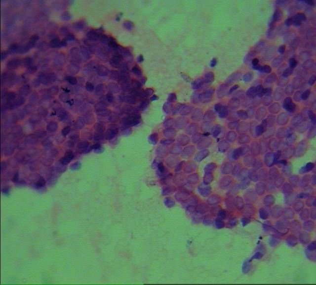
Fibroadenoma 14 Fibroadenoma 14FNAC IMAGES Fibroadenoma low power 10x
Fibroadenoma High power (40x)
Ductal carcinoma low power(10x)
Ductal Carcinoma High power(40x)
Discussion:

Breast Cancer is the commonest Cancer of Urban women and second commonest in the Rural
women next to cervical cancer. Owing to lack of awareness of this disease and in absence of
Breast Cancer Screening Programme,the majority of Breast Cancer are diagnosed at a relatively
advanced stage11,12.We had 44 cases of Ductal Carcinoma, Out of which 19 were in age group
of41-50,3inage group of 51-60 &17 patients above 61 years.
During the study the Breast lesions were classified as Inflammatory,Benign Lesion with No Risk
of Cancer,Benign Lesion with Mild to Moderate Risk of Cancer,Suspicious for Malignancy and
Malignant.
Following observations were made
 1) In present study,.02% cases were unsatisfactory for reporting.4.1% patients were males and
95.8% patients were females.

2) In the present study, Fibroadenoma is the commonest Benign Breast lesion followed by
Benign Proliferative lesion of Breast which is comparable to study by Shrestha et al.
Benign Breast lesion was common in the age group of 21- 40 which is comparable to the study
by Shrestha et al12(21-30 years) and Jarwani Puja2 et al (31-40 years).

3) Invasive Duct Carcinoma was commonest Malignant Breast lesion in the age group above 41
years which was comparable to Shrestha et al12 .
 4) In Comparitive Analysis of FNAC and Histopathology, Out of 6 Cases of FNAC suspicious
for Malignancy,5 were positive for Malignancy, 1 did not turn up.

5) Out of 44 Malignant cases in FNAC ,22 were confirmed Malignant Histopathologically in our
Institute.

6)2 Cases were Reported as false negative, 1 reported as Benign Proliferative leision with
Epithelial Hyperplasia turned out to be Malignant, & another Reported as Benign Proliferative
lesion turned out to be Tubular Adenoma.

CONCLUSION: This study like other studies also suggests that diagnosis of atypia is clinically
significant because it is associated with increased likelihood of Malignancy & such cases should
be evaluated for Histology511.FNAC Breast is low cost and the ability to render a diagnosis to the
clinician and patient at the time of procedure thus allowing treatment to be made
immediately.FNAC is minimally invasive produces speedy results and is inexpensive. All cases
of Proliferative lesion of Breast with atypia and suspicious Malignant cases should be followed
by histopathological examination.

 12. REFERENCES:
1Rosai and Ackermans, Surgical Pathology Juan Rosai M.D,Ninth edition, vloume2, P-1787.

2.KOSS Diagnostic Cytology and Iis Histopathologic basis, Vol II,Editor, Leopold,D, Koss.
CoEditor: Myron R Melame D,Lippincott Williams &Wikins,P-1083.

3.Puja Jarwani,Daxita C Patel,Shantibhai M Patel,Anupama Dayal. Fine Needle Aispiration
Cytology in Palpable Breast Lump GCSMC.J.MedSCI VOL(II) July –December 2013,P-12to16.

4.Joan Can Giarella & Aylin Simsir,Orell Sterett’s FNAC, 5th edition, P-158.
5.AyatG.,Abu-JawdehGM.,Fraser       J.L.,       GarciaLW.,UptonMP.,WangHH.,-Accuracy
&Consistency in Applicationof Probabilistic approach toreporting fine Needle aispiration
ActaCytol.47(6):973-978,2003.

6.OzakaraS.K.,UstunMO,       PaksoyN.,The       grey     zone     inbreast   fine     Needle
aispirationCytology.How to repot on it.ActaCytol 46(3):513-518,2002.

7.Amrish N. Pandya, Neelam P. Shah, Breast Fine Needle Aspiration Cytology, A study of
Application of Probabilistic Approach, Indian Medical Gazette, February 2013, pg 54-59

   8.Wang and Ducatman.B.S.,Fine Needle Aispiration of the Breast; a probabilistic approach
   to the diagnosis of carcinoma, Acta Cytol.42(2);285-289,1998.

9.Hughes J E,RoyleGT,BuchananR,TaylorI,Depression and social stress among patients with
beningn Breast lesion.Br K .Surgery 1986,73;997-9.

10.EllmanR, Angeli.N.Moss.S.Chamberlain.J,WaguireP.,Psychiatrc Morbidity associated with
screening of Breast Cancer. Br .J.Cancer 1989,60;781-4.

   11.Agarwal G. Ramakant P. – Breast Cancer in India The Current Scerlario and The
   Challenges for the future, Breast Care, 3 (1) : 21-27,2008.

12.Fine Needle aspiration Cytology in a palpable breast Lession, Shrestha A, Chahse S et al
Journal of Pathology of Nepal (2011) Vol 1 pg 131-135
You can also read