Differential Expression Study of Lysine Crotonylation and Proteome for Chronic Obstructive Pulmonary Disease Combined with Type II Respiratory ...
←
→
Page content transcription
If your browser does not render page correctly, please read the page content below
Hindawi
Canadian Respiratory Journal
Volume 2021, Article ID 6652297, 12 pages
https://doi.org/10.1155/2021/6652297

Research Article
Differential Expression Study of Lysine Crotonylation and
Proteome for Chronic Obstructive Pulmonary Disease
Combined with Type II Respiratory Failure

          Qing Gan,1 Donge Tang,2 Qiang Yan,3 Jiejing Chen,1 Yong Xu,2 Wen Xue,1 Lu Xiao,4
          Fengping Zheng,2 Huixuan Xu,2 Yingyun Fu ,4 and Yong Dai 1,2
          1
            Guangxi Key Laboratory of Metabolic Disease Research, Department of Clinical Laboratory of Guilin No. 924 Hospital,
            Guilin 541002, Guangxi, China
          2
            Clinical Medical Research Center, The Second Clinical Medical College of Jinan University, Shenzhen People’s Hospital,
            Shenzhen, Guangdong 518020, China
          3
            Organ Transplantation Center of Guilin No. 924 Hospital, Guilin 541002, Guangxi, China
          4
            Key Laboratory of Shenzhen Respiratory Diseases, Department of Pulmonary and Critical Care Medicine,
            Shenzhen Institute of Respiratory Disease, The First Affiliated Hospital of Southern University of Science and Technology,
            The Second Clinical Medical College, Jinan University, Shenzhen People’s Hospital, Shenzhen 518020, China

          Correspondence should be addressed to Yingyun Fu; yingyunf2013@163.com and Yong Dai; daiyong22@aliyun.com

          Received 29 December 2020; Revised 1 May 2021; Accepted 27 May 2021; Published 16 June 2021

          Academic Editor: Theodoros I. Vassilakopoulos

          Copyright © 2021 Qing Gan et al. This is an open access article distributed under the Creative Commons Attribution License,
          which permits unrestricted use, distribution, and reproduction in any medium, provided the original work is properly cited.
          Introduction. The modification of lysine crotonylation (Kcr) is another biological function of histone in addition to modification
          of lysine acetylation (Kac), which may play a specific regulatory role in diseases. Objectives. This study compared the expression
          levels of Kcr and proteome between patients with chronic obstructive pulmonary disease (COPD) combined with type II re-
          spiratory failure (RF) to study the relationship between Kcr, proteome, and COPD. Methods. We tested the Kcr and proteome of
          COPD combined with type II RF and normal control (NC) using croton acylation enrichment technology and liquid chro-
          matography tandem mass spectrometry (LC-MS/MS) with high resolution. Results. We found that 32 sites of 23 proteins were
          upregulated and 914 sites of 295 proteins were downregulated. We performed Kyoto Encyclopedia of Genes and Genomes
          (KEGG), protein domain, and Gene Ontology (GO) analysis on crotonylated protein. In proteomics research, we found that 190
          proteins were upregulated and 151 proteins were downregulated. Among them, 90 proteins were both modified by differentially
          expressed crotonylation sites and differentially expressed in COPD combined with type II RF and NC. Conclusion. Differentially
          expressed crotonylation sites may be involved in the development of COPD combined with type II RF. 90 proteins modified by
          crotonylation and differentially expressed in COPD combined with type II RF can be used as markers for the study of the
          molecular pathogenesis of COPD combined with type II RF.

1. Introduction                                                        functions. The cause is hypoxemia caused by lung failure or
                                                                       hypoxemia caused by pump failure or alveolar hypo-
Chronic obstructive pulmonary disease (COPD) is a com-                 ventilation and hypercapnia caused by pump failure [3]. RF
mon respiratory disease caused by incomplete reversible and            is divided into type I and type II. RF is a serious complication
progressive development of airflow limitation [1]. The main             of COPD, and the main cause of death from COPD is type II
causes of COPD are inhalation of smoke, sensitization of               RF. Our national statistics of research found that patients
respiratory tract, occupational exposure, and air pollution            with COPD combined with type II are older than type I. At
[2]. Respiratory failure (RF) is a condition in which the              present, the clinical treatment methods for COPD patients
respiratory system fails in one or both of its gas exchange            mainly include smoking cessation, drugs, exercise
2 Canadian Respiratory Journal
rehabilitation, mechanical ventilation, and lung volume Table 1: Detail clinical information for all COPD combined with
reduction surgery. However, due to delayed patient visits, type II RF.
the rate of pulmonary function tests is low, and patients PaCO2 PaO2 Smoke (pack-
often do not receive timely and effective treatment. No. Gender Age
(mmHg) (mmHg) years)
Therefore, early diagnosis and treatment of COPD com- P1 Male 79 64.3 57.1 20
bined with type II RF is of great significance to patients and P2 Male 66 89.9 127.0 30
families. P3 Male 50 30.0 74.8 10
Lysine acetylation (Kac) is the earliest and most studied P4 Male 82 49.0 74.7 20
type of modification [4]. The lysine crotonylation (Kcr) is a P5 Male 87 33.5 82.0 20
newly identified biological function of posttranslational P6 Female 79 28.9 108.0 —
modification, which mainly occurs in the lysine residues of P7 Female 82 77.2 62.0 —
histones [5]. The Kcr is similar to the Kac modification in the P8 Female 81 50.9 60.0 —
structure, the regulatory enzyme system, and the recognition
protein. However, the crotonylation modification is more
blood was obtained when the patients were admitted to the
potent for gene expression than the acetylation modification,
hospital because of the deterioration of the condition. This
and the balance of histone crotonylation modification and
study was performed according to the guidelines of the
acetylation modification has an effect on gene expression [6].
Shenzhen People’s Hospital, which abided by the Helsinki
At present, studies have confirmed that Kcr has the functions
Declaration on ethical principles for medical research in-
of regulating gene transcription, participating in sperm
volving human subjects. Then, peripheral blood mononu-
formation and stress protection of acute kidney injury
clear cells (PBMCs) were isolated by density gradient
[6–10]. Montellier et al. found that the Kcr modification had
centrifugation using Ficoll-Hypaque (GE Healthcare Bio-
a high level in the round sperm chromosome, and the Kcr
Sciences AB, Uppsala, Sweden). PBMCs were lysed with
modification sites were abundantly present at the tran-
TRIzol reagent (Invitrogen, Carlsbad, CA) and stored at
scription initiation site, thereby promoting the expression
−80°C.
levels of the gene [11]. Xu et al. found that crotonylated
modified nonhistones in H1299 cells may be involved in
multiple signaling pathways and cellular functions, such as 2.2. Quantitative Analysis of Kcr and Proteomics
involvement in transport, formation of ribosomes, and
Parkinson’s disease pathways [12]. Recently, a high degree of 2.2.1. Protein Extraction. Samples were removed from
agreement between crotonylated proteins and crotonylation −80°C and added to 4 volumes of lysis buffer (8 M urea, 1%
sites in zebrafish embryos and humans was reported, and protease inhibitor cocktail, 3 μM TSA, 50 mM NAM, and
Kcr regulates muscle contraction and protein synthesis [13]. 2 mM EDTA) for sonication. After centrifugation at 12,000 g
Therefore, Kcr may have an effect on many other diseases for 10 min at 4°C, the cell debris was removed, the super-
and may be a novel class of biomarkers for diagnosis, natant was transferred to a new centrifuge tube, and the
evaluation, and treatment targets. This study hopes to protein concentration was determined using a BCA kit.
identify some Kcr of new and reliable biomarkers, so as to
provide a data basis for the clinical research of COPD
combined with type II RF. 2.2.2. Trypsin Digestion. Dithiothreitol was added to the
protein solution to a final concentration of 5 mM and re-
duced at 56°C for 30 min. Iodoacetamide was then added to a
2. Materials and Methods final concentration of 11 mM and incubated for 15 min at
2.1. Samples Collection. Peripheral blood samples were room temperature in the dark. Finally, the urea concen-
collected from 8 patients with COPD combined with type II tration of the sample is diluted to less than 2 M. Trypsin was
RF and 36 healthy individuals from the Shenzhen People’s added at a mass ratio of 1 : 50 (pancreatin: protein), digested
Hospital (Shenzhen, China). 36 healthy individuals belonged overnight at 37 °C, added at a mass ratio of 1 : 100 (pan-
to the normal controls (NC) in this study. All 8 patients (5 creatin: protein), and continued to digest for 4 h.
males and 3 females; mean age, 75.75 ± 12.02) had long-term
symptoms of cough, shortness of breath, and lung infection. 2.2.3. Relative and Absolute Quantitation (TMT/iTRAQ)
The mean value of PaCO2 (mmHg) in blood gas analysis is Labeling. The trypsin-digested peptide was desalted with
52.96 ± 22.63, and PaO2 (mmHg) in blood gas analysis is Strata X C18 (Phenomenex) and vacuum-dried. The peptide
80.70 ± 24.78. PaCO2 (mmHg) > 50 mmHg and PaO2 was solubilized with 0.5 M TEAB and the peptide was labeled
(mmHg) < 60 mmHg were considered type II RF. Among according to the TMT kit instructions.
them, 5 males have mean pack-years of smoking 20.00 ± 7.07
years and quit smoking. They did not have severe liver and
kidney disease and family genetic history and had not re- 2.2.4. High-Performance Liquid Chromatography (HPLC)
ceived hormone therapy recently. The clinical characteristics Fractionation. For proteome experiment, the tryptic pep-
of patients are summarized in Table 1. All of the peripheral tides were fractionated into fractions by high pH reverse-
blood samples were obtained after receiving informed phase HPLC using Agilent 300Extend C18 column (5 μm
consent from the participating subject. And the peripheral particles, 4.6 mm ID, and 250 mm length).Canadian Respiratory Journal 3
2.2.5. The Kcr Modification Enrichment. The peptides were based on three categories: biological process, cellular
dissolved in IP buffer solution (100 mM NaCl, 1 mM EDTA, component, and molecular function. GO with a corrected
50 mM Tris-HCl, 0.5% NP-40, and pH 8.0), and the su- value of P < 0.05 was considered significant.
pernatant was transferred to the prewashed crotonylated
resin (PTM503, from Hangzhou Jingjie Biotechnology Co.,
2.2.9. Domain Annotation. The protein domain annotations
Ltd., PTM Bio) and placed on a rotary shaker at 4 °C. The
were performed on the identified proteins using the protein
resin was washed 4 times with IP buffer solution and twice
sequence algorithm-based software InterProScan and the
with deionized water after incubation. At that time, the 0.1%
corresponding InterPro domain database. The InterPro
trifluoroacetic acid eluent eluted the resin-bound peptide
domain database (http://www.ebi.ac.uk/interpro/) is a free
three times. Finally, the eluate was collected and dried in
web database that integrates information including protein
vacuo. After draining, the salt of peptides was removed
family classification, protein domain classification, and
according to C18 ZipTips instructions for liquid chroma-
protein functional site classification. Protein domains with a
tography tandem mass spectrometry (LC-MS/MS) analysis.
corrected value of P < 0.05 were considered significant.
2.2.6. LC-MS/MS Analysis. The peptide was dissolved in 2.2.10. Kyoto Encyclopedia of Genes and Genomes (KEGG)
liquid phase A phase (0.1% aqueous formic acid) and sep- Pathway Annotation. We used the KEGG pathway database
arated using an EASY-nLC 1000 ultrahigh-performance to annotate the protein pathway. First, the submitted protein
liquid chromatography (UPLC) system. The gradient was was annotated using the KEGG online service tool KAAS
comprised of solvent B. Liquid phase gradient setting was (http://www.genome.jp/tools/kaas/), and the annotated
0∼38 min, 8%–22% solvent B; 38∼52 min, 22%–35% solvent protein was then matched into the corresponding pathway
B; 52∼56 min, 35%–80% solvent B; 56∼60 min, 80% solvent in the database by the KEGG mapper. The pathway with a
B. corrected value of P < 0.05 was considered significant. These
The peptides were dissociated by a UPLC system and pathways were classified into hierarchical categories
injected into an NSI ion source for ionization and analyzed according to KEGG website.
™
by Q Exactive Plus (Thermo).
2.2.11. Subcellular Localization. We used WoLF PSORT, a
2.2.7. Database Search. Secondary mass spectral data was subcellular localization prediction software, to predict
retrieved using MaxQuant (v1.5.2.8). subcellular localization.
For searching Kcr database, the effect was set to trypsin/P
(the number of missed sites is set to 4). The minimum length 3. Result
of the peptide is set to 7 amino acid residues, and the
maximum number of modifications of the peptide is set to 5. 3.1. Quantitative Analysis of Kcr Modification in COPD and
The mass tolerance for precursor ions of first search and NC. A total of 946 crotonylation sites containing quanti-
main search was set to 20 ppm and 5 ppm, respectively, and tative information of differential expression were identified
the mass error tolerance of the secondary fragment ions was by LC-MS/MS in 318 proteins between COPD combined
0.02 Da. Carbamidomethyl on Cys was designated as fixed with type II RF and NC The fold change was more than 1.2
modification. Acetylation modification, Kcr modification, times as a significant upregulation and less than 1/1.2 as a
and oxidation on Met were designated as variable modifi- criterion for significant downregulation. Among them, 32
cations. FDR was adjusted to 40. proteins were downregulated. The quality control test
For searching proteome database, the effect was set to standard for mass spectrometry (MS) data is that the mass
trypsin/P (the number of missed sites is set to 2). The error is concentrated below 0 and 10 ppm, indicating that the
minimum length of the peptide is set to 7 amino acid mass accuracy of the MS data meets the requirements. Most
residues, and the maximum number of modifications of the peptides ranged in length from 8 to 20 amino acids, which
peptide is set to 5. The mass tolerance for precursor ions of were consistent with the rule of trypsin-digesting peptides,
first search and main search was set to 20 ppm and 5 ppm, indicating that sample preparation is up to standard. The
respectively, and the mass error tolerance of the secondary quality control of the peptides of all samples in this study
fragment ions was 0.02 Da. Carbamidomethyl on Cys was reached the standard.
designated as fixed modification and oxidation on Met was
designated as variable modifications. FDR was adjusted to
40. 3.2. GO Functional Annotation and Subcellular Localization of
Crotonylation Modification Sites Corresponding to Proteins.
We performed a GO functional classification of biological
2.2.8. Gene Ontology (GO) Annotation. GO annotation processes, cellular components, and molecular functions of
proteome was derived from the UniProt-GOA database quantitative proteins. In upregulated crotonylated proteins,
(www. http://www.ebi.ac.uk/GOA/). Firstly, convert iden- most crotonylated proteins were involved in response to
tified protein ID to UniProt ID and then map it to GO IDs by stimulus, single-organism process, organelle, cell, and
protein ID. Then proteins were classified by GO annotation binding, accounting for 11%, 11%, 18%, 18%, and 48% of all4 Canadian Respiratory Journal
Cellular component Immune system process,
organization or 6%
Multicellular organismal
biogenesis, 8% Signaling, process, 6%
7%
Metabolic process,
6%
Localization, 9%
Multi-organism process,
5%
Biological regulation,
10% Developmental process,
3%
Biological adhesion, 3%
Cellular process,
10% Locomotion, 3%
Single-organism
process, 10% Others, 2%
Response to
stimulus, 10%
(a)
Membrane, 15%
Membrane-
enclosed
lumen, 11%
Macromolecular
Extracellular region, 17% complex, 10%
Cell junction, 7%
Organelle, 18%
Supramolecular
complex, 3%
Cell, 18%
Others, 1%
(b)
Figure 1: Continued.Canadian Respiratory Journal 5
Catalytic activity,
13%
Molecular function
regulator, 13%
Transporter activity,
13%
Signal transducer activity,
5%
Molecular transducer
activity, 4%
Binding, 48%
Structural molecule
activity, 4%
(c)
Cellular component
organization or
biogenesis, 7%
Metabolic process, 7%
Multicellular organismal
process, 6%
Immune system
process, 6%
Localization, 9%
Signaling, 6%
Response to stimulus,
Developmental process,
10%
5%
Biological Multi-organism
regulation, 11% process, 3%
Locomotion, 2%
Cellular process, Biological adhesion, 2%
Single-organism
process, 12% 13%
Reproduction, 1%
Others, 1%
(d)
Figure 1: Continued.6 Canadian Respiratory Journal
Macromolecular complex,
Membrane, 14%
10%
Extracellular region,
16%
Membrane-enclosed
lumen, 10%
Cell junction,
5%
Organelle, 20%
Supramolecular
complex, 3%
Synapse, 2%
Cell, 20%
Others, 0%
(e)
Catalytic activity, 23%
Molecular function
regulator, 7%
Structural molecule
activity, 6%
Transporter activity, 5%
Antioxidant activity, 2%
Binding, 52%
Signal transducer
activity, 2%
Molecular transducer
activity, 1%
Other, 2%
(f )
Figure 1: GO functional annotation of crotonylated proteins. GO analysis of upregulated crotonylated proteins shows biological processes
(a), cellular component (b), and molecular function (c). GO analysis of downregulated crotonylated proteins shows biological processes
(d), cellular component (e), and molecular function (f ).Canadian Respiratory Journal 7
Mitochondria,
Nucleus, 9% Nucleus, 12% 10%
Extracellular, 22%
Extracellular,
Mitochondria, 10%
9% Plasma
Endoplasmic membrane, 5%
reticulum, 4%
Cyto_nucl, 4%
Peroxisome, 4%
Endoplasmic
Cytoplasm, 54% reticulum, 3%
Cyto_nucl, 4% Cytoskeleton, 1%
Cytoplasm, 48%
Cyto_mito, 0%
Peroxisome, 0%
(a) (b)
Figure 2: Subcellular localization of upregulated (a) and downregulated (b) crotonylated proteins.
crotonylated proteins, respectively (Figures 1(a)–1(c)). In binding, and enzyme binding. The enrichment results of the
downregulated crotonylated proteins, most crotonylated biological processes showed that the upregulated crotony-
proteins were involved in cellular process, single-organism lated proteins were highly enriched in the regulation of
process, cell, organelle, extracellular region, binding, and protein stability, cell activation, regulation of nitric oxide
catalytic activity, accounting for 13%, 12%, 20%, 20%, 16%, biosynthetic process, positive regulation of nitric oxide
52%, and 23% of all crotonylated proteins, respectively metabolic process, and positive regulation of nitric oxide
(Figures 1(d)–1(f )). The upregulated crotonylated proteins biosynthetic process (Figure 3(a)). The KEGG pathway
and downregulated crotonylated proteins most were in- enrichment analysis showed that the upregulated crotony-
volved in organelle, cell, and binding. lated proteins were mainly enriched in the hsa04657 IL-17
We used WoLF PSORT to predict the subcellular signaling pathway and hsa04612 antigen processing and
localization of crotonylated proteins. Most upregulated presentation (Figure 3(b)). The upregulated crotonylated
crotonylated proteins were distributed in the cytoplasm protein domains were significantly enriched in the serpin
(48%) and extracellular (22%). Most downregulated domain, histidine kinase-like ATPase, C-terminal domain,
proteins were distributed in the cytoplasm (54%), nucleus heat shock protein Hsp90, N-terminal, and ribosomal
(12%), and mitochondria (10%) (Figures 2(a) and 2(b)). protein S5 domain 2-type fold (Figure 3(c)). It is suggested
The upregulated crotonylated proteins and downregulated that the Kcr plays an important role in these processes.
crotonylated proteins most were distributed in the However, there is no downregulation of crotonylation
cytoplasm. protein enrichment in GO analysis, KEGG pathway, and
protein domains.
3.3. Functional Enrichment Analysis of Crotonylation
Modification Sites Corresponding to Proteins. In order to 3.4. Interaction between Differential Expression of Kcr and
detect whether the differential expression has a significant Proteins. In this project experiment, 341 proteins’ quanti-
enrichment trend in some functional types, we performed tative information was obtained by LC-MS/MS between
enrichment analysis of functional annotation types such as COPD combined with type II RF and NC. The fold change
GO, KEGG, and protein domains for the corresponding was more than 1.2 times as a significant upregulation and
proteins of crotonylation modification sites. The P-value less than 1/1.2 as a criterion for significant downregulation.
obtained by the enrichment test (Fisher’s exact test) is Among them, 190 proteins were upregulated and 151
converted by negative logarithm (−log10). The larger the proteins were downregulated.
converted value is, the more significant the richness of this We analyzed the expression profiles of differentially
functional type is. expressed crotonylation site corresponding protein and
The enrichment analysis of the cellular components proteome and conducted a correlation analysis between Kcr
revealed that the upregulated crotonylated proteins were and proteome. As a result, there were 90 identical proteins in
highly enriched in the extracellular space, cytoplasmic the crotonylated proteins and proteome with quantitative
vesicle part, vesicle lumen, and cytoplasmic vesicle lumen. information. This result showed that these 90 proteins were
The enrichment results of the molecular function category both modified by differentially expressed crotonylation sites
showed that the upregulated crotonylated proteins were and differentially expressed in COPD combined with type II
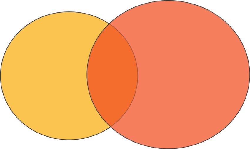
highly enriched in the iron ion binding, glycoprotein RF and NC (Figure 4 and Table 2).8 Canadian Respiratory Journal
–log10 (Fisher’s exact test p value)
0 0.5 1 1.5 2 2.5 3 3.5 4 4.5 5
Extracellular space 3.75
Cytoplasmic vesicle part 3.19
Cellular component
Vesicle lumen 2.99
Cytoplasmic vesicle lumen 2.99
Cytoplasmic vesicle 2.36
Intracellular vesicle 2.36
Secretory granule 2.34
Secretory vesicle 2.21
Iron ion binding 2.75
Glycoprotein binding 2.57
Molecular function
Enzyme binding 2.54
MHC protein complex binding 2.45
MHC class II protein complex binding 2.45
DNA polymerase binding 2.45
TPR domain binding 2.45
Identical protein binding 2.4
Regulation of protein stability 4.36
Cell activation 4.15
Regulation of nitric oxide biosynthetic process 3.69
Positive regulation of nitric oxide metabolic process 3.69
Positive regulation of nitric oxide biosynthetic process 3.69
Biological process
Regulated exocytosis 3.47
Exocytosis 3.42
Cell activation involved in immune response 3.36
Leukocyte activation involved in immune response 3.36
Metal ion homeostasis 3.36
Cellular metal ion homeostasis 3.36
Homeostatic process 3.33
Response to stress 3.27
Secretion by cell 3.22
(a)
–log10 (Fisher’s exact test p value)
0 0.5 1 1.5 2 2.5 3 3.5
hsa04657 IL-17 signaling pathway 3.01
hsa04612 Antigen processing and presentation 1.98
hsa04914 Progesterone-mediated oocyte maturation 1.46
hsa04659 Th17 cell differentiation 1.46
(b)
Figure 3: Continued.Canadian Respiratory Journal 9
–log10 (Fisher’s exact test p value)
0 0.5 1 1.5 2 2.5 3
Serpin domain 2.41
Histidine kinase-like ATPase, C-terminal domain 1.95
Heat shock protein Hsp90, N-terminal 1.95
Ribosomal protein S5 domain 2-type fold 1.66
(c)
Figure 3: Functional enrichment analysis of crotonylation modification sites corresponding to proteins. (a) Enrichment of GO analysis of
upregulated crotonylated protein. (b) Enrichment of KEGG pathway analysis of upregulated crotonylated protein. (c) Enrichment of protein
domain analysis of upregulated crotonylated protein.
318 90 341
Crotonylation site
corresponding proteins Proteome
Figure 4: Common proteins of differentially expressed crotonylation site corresponding proteins and proteome between COPD and NC.
Table 2: 90 protein expression fold change value.
Protein fold Protein-regulated
Protein description
change type
FYN-binding protein OS � Homo sapiens GN � FYB 0.788 Down
Flotillin-1 OS � Homo sapiens GN � FLOT1 1.452 Up
Haptoglobin OS � Homo sapiens GN � HP 0.462 Down
Carbonic anhydrase 1 OS � Homo sapiens GN � CA1 2.024 Up
Carbonic anhydrase 2 OS � Homo sapiens GN � CA2 1.39 Up
Alpha-1-antitrypsin OS � Homo sapiens GN � SERPINA1 0.475 Down
Ig kappa chain C region OS � Homo sapiens GN � IGKC 0.411 Down
Ig gamma-1 chain C region OS � Homo sapiens GN � IGHG1 0.386 Down
Ig gamma-2 chain C region OS � Homo sapiens GN � IGHG2 0.468 Down
“Spectrin alpha chain, erythrocytic 1 OS � Homo sapiens GN � SPTA1” 1.522 Up
Apolipoprotein A-I OS � Homo sapiens GN � APOA1 0.299 Down
Apolipoprotein A-II OS � Homo sapiens GN � APOA2 0.197 Down
Fibrinogen alpha chain OS � Homo sapiens GN � FGA 0.803 Down
Fibrinogen beta chain OS � Homo sapiens GN � FGB 0.819 Down
Fibrinogen gamma chain OS � Homo sapiens GN � FGG 0.791 Down
Platelet basic protein OS � Homo sapiens GN � PPBP 0.715 Down
Serotransferrin OS � Homo sapiens GN � TF 0.271 Down
Lactotransferrin OS � Homo sapiens GN � LTF 1.463 Up
Catalase OS � Homo sapiens GN � CAT 1.416 Up
Apolipoprotein B-100 OS � Homo sapiens GN � APOB 0.668 Down
Cytochrome b-245 heavy chain OS � Homo sapiens GN � CYBB 1.361 Up
Protein S100-A8 OS � Homo sapiens GN � S100A8 1.42 Up
Myeloperoxidase OS � Homo sapiens GN � MPO 1.348 Up
Glucose-6-phosphate isomerase OS � Homo sapiens GN � GPI 1.307 Up
Protein disulfide-isomerase OS � Homo sapiens GN � P4HB 1.531 Up
Annexin A2 OS � Homo sapiens GN � ANXA2 0.792 Down10 Canadian Respiratory Journal
Table 2: Continued.
Protein fold Protein-regulated
Protein description
change type
Annexin A6 OS � Homo sapiens GN � ANXA6 0.782 Down
Heat shock protein HSP 90-beta OS � Homo sapiens GN � HSP90AB1 1.316 Up
Leukotriene A-4 hydrolase OS � Homo sapiens GN � LTA4H 1.264 Up
Heat shock 70 kDa protein 1B OS � Homo sapiens GN � HSPA1B 1.67 Up
78 kDa glucose-regulated protein OS � Homo sapiens GN � HSPA5 1.266 Up
“Solute carrier family 2, facilitated glucose transporter member 1 OS � Homo sapiens
1.919 Up
GN � SLC2A1”
“C-1-Tetrahydrofolate synthase, cytoplasmic OS � Homo sapiens GN � MTHFD1” 1.365 Up
Alcohol dehydrogenase class-3 OS � Homo sapiens GN � ADH5 0.825 Down
Annexin A3 OS � Homo sapiens GN � ANXA3 2.256 Up
X-ray repair cross-complementing protein 5 OS � Homo sapiens GN � XRCC5 0.777 Down
“HLA class I histocompatibility antigen, A-11 alpha chain OS � Homo sapiens GN � HLA-A” 1.296 Up
Endoplasmin OS � Homo sapiens GN � HSP90B1 1.214 Up
Bactericidal permeability-increasing protein OS � Homo sapiens GN � BPI 1.773 Up
Phosphoglycerate mutase 1 OS � Homo sapiens GN � PGAM1 1.257 Up
Nucleolin OS � Homo sapiens GN � NCL 0.748 Down
Neutrophil cytosol factor 2 OS � Homo sapiens GN � NCF2 1.415 Up
“V-type proton ATPase subunit B, brain isoform OS � Homo sapiens GN � ATP6V1B2” 1.262 Up
Nonspecific lipid-transfer protein OS � Homo sapiens GN � SCP2 1.359 Up
Nucleoside diphosphate kinase B OS � Homo sapiens GN � NME2 1.328 Up
cAMP-dependent protein kinase catalytic subunit beta OS � Homo sapiens GN � PRKACB 0.649 Down
Cofilin-1 OS � Homo sapiens GN � CFL1 0.77 Down
Cathepsin S OS � Homo sapiens GN � CTSS 1.219 Up
Protein S100-P OS � Homo sapiens GN � S100P 3.417 Up
Erythrocyte band 7 integral membrane protein OS � Homo sapiens GN � STOM 1.392 Up
Calreticulin OS � Homo sapiens GN � CALR 1.243 Up
Transketolase OS � Homo sapiens GN � TKT 1.392 Up
Flavin reductase (NADPH) OS � Homo sapiens GN � BLVRB 1.609 Up
“Peptidyl-prolyl cis-trans isomerase F, mitochondrial OS � Homo sapiens GN � PPIF” 0.812 Down
Coronin-1A OS � Homo sapiens GN � CORO1A 0.822 Down
Protein S100-A11 OS � Homo sapiens GN � S100A11 1.698 Up
Peroxiredoxin-2 OS � Homo sapiens GN � PRDX2 1.967 Up
Transaldolase OS�Homo sapiens GN � TALDO1 1.25 Up
Macrophage-capping protein OS � Homo sapiens GN � CAPG 1.339 Up
“Malate dehydrogenase, cytoplasmic OS � Homo sapiens GN � MDH1” 0.702 Down
Myeloid cell nuclear differentiation antigen OS � Homo sapiens GN � MNDA 1.259 Up
LIM and senescent cell antigen-like-containing domain protein 1 OS � Homo sapiens
0.799 Down
GN � LIMS1
“6-Phosphogluconate dehydrogenase, decarboxylating OS � Homo sapiens GN � PGD” 1.609 Up
Histone H4 OS � Homo sapiens GN � HIST1H4A 0.309 Down
Tubulin alpha-4A chain OS � Homo sapiens GN � TUBA4A 0.721 Down
Hemoglobin subunit beta OS � Homo sapiens GN � HBB 1.475 Up
Neutrophil gelatinase-associated lipocalin OS � Homo sapiens GN � LCN2 1.714 Up
“Phosphate carrier protein, mitochondrial OS � Homo sapiens GN � SLC25A3” 0.719 Down
14-3-3 protein eta OS � Homo sapiens GN � YWHAH 1.271 Up
Selenium-binding protein 1 OS � Homo sapiens GN � SELENBP1 1.342 Up
Integrin-linked protein kinase OS � Homo sapiens GN � ILK 0.819 Down
Stromal interaction molecule 1 OS � Homo sapiens GN � STIM1 0.764 Down
Four and a half LIM domains protein 1 OS � Homo sapiens GN � FHL1 0.653 Down
Protein disulfide-isomerase A6 OS � Homo sapiens GN � PDIA6 1.223 Up
Septin-7 OS � Homo sapiens GN � SEPT7 0.822 Down
Putative elongation factor 1-alpha-like 3 OS � Homo sapiens GN � EEF1A1P5 1.222 Up
Phospholipase B-like 1 OS � Homo sapiens GN � PLBD1 1.748 Up
Olfactomedin-4 OS � Homo sapiens GN � OLFM4 3.945 Up
Fermitin family homolog 3 OS � Homo sapiens GN � FERMT3 0.772 Down
Histone H2A type 1-C OS � Homo sapiens GN � HIST1H2AC 0.592 Down
Sarcoplasmic/endoplasmic reticulum calcium ATPase 3 OS � Homo sapiens GN � ATP2A3 0.833 Down
WW domain-binding protein 2 OS � Homo sapiens GN � WBP2 1.24 Up
EF-hand domain-containing protein D2 OS � Homo sapiens GN � EFHD2 0.78 DownCanadian Respiratory Journal 11
Table 2: Continued.
Protein fold Protein-regulated
Protein description
change type
Histone H2A type 1-J OS � Homo sapiens GN � HIST1H2AJ 0.732 Down
Beta-parvin OS � Homo sapiens GN � PARVB 0.815 Down
Alpha-hemoglobin-stabilizing protein OS � Homo sapiens GN � AHSP 1.824 Up
Bridging integrator 2 OS � Homo sapiens GN � BIN2 0.824 Down
Coronin-1C OS � Homo sapiens GN � CORO1C 0.808 Down
Protein kinase C and casein kinase substrate in neurons protein 2 OS � Homo sapiens
1.23 Up
GN � PACSIN2
Voltage-dependent anion-selective channel protein 3 OS � Homo sapiens GN � VDAC3 0.694 Down
The fold change was more than 1.2 times as a significant upregulation and less than 1/1.2 as a criterion for significant downregulation.
4. Discussion proteomics. We found that 90 proteins were both modified by
differentially expressed crotonylation sites and differentially
Since COPD has been defined as a heterogeneous disease expressed in COPD and NC. These 90 proteins were differ-
[14], more and more researchers have conducted molecular entially expressed between COPD combined with type II RF
biology research, including genomics [15–19] and metab- and NC and may be biomarkers for the diagnosis of COPD
olomics [20, 21]. For proteomics, Braido et al. [22] found combined with type II RF.
that there is a possible link between the innate immune The present study has some limitations. Firstly, the small
system and the worsening of COPD infectivity by discov- number of samples may cause certain errors in the research
ering that the immune-associated Clara cell 16 (CC-16) is results. Secondly, this research provides a data basis for the
differentially expressed in COPD. Leuzzi et al. [23] found correlation between Kcr and COPD combined with type II
that high baseline C-reactive protein (CRP) levels are sig- RF, which is only the initial stage. Therefore, we will consider
nificantly associated with late mortality in COPD patients. collecting more samples for subsequent research. In a later
Recently, Xiang et al. [24] found that YKL-40 proteins were study, we will consider performing site-directed mutagenesis
differentially expressed in COPD, suggesting that YKL-40 in vitro and introduce site mutation for proteins and sites
may be involved in bronchial inflammation and remodeling that modify levels of difference. Immunofluorescence (IF),
of COPD and may be a useful biomarker for the diagnosis fluorescent resonance energy transfer (FRET), and other
and monitoring of COPD. Although the relationship be- fluorescent labeling methods will be used to analyze the
tween proteomics and COPD has been the focus of research, subcellular localization and real-time dynamic changes of
the relationship between Kcr and COPD combined with type the target molecule, which will provide a basis for functional
II RF is largely unknown. mechanism research and protein interaction.
In the GO enrichment, the upregulated crotonylated
proteins were enriched in biological processes, cell structural
components, and cell-binding functional types, indicating 5. Conclusion
that Kcr participated in pathologically related cell and
molecular life activities in COPD combined with type II RF. We found that differentially expressed Kcr may be involved
The functional enrichment of Kcr in KEGG analysis revealed in the modification of COPD-related proteins and is in-
IL-17 signaling pathway, antigen processing, and presen- volved in the pathogenesis of COPD combined with type II
tation, all of which have an association with immunology RF. Our research provides an idea for studying the asso-
[25, 26]. Mario and Maria [27] have also shown that IL-17 ciation of Kcr, proteins, and COPD to identify biomarkers of
plays an important proinflammatory effect in COPD. The COPD combined with type II RF.
lung infection of COPD is mainly in the large and small
airway position, with bronchial mucosal edema, inflam- Data Availability
mation, and hypersecretion of gland as the main clinical
manifestations [28]. It is suggested that the differentially The raw datasets used to support the findings of this study
expressed Kcr sites enriched in IL-17 signaling pathway and are available from the corresponding author upon request.
antigen processing and presentation may regulate the ex-
pression of related proteins in the immune response of
COPD lung infection. Disclosure
We used TMT labeling, crotonylation enrichment tech- Authors’ institution 1 and institution 2 contributed equally
nology, and the quantitative proteomics research strategy of to this work.
high-resolution LC-MS/MS to quantitatively study Kcr and
proteomics in COPD combined with type II RF and NC. We
found 32 and 914 sites, corresponding to 23 and 295 proteins, Conflicts of Interest
which were significantly upregulated and downregulated, re-
spectively. We also conducted differential expression studies on The authors declare no conflicts of interest.12 Canadian Respiratory Journal
Authors’ Contributions [13] O. K. Kwon, S. J. Kim, and S. Lee, “First profiling of lysine
crotonylation of myofilament proteins and ribosomal proteins
Qing Gan and Donge Tang contributed equally to this work. in zebrafish embryos,” Scientific Reports, vol. 8, no. 1, p. 3652,
2018.
[14] A. Agusti and D. D. Sin, “Biomarkers in COPD,” Clinics in
Acknowledgments Chest Medicine, vol. 35, no. 1, pp. 131–141, 2014.
[15] A. R. Bentley, P. Emrani, and P. A. Cassano, “Genetic vari-
The authors gratefully acknowledge the financial support by the ation and gene expression in antioxidant related enzymes and
Fund of Guangxi Key Laboratory of Metabolic Diseases risk of COPD: a systematic review,” Thorax, vol. 63, no. 11,
Research (No. 20-065-76), Shenzhen Science and Technology pp. 956–961, 2008.
Project (Nos. JCYJ20170307095606266, JCYJ20170413093 [16] P. J. Castaldi, M. H. Cho, M. Cohn et al., “The COPD genetic
032806, and JCYJ20150403101146305), and Science and association compendium: a comprehensive online database of
Technology Planning Project of Guangdong Province, China COPD genetic associations,” Human Molecular Genetics,
(No. 2017A020214016). The authors thank the patients and vol. 19, no. 3, pp. 526–534, 2010.
healthy volunteers who participated in this study. [17] A. O. Nielsen, C. S. Jensen, M. S. Arredouani et al., “Variants
of the ADRB2 gene in COPD: systematic review and meta-
analyses of disease risk and treatment response,” COPD
References Journal of Chronic Obstructive Pulmonary Disease, vol. 14,
no. 4, pp. 1–10, 2017.
[1] M. Minas, K. Dimitropoulos, C. Pastaka, D. Papadopoulos, [18] Y. V. Durme, M. Eijgelsheim, G. F. Joos et al., “Hedgehog-
N. Markoulis, and K. Gourgoulianis, “Global initiative for interacting protein is a COPD susceptibility gene: the Rot-
chronic obstructive lung disease for chronic obstructive terdam study,” European Respiratory Journal, vol. 36, no. 1,
pulmonary disease: GOLD opportunity for lung disorders,” p. 89, 2010.
Preventive Medicine, vol. 40, no. 3, pp. 274–277, 2005. [19] Z. H. Chen, H. P. Kim, S. W. Ryter et al., “Identifying targets
[2] D. M. Mannino and A. S. Buist, “Global burden of COPD: risk for COPD treatment through gene expression analyses,”
factors, prevalence, and future trends,” Lancet (London, En- International Journal of Chronic Obstructive Pulmonary
gland), vol. 370, no. 9589, pp. 765–773, 2007, North American Disease, vol. 3, no. 3, pp. 359–370, 2008.
Edition. [20] Q. Chen, R. S. Deeb, Y. Ma et al., “Serum metabolite bio-
[3] T. S. Lamba, R. S. Sharara, A. C. Singh, and M. Balaan, markers discriminate healthy smokers from COPD smokers,”
“Pathophysiology and classification of respiratory failure,” PLoS One, vol. 10, no. 12, Article ID e0143937, 2015.
Critical Care Nursing Quarterly, vol. 39, no. 2, pp. 85–93, 2016. [21] E. D. Watson, C. R. Sweeney, and K. A. Steensma, “Arach-
[4] D. Phillips, “The presence of acetyl groups in histones,” idonate metabolites in bronchoalveolar lavage fluid from
Biochemical Journal, vol. 87, no. 2, pp. 258–263, 1963. horses with and without COPD,” Equine Veterinary Journal,
[5] M. Tan, H. Luo, S. Lee et al., “Identification of 67 histone vol. 24, no. 5, pp. 379–381, 1992.
marks and histone 1ysine crotonylation as a new type of [22] F. Braido, A. M. Riccio, L. Guerra et al., “Clara cell 16 protein
histone modifcation,” Cell, vol. 146, no. 6, pp. 1016–1028, in COPD sputum: a marker of small airways damage?” Re-
2011. spiratory Medicine, vol. 101, no. 10, pp. 2119–2124, 2007.
[6] B. R. Sabari, Z. Tang, H. Huang et al., “Intracellular crotonyl- [23] G. Leuzzi, C. Galeone, F. Taverna, P. Suatoni, D. Morelli, and
CoA stimulates transcription through p300-catalyzed histone U. Pastorino, “C-reactive protein level predicts mortality in
crotonylation,” Molecular Cell, vol. 58, no. 2, pp. 203–215, COPD: a systematic review and meta-analysis,” European
2015. Respiratory Review, vol. 26, no. 143, p. 160070, 2017.
[7] B. R. Sabari, D. Zhang, C. D. Allis et al., “Metabolic regulation [24] T. Xiang, W. Dongguang, L. Sitong et al., “The YKL-40 protein
of gene expression through histone acylations,” Nature Re- is a potential biomarker for COPD: a meta-analysis and
views Molecular Cell Biology, vol. 18, no. 2, p. 90, 2017. systematic review,” International Journal of Chronic Ob-
[8] R. A. Olga, S. N. M. Dolores, C. O. Pablo et al., “Histone lysine structive Pulmonary Disease, vol. 13, pp. 409–418, 2018.
crotonylation during acute kidney injury in mice,” Disease [25] T. Korn, E. Bettelli, M. Oukka, and V. K. Kuchroo, “IL-17 and
Models & Mechanisms, vol. 9, no. 6, pp. 633–645, 2016. Th17 cells,” Annual Review of Immunology, vol. 27, no. 1,
[9] X. Liu, W. Wei, Y. Liu et al., “MOF as an evolutionarily pp. 485–517, 2009.
conserved histone crotonyltransferase and transcriptional [26] R. N. Germain and D. H. Margulies, “The biochemistry and
activation by histone acetyltransferasedefi cient and croto- cell biology of antigen processing and presentation,” Annual
nyltransferase-competent CBP/p300,” Cell Discovery, vol. 23, Review of Immunology, vol. 11, no. 1, pp. 403–450, 1993.
no. 3, p. 17016, 2017. [27] C. Mario and G. M. Maria, “IL-17 in chronic obstructive
[10] W. Wei, X. Liu, J. Chen et al., “Class I histone deacetylases are pulmonary disease,” Expert Review of Respiratory Medicine,
major histone decrotonylases: evidence for critical and broad vol. 6, no. 2, pp. 135–138, 2012.
function of histone crotonylation in transcription,” Cell [28] M. H. Gotfried, “Macrolides for the treatment of chronic
Discovery, vol. 27, no. 7, pp. 898–915, 2017. sinusitis, asthma, and COPD,” Chest, vol. 125, no. 2,
[11] E. Montellier, S. Rousseaux, Y. Zhao, and S. Khochbin, pp. 52S–61S, 2004.
“Histone crotonylation specifically marks the haploid male
germ cell gene expression program: post-meiotic male-spe-
cific gene expression,” Bioessays News & Reviews in Molecular
Cellular & Developmental Biology, vol. 34, no. 3, pp. l87–93,
2012.
[12] W. Xu, J. Wall, J. Zhan et al., “Global profiling of crotony-
lation on non-histone proteins,” Cell Research, vol. 27, no. 7,
pp. 946–949, 2017.You can also read