Effects of Long-Term Vagus Nerve Electrical Stimulation Therapy on Acute Cerebral Infarction and Neurological Function Recovery in Post MCAO Mice
←
→
Page content transcription
If your browser does not render page correctly, please read the page content below
Hindawi
Oxidative Medicine and Cellular Longevity
Volume 2022, Article ID 8131391, 9 pages
https://doi.org/10.1155/2022/8131391

Research Article
Effects of Long-Term Vagus Nerve Electrical Stimulation Therapy
on Acute Cerebral Infarction and Neurological Function
Recovery in Post MCAO Mice

 Li Du ,1 Zhenxing Yang ,2 Huaxin Sheng ,3 Min Liu ,1 and Qian Sun 1

 1
 Department of Anesthesiology, Renmin Hospital of Wuhan University, Wuhan, China
 2
 Department of Neurosurgery, Renmin Hospital of Wuhan University, Wuhan Hubei, China
 3
 Multidisciplinary Neuroprotection Laboratory, Department of Anesthesiology, Duke University Medical Center, Durham, USA

 Correspondence should be addressed to Zhenxing Yang; neuroyzx@whu.edu.cn

 Received 24 August 2021; Revised 28 February 2022; Accepted 8 March 2022; Published 29 March 2022

 Academic Editor: Alin Ciobica

 Copyright © 2022 Li Du et al. This is an open access article distributed under the Creative Commons Attribution License, which
 permits unrestricted use, distribution, and reproduction in any medium, provided the original work is properly cited.

 Background. Vagus nerve stimulation therapy is proven to produce neuroprotective effects against central nervous system diseases
 and reduce neurological injury, having a positive effect on the recovery of neurological functions in mouse model of stroke.
 Objective. This study was aimed at exploring a wider time window for VNS treatment, investigating the long-term behavioral
 improvement of long-term VNS in mice after pMCAO, and exploring the antiapoptotic properties of VNS and its role in
 autophagy, all of which may be a permanent deficiency potential mechanism of neuroprotection in hemorrhagic stroke.
 Methods. Permanent focal cerebral ischemia and implantation of vagus nerve stimulator were performed through intracavitary
 occlusion of the right middle cerebral artery (MCA). The vagus nerve stimulation group received five times vagus nerve
 stimulation from 6 h after surgery for 5 days. Adhesive removal test and NSS neurological score were used to evaluate the
 neurological deficit of mice. TTC staining of mouse brain tissue was performed one week after surgery in order to assess the
 area of cerebral infarction. Additionally, frozen sections were stained with Fluoro-Jade B to observe the apoptotic cells in the
 ischemic penumbra of brain tissue. Finally, Western blot was used to detect the changes in the levels of apoptosis-related
 proteins such as cleaved-caspase3 and Bcl-2 and autophagy-related proteins such as mTOR, Beclin-1, and LC3-II in brain
 tissue. Results. VNS can effectively reduce the behavioral score of pMCAO mice; TTC results showed that VNS could
 effectively reduce the infarct area after pMCAO (P < 0:05). After VNS intervention of the pMCAO group compared with the
 pMCAO+VNI group, the FJB-positive cells in the VNS group were significantly decreased (P < 0:05); Western Blot analysis
 showed that the expression of cleaved-caspase3 in the brain tissue of mice increased after pMCAO (P < 0:05), and the
 expression of Bcl-2 decreased (P < 0:05). This change could be effectively reversed after VNS intervention (P < 0:05).
 Conclusion. VNS could effectively improve the behavioral performance of mice after permanent stroke in addition to
 significantly reducing the infarct size of the brain tissue. The mechanism may be related to the effective reduction of cell
 apoptosis and excessive autophagy after pMCAO by VNS.

1. Introduction treatment worldwide. However, due to the limitation of the
 interventional treatment time window, the proportion of
Stroke is the second leading cause of death globally and is one patients treated within a suitable time frame is limited to
of the main causes of disability. Due to the aging among the 10% or less. Drug thrombolytic therapy has serious complica-
general population, the incidence is increasing on an annual tions such as hemorrhage, and the treatment effect for stroke is
basis [1]. Meanwhile, as the proportion of unhealthy eating not ideal. Nerve stimulation is an alternative or auxiliary
habits increases, the proportion of young people suffering method for the treatment of failed thrombolysis, which in turn
from stroke is also increasing [2]. Intravascular interventional can protect the brain during acute ischemia. In addition, nerve
thrombolysis and anticoagulants are still the backbone of stimulation can also repair the nerve function of patients
2 Oxidative Medicine and Cellular Longevity

(treated or untreated) with severe functional impairment in trunk was lifted with an 8-0 needle and permanently ligated
the poststroke stage in addition to promoting the recovery of with silk suture proximal to the cortical branch to the frontal
neuronal function. In the past few decades, various animal cortex. The muscle and skin were then closed separately.
stroke models have shown the efficacy and safety of many can- Animals that did not show an infarct lesion were excluded.
didate neuroprotective agents, but in clinical trials, promising 80 male C57BL/6J mice were randomly divided into four
preclinical studies have not yet been translated into positive groups: (1) sham+VNS group (sham operation+vagus nerve
results [3]. stimulation group), (2) pMCAO+VNS group (pMCAO
 Ideally, the future treatment plan for ischemic stroke +vagus nerve stimulation group), (3) pMCAO+VNI group
should include thrombolysis, antithrombosis (secondary (pMCAO+vagus nerve isolation group), and (4) sham group
prevention), neuroprotection (stabilizing penumbra), and (sham operation group).
strategies to enhance neuroplasticity.
 Vagus nerve stimulation is proven to play a potential 2.3. Vagus Nerve Electrode Implantation. An incision was
protective role in central nervous system diseases such as made in the middle of the neck of the mouse to separate the
Alzheimer’s disease and traumatic brain injury [4, 5]. It sternohyoid and sternomastoid muscles longitudinally and is
has been suggested to provide protection against ischemic then pulled to the side to expose the common carotid artery
brain injury. So far, however, the vagus nerve stimulation and the right vagus nerve located outside the carotid sheath.
experiment reported in the relevant paper was a single stim- In order to reduce the collateral circulation compensation
ulation during or after surgery, and there are no further and ensure the success rate of the middle cerebral artery ische-
researches on the long-term stimulation of the vagus nerve mia model, we ligated the common carotid artery with silk
on the recovery of nerve function after cerebral ischemia. thread. The vagus nerve is carefully separated from the sur-
We have designed a new vagus nerve stimulation therapy rounding connective tissue and sympathetic nerve trunk. After
with a wider time window that can observe the effect and the 5 mm long VN is exposed, the exposed end of the VN
the change of the electrical stimulation of vagus nerve after stimulation electrode is wrapped around the nerve in a spiral
brain stroke, which is more in line with clinical needs of form and separated from the surrounding tissue by a rubber
achieving neuroprotection. sheet. Through a continuity test using an ohmmeter, the con-
 Ischemic neurons showed both necrotic and apoptotic tact between the electrode and the exposed VN was intact. The
features [6]. The same neurons also exhibited caspase-3 electrode lead is then passed from the tunnel through the fas-
cleavage and cytochrome-C release as markers of apoptosis. cia to the back of the skin on the neck. In order to prevent the
It seems that Bcl-2 family protein members play a key role in nerve or electrode from shifting, a suture outside of the elec-
mediating necroptosis by inducing necrotic mitochondrial trode was done on the skin of the neck (image A-C).
damage [7]. The signaling pathway of autophagy is driven Mice in the pMCAO+VNS group were stimulated with
on mechanistic target of rapamycin (mTOR), which leads 500 μs width 1 mA electric pulses for 10 min at 5 Hz every
to autophagy initiation, respectively [8]. We plan to evaluate day for 10 mins, from 6 h after surgery and each day for 5 days,
the protective effect of vagus nerve stimulation on cerebral 5 times in total [10]. The criterion for a successful vagus nerve
ischemia injury in mice by analyzing the changes in the stimulation is an increase in heart rate variability. Animals
above proteins and signaling pathways. that did not show an increased heart rate variability were
 excluded. Mice returned to standard diet after five days of
 liquid diet.
2. Materials and Methods
 2.4. Behavioral Test. All evaluations were performed by
2.1. Ethics Statement and Animals. Animal experiments were
 observers who were blinded to group assignment.
carried out in accordance with the Duke University Animal
Care and Use Committee. All studies were approved by the 2.5. Adhesive Removal Test. All mice were pretrained on the
United States Public Health Service’s Policy on Humane Care adhesive removal tasks 3 days prior to the VN surgery. The
and Use of Laboratory Animals. Inbred male C57BL6/J mice, adhesive removal test measures sensorimotor function as pre-
12-14 weeks old (Jackson Laboratories, Bar Harbor, ME). viously described [11]. Two small adhesive dots were placed
The online tool QuickCalcs (http://www.graphpad.com/ on forepaws, and the amount of time (seconds) needed to con-
quickcalcs/) was used to randomly assign animals to groups. tact and remove the sticker from each forepaw was recorded.
 Recording was then stopped if the animal failed to contact
2.2. pMCAO Model Establishment. The pMCAO mice model the sticker within 2 min. The test was performed 3 times per
are made as the article described as follows [9]. The mice mouse, and the average time was used in the analysis before
were anesthetized with isoflurane, intubated, and ventilated. stroke in addition to 7, 14, and 21 days after stroke.
Their rectal temperature was maintained at 37° C ± 0:2° C
throughout the procedure. Mice were placed in left lateral 2.6. mNSS Test. Mice were tested and scored for neurological
position, and a small skin incision was made between the deficits using a modified Neurological Severity Score (mNSS)
right eye and ear. The temporal muscle was cut slightly using 7, 14, and 21 days after the onset of ischemia. An 18-point
a high temperature loop, and a 3 mm long segment of the neurological score was employed with slight modifications
zygomatic arch was removed. After exposing the skull base described before [12]. The score consists of 5 individual clini-
and trigeminal nerve branch, a small bone window (3- cal parameters, including tasks on motor function, alertness,
4 mm2) was drilled on the skull above the MCA. The MCA and physiological behavior, whereby 1 point is given for
Oxidative Medicine and Cellular Longevity 3

failure. A maximum NSS of 18 point indicates severe neuro- 3. Results
logical dysfunction with failure at all tasks.
 3.1. VNS Treatment Improves Long-Term Behavior Performance
 in pMCAO Mice. To determine whether VNS treatment pro-
2.7. TTC Staining. Infarct volume was measured using the
 moted neurological recovery in pMCAO mice, mNSS and
2,3,5-triphenyltetrazolium chloride (TTC) staining method.
 adhesion removal tests were performed. Compared with the
Brain tissue was removed and frozen at −20°C for 30 min and
 sham operation group and the sham operation+VNS group,
then cut into 1 mm thick coronal sections (7 slices) and incu-
 ischemic mice (pMCAO+VNS and pMCAO+VNI) performed
bated in 2% TTC (Sigma-Aldrich, Saint Louis, MO, USA) at
 poorly in behavioral tests at different time points after pMCAO
37°C for 30 min. Each section was soaked in 4% paraformalde-
 (P < 0:05, Figures 1(a)–1(c)). The mNSS score of the sham
hyde for 24 h and was then scanned. ImageJ software was used
 operation group was zero at each time point (Figure 1(a)), but
to analyze the infarct area [13]. All evaluations were performed
 at 7, 14, and 21 days after pMCAO, the mNSS score of mice
by observers who were blinded to group assignment.
 in the pMCAO+VNS group was lower than that of the
 pMCAO+VNI group score (P < 0:05, Figure 1(a)). The
2.8. Fluoro-Jade B Staining. The procedure of Fluoro-Jade B pMCAO+VNS group had a significantly less time in both con-
stain is as described in the previous article [14]. Brain sec- tact and remove time (Figures 1(b) and 1(c)) at 7, 14, and 21
tions were mounted on microscope slides and placed in days after pMCAO compared to the pMCAO+VNI group
70% ethanol and ultrapure water for 3 min followed by 3 (P < 0:05). In general, these results suggest that VNS treatment
washes in ultrapure water for 1 min each rinse. Sections were promoted the recovery of neurological function in mice after
oxidized by soaking in a solution of 0.06% KMnO4 for stroke and significantly improved their limb gratitude and
15 min and then washed 3 times in ultrapure water 1 min motor function (P < 0:05).
each. Sections were subsequently stained in 0.001% Fluoro-
Jade B (Sigma-Aldrich, Saint Louis, MO, USA) in 0.1% ace- 3.2. VNS Treatment Reduces Cerebral Infarction Volume and
tic acid for 20 min. Slides were subsequently washed 3 times Promotes Neuron Survival in pMCAO Mice. As the result of
in ultrapure water for 1 min each and dried overnight at improved neurological function may be attributed to the
room temperature. Dried slides were cleared in xylene and decrease of the damaged area of brain tissue, the cerebral
coverslips were mounted using paramount. infarct volume was evaluated by TTC staining (Figures 2(a)
 In this study, four fields of view were randomly selected and 2(b)). The results showed that VNS treatment signifi-
and photographed in the cerebral cortex infarcted ischemic cantly reduced the infarct volume compared to the
area in each slice by a fluorescence microscope (Zeiss, Ger- pMCAO+VNI group (P < 0:05, Figures 2(a) and 2(b) ). No
many). Then, the number of positively stained cells in each infarct change was found in the sham group.
field of view was calculated at a higher magnification (200x)
by researchers that were blinded to experiment design. 3.3. VNS Treatment Decreased Neuronal Apoptosis in the Peri-
 infarct Cortex. Fluoro-Jade B was used as a high affinity fluo-
2.9. Western Blotting. Western blot analysis was performed as rescent marker for the localization of neuronal apoptosis to
previously described [15]. The concentration of proteins was observe in the peri-infarct cortex. In the pMCAO+VNS group,
determined by using a protein assay kit (Bio-Rad, USA). the number of FJB-positive cells significantly increased com-
40 μg of proteins was electrophoresed on SDS-PAGE gels pared with the pMCAO+VNI group (P < 0:05). While the
and then transferred onto polyacrylamide difluoride (PVDF) sham group had no significant positive cells (Figures 3(a)
membrane (Millipore, Massachusetts, USA). Membranes were and 3(b)). These results suggested that VNS treatment reduces
incubated with respective primary antibodies (cleaved-cas- apoptosis in pMCAO injury.
pase-3, 1 : 1000, Abcam; Bcl-2, 1 : 1000, Thermo Fisher; 3.4. Apoptosis Protein Levels in Brain Tissues of Four Groups
mTOR, 1 : 2000, Abcam; beclin-1, 1 : 1000, Thermo Fisher; of Mice. In order to further investigate whether VNS treat-
LC3II, 1 : 1000, Thermo Fisher) overnight at 4°C, followed by ment inhibits neuronal apoptosis in the peri-infarct cortex,
incubation with horseradish peroxidase- (HRP-) conjugated we tested the expression levels of cleaved caspase-3 and
goat anti-rabbit (1 : 5000, Thermo Fisher) or goat anti-mouse Bcl-2 in the peri-infarct cortex of the four groups of mice.
IgG secondary antibodies (1 : 10000, Thermo Fisher) for 1 Statistical analysis was assessed by unpaired t-test. Western
hour at room temperature. Protein bands were visualized by blot results showed that the Bcl-2 protein level of the VNS-
an enhanced chemiluminescence system (ECL kit, GE treated group was significantly increased, while the expres-
Healthcare, USA). Quantification of band intensity was ana- sion of cleaved caspase3 decreased compared with the
lyzed using ImageJ software. β-Actin (1 : 5000, Thermo Fisher) pMCAO+VNI group (P < 0:05, Figures 4(a) and 4(b)).
served as loading condition.
 3.5. Autophagy Protein Levels in Brain Tissues of Four Groups
2.10. Statistical Analysis. All data analyses were performed of Mice. To further confirm the role of VNS treatment in
with Prism 8. Statistical analysis was assessed by unpaired Stu- autophagy-related pathways, we tested the autophagy-related
dent’s t-test (infarct volumes and protein levels) or Mann- proteins including mTOR, Beclin-1, and LC3II in the peri-
Whitney U test (neurologic scores and FJC positive cells). infarct cortex. Statistical analysis was assessed by unpaired t
Data are presented as mean ± SEM, mean ± SD, or the -test. Western blot results showed that Beclin-1 and LC3II
median. The level of significance was set at P < 0:05. levels were significantly decreased following VNS treatment
4 Oxidative Medicine and Cellular Longevity

 mNSS score Adhesive removal test
 20 25 ⁎
 &

 ⁎& ⁎&
 20

 Contact time (sec)
 15 ⁎
 &
 Score (18)

 15 ⁎
 &
 10
 10

 5 5

 0 0
 0 7 14 21 0 Day 7 Day 14 Day 21
 Days after pMCAO Days after pMCAO
 Sham+VNS pMCAO+VNI
 Sham+VNS pMCAO+VNI
 pMCAO+VNS Sham
 pMCAO+VNS Sham
 (a) (b)
 Adhesive removal test

 40
 &⁎
 Removal time (sec)

 30
 &⁎
 20 &⁎

 10

 0
 0 7 14 21
 Days after pMCAO

 Sham+VNS pMCAO+VNI
 pMCAO+VNS Sham
 (c)

Figure 1: Long-term VNS treatment improved neurological deficits of the mice at 7, 14, and 21 days after pMCAO. The results of
quantitative analysis of mNSS (a) and adhesive removal test (b and c) were expressed as mean ± SD, n = 15 per group. ∗ P < 0:05 vs.
sham group and sham+VNS group and &P < 0:05 vs. pMCAO+VNS group.

compared with the pMCAO+VNI group (P < 0:05, Figures 5 inhibition of neuronal apoptosis in the ischemic penumbra
(a)–5(c)), while mTOR expressions were increased compared and suggested that reduction of neuronal apoptosis and a
with the pMCAO+VNI group (P < 0:05). moderate activation of autophagy in the peri-infarct cortex
 induced by VNS treatment may be the possible mechanism
4. Discussion underlying its neuroprotective effect. The behavioral improve-
 ment of pMCAO mice by VNS may be related to its reduction
In our research, vagus nerve electrode stimulators were of cell apoptosis with a moderate increase in autophagy.
implanted to stimulate the vagus nerve 6 hours after acute At present, a relevant paper reports that electrical stimula-
cerebral infarction in the mouse model, once a day for five tion of the vagus nerve can in fact reduce the inflammatory
days, which can be used to observe the effects of electrical response and brain edema in the brain tissue of the mouse after
stimulation of the vagus nerve in mice for a longer period acute stroke and promote the recovery of the neurological func-
of time. We found that VNS decreased the number of apo- tion of the mouse with chronic stroke in addition to improving
ptotic cells in the penumbra compared with the pMCAO the living ability of the mouse [16, 17]. However, the current
+VNI group in FJB results. We have also found that VNS electrical stimulation in the paper is a single electrical stimula-
treatment increased the Bcl-2 expression and decreased tion that is given immediately after the operation and cannot
cleaved caspase-3 expression in the peri-infarct cortex. Our interfere with the subsequent stroke development process in
results also indicated a decreased Beclin-1 and LC3II, while mice. The inflammatory progression and edema of acute stroke
the expression of mTOR increased compared with the are usually developed gradually a few hours after acute brain
pMCAO+VNI group. The results above indicated that neu- stroke. At the same time, long-term animal experiment obser-
roprotection induced by VNS is partly attributable to the vation can also provide scientific data and theoretical basis for
Oxidative Medicine and Cellular Longevity 5

 pMCAO+VNS pMCAO+VNI

 ⁎

 Infarct volume (mm3) Group pMCAO+VNS
 Group pMCAO+VNI
 (a) (b)

Figure 2: Long-term VNS treatment can reduce the volume of cerebral infarction in mice and increase the number of surviving neurons. (a
and b) TTC-stained brain slices and quantitative analysis of the percentage of cerebral infarction volume in each group showed that
compared with the pMCAO+VNI group, VNS treatment significantly reduced the infarct volume (∗ P < 0:05). Data were presented as
mean ± SD, n = 5/group.

 Sham+VNS

 pMCAO+VNS

 FJB in ischemic penumbra
 40 ⁎
 #
 Apoptotic cell/field of view

 pMCAO+VNI 30

 20 ⁎

 10
 #
 Sham
 0

 −10
 Sham+VNS

 pMCAO+VNS

 pMCAO+VNI

 Sham

 (a) (b)

Figure 3: VNS treatment decreased the apoptosis-positive cells in pMCAO. (a) FJB-positive cells in four groups of ischemic penumbra. (b)
FJB-positive cell number comparison in four groups. Data were presented as mean ± SD, n = 5/group. ∗ P < 0:05 vs. sham+VNS group and
#
 P < 0:05 vs. pMCAO+VNS group.
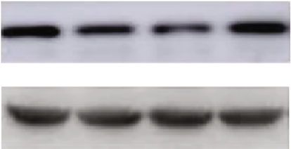
6 Oxidative Medicine and Cellular Longevity

 Cleaved- Bcl-2
 caspase3

 -actin -actin

 1.5

 Relative Bcl-2 protein level
 cleaved-caspas3 protein level
 1.5
 ⁎# #
 1.0
 1.0 ⁎ ⁎
 Relative

 # ⁎#
 0.5
 0.5

 0.0 0.0

 Sham+VNS

 pMCAO+VNS

 pMCAO+VNI

 Sham+VNI
 Sham+VNS

 pMCAO+VNS

 pMCAO+VNI

 Sham+VNI
 (a) (b)

Figure 4: VNS treatment can reduce neuronal damage by regulating the expression of apoptosis-related proteins in the cortex around the
infarct. (a) Western blots and quantitative analysis for cleaved-caspase3 and Bcl-2. (b) VNS treatment downregulated the expression of
cleaved-caspase3 and upregulated the expression of Bcl-2 compared with the pMCAO+VNI group. Data were presented as mean ± SD, n
 = 5/group. ∗ P < 0:05 vs. sham+VNS group and #P < 0:05 vs. pMCAO+VNS group.

clinical development of vagus nerve electrical stimulation for VNS, reducing the occurrence of inflammation, reducing neu-
the treatment of human acute stroke. In this study, the time ronal cell apoptosis, and inhibiting glial cell overreaction are
window of vagus nerve stimulation therapy was innovatively potential target therapeutic approaches to reduce brain injury
extended to three days after stroke. From a clinical application after cerebral ischemia.
perspective, the treatment time window has been effectively Another current explanation for the neuroprotective mech-
extended, and the scope of adaptation is wider, which can allow anism induced by VNS is the regulation of the afferent vagus
more strokes to be treated and is expected to restore nerve nerve pathway. The afferent nerve activity is relayed to the
function to the greatest extent possible. nucleus tractus solitarius which has projections to the locus
 Previous studies have shown that the appropriate activa- coeruleus (LC) [22] and is likely to play an important role in
tion of autophagy appears to be beneficial in poststroke con- VNS therapy through the release of norepinephrine (NE) and
ditions, which can remove necrotic materials in tissues. 5-hydroxytryptamine (5-HT) [23]. VNS can activate NE to
However, the decrease or increase of autophagy protein indi- play the anti-inflammatory effects, which can stimulate the 5-
cates that the autophagy response is excessive, which will HT releasement [24, 25]. The influence of these afferent nerve
increase the prodeath effect on neuronal cells. Several studies pathways may contribute to the effectiveness of VNS in
have revealed that autophagy is activated in the penumbra cerebral ischemia. Another suggestive mechanism of neuropro-
[18, 19]. Here, our results demonstrate that VNS can indeed tection induced by VNS is by affecting the efferent vagus nerve
induce the regulation of autophagy activation in pMCAO pathway. Studies have found that the cholinergic anti-
mice. Some studies have reported that autophagy is an intra- inflammatory pathway (CAP) can be activated by the central
cellular protective mechanism, which has been shown to be cholinergic system of the brain through the stimulation of the
closely related to inflammation in mice with cerebral ische- efferent fibers of the vagus nerve [26], which is mediated by
mia. The activation of autophagy can help the body effectively α7 nicotinic acetylcholine receptors. Therefore, CAP plays a
remove necrotic cells and substances. After ischemia, a large key role in the inhibition of inflammation [27], and repeated
number of neurons and glial cells are ischemic necrosis, and electrical stimulation of the efferent fibers of the vagus nerve
autophagy regulates the inflammatory response. Both respond in our experiment may activate the cholinergic anti-
to the stimulation caused by cerebral ischemia injury [20]. They inflammatory pathway and reduce the inflammatory response
found that inhibition of autophagy with Beclin-1 siRNA caused by stroke, thereby achieving brain protection The effect
increased the inflammatory response of the main microglia of of this is consistent with our experimental results, but the spe-
cells cultured with GSK-3β inhibitors in vitro, thereby increas- cific mechanism needs to be further studied and confirmed.
ing neuronal damage. Other researchers treated pMCAO mice Although regulatory mechanisms of neuroprotection after
with the autophagy inhibitor 3-MA and found that the inflam- ischemic stroke are diverse, apoptosis and autophagy can
matory response caused by ischemia was alleviated, while the explain the destruction and regeneration of neurons relatively
autophagy inducer rapamycin significantly promoted the stable. In our study, we downregulated cleaved-caspase3 and
inflammatory response [21]. In conclusion, our research shows upregulated Bcl-2 and mTOR in pMCAO through long-term
that through the regulation of phagocytic response in mice by stimulation of VNS, suggesting that VNS can achieve ischemic
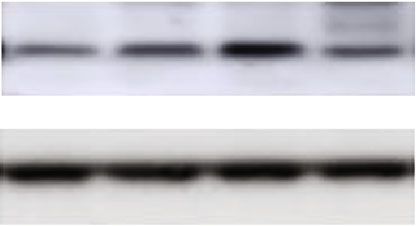
Oxidative Medicine and Cellular Longevity 7

 mTOR Beclin-1

 -actin -actin

 1.5

 Relative mTOR protein level

 Relative beclin-1 protein level
 1.0 ⁎#
 # 0.8
 1.0 ⁎ ⁎
 ⁎# #
 0.6

 0.4
 0.5
 0.2

 0.0 0.0

 Sham+VNS

 pMCAO+VNS

 pMCAO+VNI

 Sham
 Sham+VNS

 pMCAO+VNS

 pMCAO+VNI

 Sham

 (a) (b)

 LC3-II

 -actin

 0.8 ⁎#
 Relative LC3II protein level

 ⁎
 0.6
 #
 0.4

 0.2

 0.0
 Sham+VNS

 pMCAO+VNS

 pMCAO+VNI

 Sham

 (c)

Figure 5: VNS treatment can reduce brain damage and protect ischemic neurons by regulating the expression of autophagy-related proteins
in the peri-infarct cortex. (a–c) Western blots and quantitative analysis for mTOR, Beclin-1, and LC3-II. VNS treatment downregulated the
expression of Beclin-1 and LC3II and upregulated the expression of mTOR compared with the pMCAO+VNI group. Data were presented as
mean ± SD, n = 5/group. ∗ P < 0:05 vs. sham+VNS group and #P < 0:05 vs. pMCAO+VNS group.

brain tissue in mice by inhibiting cell apoptosis and regulating accurately, it also causes certain trauma, which is also a lim-
autophagy. This has been shown to have a protective effect on itation in this study. Yet, the research group is committed to
the long-term neurological behavior of mice after stroke. developing noninvasive vagus nerve stimulation methods,
 This study has preliminarily determined that vagal nerve which will further improve its clinical application.
stimulation therapy for stroke treatment is effective in
improving neurological function, but we failed to further 5. Conclusion
study its downstream signaling pathway and its key target
proteins. But we will continue to further explore these in The results provided in this article are the first evidence for
the next step. The vagus nerve stimulation method used in the concept of a new strategy for using long-term VNS to
this study is the implantation of vagus nerve electrical stim- achieve neuroprotection from ischemic stroke. After long-
ulator. Although it is to stimulate the vagus nerve more term VNS treatment on mice after cerebral ischemic stroke,
8 Oxidative Medicine and Cellular Longevity

we found that the neurological dysfunction of the mice was [7] J. Hitomi, D. E. Christofferson, A. Ng et al., “Identification of a
significantly improved. At the same time, histological studies molecular signaling network that regulates a cellular necrotic
found that the infarct volume of brain tissue was reduced cell death pathway,” Cell, vol. 135, no. 7, pp. 1311–1323, 2008.
and neuronal apoptosis was inhibited. Through changes in [8] S. Alers, A. S. Löffler, S. Wesselborg, and B. Stork, “Role of
the expression of inflammatory proteins in brain tissue, we AMPK-mTOR-Ulk1/2 in the regulation of autophagy: cross
speculate that the protective effect of VNS on brain tissue is talk, shortcuts, and feedbacks,” Molecular and Cellular Biology,
related to reducing cell apoptosis and regulating autophagy. vol. 32, no. 1, pp. 2–11, 2012.
Our research shows that long-term electrical stimulation of [9] M. Jiang, S. Yu, Z. Yu et al., “XBP1 (X-box-binding protein-1)-
the vagus nerve can be used as a new and promising therapy dependent O-GlcNAcylation is neuroprotective in ischemic
for the treatment of human acute and chronic stroke. It has stroke in young mice and its impairment in aged mice is res-
 cued by thiamet-G,” Stroke, vol. 48, no. 6, p. 1646, 2017.
broad clinical application prospects and its neuroprotective
mechanism is worthy of further study. Further signaling path- [10] H. Yuan and S. D. Silberstein, “Vagus nerve and vagus nerve
 stimulation, a comprehensive review: part II,” Headache,
ways and better vagus nerve stimulation methods we will con-
 vol. 56, no. 2, pp. 259–266, 2016.
tinue to explore in the next study.
 [11] T. Freret, V. Bouet, C. Leconte et al., “Behavioral deficits after
 distal focal cerebral ischemia in mice: usefulness of adhesive
Data Availability removal test,” Behavioral Neuroscience, vol. 123, no. 1,
 pp. 224–230, 2009.
The underlying data supporting the results of this study are [12] J. Chen, P. R. Sanberg, Y. Li et al., “Intravenous administration
stored in the university data system and are available on of human umbilical cord blood reduces behavioral deficits after
request. stroke in rats,” Stroke, vol. 32, no. 11, pp. 2682–2688, 2001.
 [13] Y. Hou, K. Wang, W. Wan, Y. Cheng, X. Pu, and X. Ye, “Res-
Disclosure veratrol provides neuroprotection by regulating the JAK2/
 STAT3/PI3K/AKT/mTOR pathway after stroke in rats,” Genes
We promise that the content and images of the manuscript & Diseases, vol. 5, no. 3, pp. 245–255, 2018.
have not been published before. [14] A. Ehara and S. Ueda, “Application of Fluoro-Jade C in acute
 and chronic neurodegeneration models: utilities and staining
 differences,” Acta Histochemica et Cytochemica, vol. 42,
Conflicts of Interest no. 6, pp. 171–179, 2009.
 [15] S. Liu, H. Sheng, Z. Yu, W. Paschen, and W. Yang, “O-linked
We declare that there is no conflict of interest.
 β-N-acetylglucosamine modification of proteins is activated
 in post-ischemic brains of young but not aged mice: implica-
Authors’ Contributions tions for impaired functional recovery from ischemic stress,”
 Journal of Cerebral Blood Flow and Metabolism, vol. 36,
All authors contributed to the important intellectual content of no. 2, pp. 393–398, 2016.
the manuscript and read and approved the final manuscript. [16] E. C. Meyers, B. R. Solorzano, J. James et al., “Vagus nerve
 stimulation enhances stable plasticity and generalization of
 stroke recovery,” Stroke, vol. 49, no. 3, pp. 710–717, 2018.
References
 [17] N. D. Engineer, T. J. Kimberley, C. N. Prudente, J. Dawson,
 [1] A. Alok, Z. Lei, N. S. Jagannathan et al., “Wnt proteins syner- W. B. Tarver, and S. A. Hays, “Targeted vagus nerve stimula-
 gize to activate β-catenin signaling,” Journal of Cell Science, tion for rehabilitation after stroke,” Frontiers in Neuroscience,
 vol. 130, no. 9, pp. 1532–1544, 2017. vol. 13, p. 280, 2019.
 [2] M. Katan and A. Luft, “Global burden of stroke,” Seminars in [18] Y. Mo, Y. Y. Sun, and K. Y. Liu, “Autophagy and inflammation
 Neurology, vol. 38, no. 2, pp. 208–211, 2018. in ischemic stroke,” Neural Regeneration Research, vol. 15,
 [3] A. Moretti, F. Ferrari, and R. F. Villa, “Neuroprotection for no. 8, pp. 1388–1396, 2020.
 ischaemic stroke: current status and challenges,” Pharmacol- [19] A. Plaza-Zabala, V. Sierra-Torre, and A. Sierra, “Autophagy
 ogy & Therapeutics, vol. 146, pp. 23–34, 2015. and microglia: novel partners in neurodegeneration and
 [4] D. C. Smith, A. A. Modglin, R. W. Roosevelt et al., “Electrical aging,” International Journal of Molecular Sciences, vol. 18,
 stimulation of the vagus nerve enhances cognitive and motor no. 3, p. 598, 2017.
 recovery following moderate fluid percussion injury in the [20] X. Zhou, J. Zhou, X. Li, C.’. Guo, T. Fang, and Z. Chen, “GSK-
 rat,” Journal of Neurotrauma, vol. 22, no. 12, pp. 1485–1502, 3β inhibitors suppressed neuroinflammation in rat cortex by
 2005. activating autophagy in ischemic brain injury,” Biochemical
 [5] R. Kaczmarczyk, D. Tejera, B. J. Simon, and M. T. Heneka, and Biophysical Research Communications, vol. 411, no. 2,
 “Microglia modulation through external vagus nerve stimula- pp. 271–275, 2011.
 tion in a murine model of Alzheimer's disease,” Journal of [21] Z. Yang, T. Z. Zhao, Y. J. Zou, J. H. Zhang, and H. Feng, “Hyp-
 Neurochemistry, vol. 146, no. 1, pp. 76–85, 2018. oxia induces autophagic cell death through hypoxia-inducible
 [6] M. Kilinc, Y. Gürsoy-Özdemir, G. Gürer et al., “Lysosomal factor 1α in microglia,” PLoS One, vol. 9, no. 5, article e96509,
 rupture, necroapoptotic interactions and potential crosstalk 2014.
 between cysteine proteases in neurons shortly after focal ische- [22] B. G. Fahy, “Intraoperative and perioperative complications
 mia,” Neurobiology of Disease, vol. 40, no. 1, pp. 293–302, with a vagus nerve stimulation device,” Journal of Clinical
 2010. Anesthesia, vol. 22, no. 3, pp. 213–222, 2010.
Oxidative Medicine and Cellular Longevity 9

[23] C. Cheyuo, A. Jacob, R. Wu, M. Zhou, G. F. Coppa, and
 P. Wang, “The parasympathetic nervous system in the quest
 for stroke therapeutics,” Journal of Cerebral Blood Flow and
 Metabolism, vol. 31, no. 5, pp. 1187–1195, 2011.
[24] J. L. Madrigal, D. L. Feinstein, and C. Dello Russo, “Norepi-
 nephrine protects cortical neurons against microglial-
 induced cell death,” Journal of Neuroscience Research, vol. 81,
 no. 3, pp. 390–396, 2005.
[25] S. Manta, J. Dong, G. Debonnel, and P. Blier, “Enhancement of
 the function of rat serotonin and norepinephrine neurons by
 sustained vagus nerve stimulation,” Journal of Psychiatry &
 Neuroscience, vol. 34, no. 4, pp. 272–280, 2009.
[26] K. J. Tracey, “Physiology and immunology of the cholinergic
 antiinflammatory pathway,” The Journal of Clinical Investiga-
 tion, vol. 117, no. 2, pp. 289–296, 2007.
[27] K. J. Tracey, “Reflex control of immunity,” Nature Reviews.
 Immunology, vol. 9, no. 6, pp. 418–428, 2009.
You can also read