CALCULATION OF SPATIAL RESPONSE OF A COLLIMATED SEGMENTED HPGE DETECTOR FOR GAMMA EMISSION TOMOGRAPHY BY MCNP SIMULATIONS - DIVA PORTAL
←
→
Page content transcription
If your browser does not render page correctly, please read the page content below
714                                                                                IEEE TRANSACTIONS ON NUCLEAR SCIENCE, VOL. 69, NO. 4, APRIL 2022

      Calculation of Spatial Response of a Collimated
      Segmented HPGe Detector for Gamma Emission
            Tomography by MCNP Simulations
                         Vikram Rathore , Lorenzo Senis , Peter Jansson , Erik Andersson Sundén,
                                         Ane Håkansson , and Peter Andersson

   Abstract— We have proposed a planar electronically segmented                  tomography (GET) of nuclear fuel. In particular, the GET
high-purity germanium (HPGe) detector concept in combination                     technique has been used in the examination of nuclear
with a multislit collimator for gamma emission tomography.                       fuel rods exposed to in-pile transient tests in material test
In this work, the spatial resolution achievable using the collimated
segmented HPGe detector was evaluated, prior to the manufac-                     reactors [5]–[7]. In such GET inspections, high spatial
ture and operation of the detector. The spatial response of a                    resolution is valuable for studying fragmentation, relocation,
collimated segmented HPGe detector concept was evaluated using                   and dispersal of fuel from a fuel element or rod. The
simulations performed with Monte Carlo N-Particle (MCNP)                         feasibility of segmented HPGe detector for GET instruments
transport code MCNP6. The full detector and multislit collimator                 was evaluated through the simulation study in [3], and the
system were modeled, and for the quantification of the spatial
response, the modulation transfer function (MTF) was chosen as                   detector geometry and segmentation pattern were optimized
a performance metric. The MTF curve was obtained through the                     in [4].
calculation of the line spread function (LSF) by analyzing the sim-                 Due to the promising results, an instrument is currently
ulated projection data. In addition, tomographic reconstructions                 being manufactured for experimental demonstration. In this
of the simulated simplified test objects were made to demonstrate                work, the demonstration device is presented, which is an elec-
the performance of the segmented HPGe detector in planned
application. For 662-keV photons, the spatial resolution obtained                tronically segmented HPGe detector similar to the proposed
was approximately the same as the collimator slit width for both                 design in [3] and [4], albeit scaled-down with fewer segments
the 100- and 150-mm-long collimators. The corresponding spatial                  and of planar geometry, to lower the costs of manufacturing.
resolution at 1596-keV photon energy was almost twice the slit                   Aside from lower manufacturing costs of the detector itself,
width for the 100-mm collimator, due to the partial penetration                  other reasons for choosing the scaled-down planar design for
of the high-energy gamma rays through the collimator bulk. For
a 150-mm-long collimator, an improved resolution was obtained.                   demonstration are its comparatively less complex manufactur-
                                                                                 ing process than the full coaxial one, less expensive associated
   Index Terms— Gamma emission tomography (GET), Monte                           data acquisition system, and less expensive multislit collima-
Carlo N-Particle (MCNP), modulation transfer function (MTF),
segmented high-purity germanium (HPGe).                                          tor. These factors will eventually help in having a full device
                                                                                 ready for demonstration in comparatively less time so that the
                                                                                 different aspects of the segmented HPGe detector for GET
                          I. I NTRODUCTION                                       measurement could be experimentally validated/evaluated such

H     IGH-PURITY germanium (HPGe) detectors are
      well-known for their excellent energy resolution and
reasonably high detection efficiency in the gamma-ray
                                                                                 as detection efficiency, data analysis methods, and count rate
                                                                                 capabilities. Even though the demonstration device is different
                                                                                 in size and shape, the principle of operation (data acquisition
spectrometry field. Because of these properties, HPGe                            and analysis) is similar to the full coaxial one. The spatial
detectors are widely used in many scientific applications.                       response of the demonstration detector was evaluated. For
In nuclear structure studies, electronically segmented HPGe                      this purpose, the modulation transfer function (MTF) was
detectors have been used for gamma-ray tracking applications                     chosen as a performance parameter [8], [9]. Simulations were
such as Advanced GAmma Tracking Array (AGATA) [1]                                performed using the particle transport code MCNP6.2 [10] to
and Gamma-Ray Energy Tracking In-beam Nuclear Array                              obtain the line spread function (LSF) whose Fourier transform
(GRETINA) [2]. A similar kind of segmented HPGe                                  constitutes the MTF curve. Furthermore, to demonstrate the
detector [3], [4] has also been proposed for gamma emission                      performance, tomographic reconstructions were also made
                                                                                 from the simulated projection data of simplified symmetric
  Manuscript received January 13, 2022; accepted February 11, 2022. Date
of publication February 15, 2022; date of current version April 19, 2022. This   test object inspections.
work was supported in part by the Swedish Research Council under Grant
2017-06448 and in part by the Swedish Foundation for Strategic Research                   II. D ESIGN AND W ORKING P RINCIPLE OF
under Grant EM-16-0031.
  The authors are with the Department of Physics and Astronomy, Uppsala                           THE P ROPOSED D ETECTOR
University, 751 20 Uppsala, Sweden (e-mail: vikram.rathore@physics.uu.se).         The proposed detector is a planar, electronically segmented
  Color versions of one or more figures in this article are available at
https://doi.org/10.1109/TNS.2022.3152056.                                        HPGe detector whose front electrode is segmented to obtain
  Digital Object Identifier 10.1109/TNS.2022.3152056                             seven segments as shown schematically in Fig. 1. The six small
      This work is licensed under a Creative Commons Attribution 4.0 License. For more information, see https://creativecommons.org/licenses/by/4.0/
RATHORE et al.: CALCULATION OF SPATIAL RESPONSE OF A COLLIMATED SEGMENTED HPGe DETECTOR FOR GET 715
in that scattering segment. Transient signals (net zero,
during the experiment) from the neighboring scattering
segments due to the induced mirror charges are ignored
for this purpose [and are not simulated with Monte Carlo
N-Particle (MCNP)]. The deposited energy information
for the event can be obtained through the back electrode
signal or summing the coincidence signals from the
scattering segment and the energy deposition segment,
which may have received a fraction of the gamma-ray
energy due to multiple scattering.
2) If there are net signals, in coincidence, from more than
one scattering segment, then such events are discarded
to avoid the ambiguity of slit localization.
III. S PATIAL R ESPONSE OF THE D ETECTOR
Fig. 1. Proposed planar electronically segmented HPGe detector. The front
electrode of the detector is segmented into a total of seven segments. The spatial response of the segmented detector–collimator
system to a photon-emitting object, e.g., a fuel rod, was studied
and the achievable spatial resolution was quantified. This will
aid in designing the experimental setup, including the design
of the collimator, and fixing other relevant parameters. The
spatial resolution was quantified with the MTF which is widely
used [8], [9], [11] to quantify the spatial resolution for imaging
systems.
The MTF describes how the spatial variations in the inten-
sity are transferred from the object to the image obtained
through an imaging device, as a function of the spatial
frequency in the object. The MTF typically has values ranging
from 1 to 0 as a function of the spatial frequency, meaning
100% contrast transfer (ideal) to 0% contrast transfer at the
respective spatial frequency. To obtain the MTF curve for the
Fig. 2. Illustration of the detector and multislit collimator setup during GET collimated detector system, the LSF method was used. This
measurement.
implies that.
1) A line source was scanned laterally and the full-energy
equal-sized segments are named “scattering segments,” and the peak counts were obtained at each position (as obtained
rest of the portion is named “energy deposition segment.” The using the data analysis method of Section II) to obtain
naming convention of the segments is similar to the one used the LSF. In the MCNP simulations, the full-energy
in [3]. The back electrode is unsegmented and a high-voltage counts were obtained as the probability of full-energy
bias is applied to this electrode. The signals from each segment deposition per source particle; after taking into consid-
of the detector and from the unsegmented back electrode eration the source activity, the counts per unit time were
are read using a high-speed digitizer. The outer dimensions provided.
(w × h × d) of the detector are 47 mm × 44 mm × 30 mm, 2) The Fourier transform of the LSF was calculated, and
and each scattering segment is 10 mm in height, 5 mm in the MTF curve was obtained.
width, and 30 mm in depth. In addition to the MTF, a few tomographic reconstructions
The experimental arrangement of the detector system dur- were also made from the simulated data (obtained from
ing a foreseeable GET measurement is illustrated in Fig. 2. scanning the test objects using MCNP) and using the filtered
As shown in the figure, a multislit collimator is used whose back projection (FBP) method to reconstruct the activity
slits are aligned with the scattering segments, and each slit distribution of the object. The test objects were selected to aid
only irradiates its respective scattering segment. Each slit of the analysis of the performance in terms of spatial resolution
the collimator defines the volume of the fuel object being of the system. The details of the simulations performed are
measured, and the collimator–detector system moves together given in Section IV.
to perform a full lateral scan of the object. The number of
counts at each measuring position is obtained through the IV. S IMULATION S TUDY
analysis of the signal data obtained from each segment and
The geometry of the segmented detector and the multi-
the back electrode. The proposed data analysis method, which
slit collimator, proposed to be made of Densimet1 (density:
is also used in the simulations, is as follows.
17.6 g/cm3 ), was modeled and simulated using the particle
1) An event is assigned to a scattering segment and, thus,
1
to the corresponding slit only if there is a net signal Registered Trademark.716 IEEE TRANSACTIONS ON NUCLEAR SCIENCE, VOL. 69, NO. 4, APRIL 2022
simulations were performed by modeling the full detector
without the collimator and irradiating only the scattering seg-
ments by a point monoenergetic gamma-ray source located at a
distance of 20 mm from the detector front face. Each scattering
segment was irradiated in separate simulations. The intrinsic
full-energy efficiency, which is defined as the probability
of full-energy deposition per incident source particle on the
scattering segment, was obtained from the simulated spectra
from each simulation. The intrinsic total spectrum efficiency
was also evaluated, obtained by summing the probabilities over
Fig. 3. Plot (horizontal plane view) of the modeled geometry in MCNP all energy bins. The number of photon tracks used in each
from one of the simulations. The source/object was located to the left of the simulation was 108 .
collimator.
B. Line Spread Function
transport code MCNP6.2. The dimensions of the detector used To obtain the LSF, simulations were performed in which
in the simulations are as shown in Fig. 1 with the addition a vertical line source was scanned by moving the collimated
of the 1-mm-thick aluminum detector enclosure. The length detector system in small steps horizontally in front of the line
of the collimator was varied in the LSF simulations, but in source and obtaining the full-energy peak counts per source
the reconstruction simulations, only one collimator length was particle at each position from the pulse height spectra.
evaluated. For reference, a cross-sectional view of the modeled The line source was modeled at a 10-mm distance from
geometry from one of the simulations is given in Fig. 3. the collimator face and in front of the slit aligned with the
In all the simulations, both photon and electron transport scattering segment-3 (see Fig. 1). The height of the line source
and the default physics options [12] were used except for was 5 mm which was 0.5 mm longer at both ends compared
bremsstrahlung photon generation which was turned off to with the slit height (4 mm). The emission angle from the line
reduce the computational time (the difference in the MCNP source was not isotropic, rather source photons were emitted
simulation output due to this was less than 2% as observed in a in the forward direction toward the collimator to save the
test simulation). The F8 pulse height tally (modified according computation time. The emission angle covered a 160◦ spread
to the coincidence and anticoincidence logic) was used in the which means the emitted photons from the line source can
simulations which is analogous to a physical detector. The irradiate all the four corners of the collimator front face.
F8 [12] tally provides the energy distribution of pulses created The energies of the photons in the simulations were 662 and
in the region that models the segmented HPGe detector along 1596 keV, which are representative of the studies of irradiated
with the statistical relative error for each energy bin. F8 tally fuel [13]–[16].
results provide only the probability per source particle for Two sets of LSF values were obtained for two different
each energy bin; to convert the tally result into counts, it has collimator lengths, 100 and 150 mm with a rectangular slit of
to be multiplied by the source activity. We do not consider size 0.1 mm (width) × 4 mm (height). The pulse height spectra
nonlinearities of the pulse height response, but this is rather were obtained from the simulations at a stepping interval of
negligible with HPGe detectors. Gaussian broadening of peaks 0.05 mm, and 2 × 10¹⁰ photon tracks were simulated at each
was not considered in the MCNP tally; however, the width of step.
the simulation energy bin was 2 keV, which corresponds well
C. Test Object Reconstructions
to a typical peak width in HPGe.
Primarily, three different simulation studies were performed, For the test object reconstructions, a few simulations were
and their details are given in Sections IV-A–IV-C. performed with the geometry modeled in a manner as shown
in Fig. 4. The test objects were modeled as concentric UO2
cylinders of density 10.98 g/cm3 and a varying gap width in
A. Intrinsic Full-Energy Efficiency between according to Table I to provide suitable test features
The main purpose of having the electronic segmentation is for tomographic reconstruction. For the illustration purpose,
to achieve simultaneous detection of photons that enter into ideal tomograms are shown in Figs. 5 and 6 for object-1 and
the scattering segments after traveling through the respective object-2, respectively.
collimator slits, as if we are using physically separate detectors The objects were scanned laterally by moving the
and counting the photons from each respective slit. In the collimator–detector system in steps (see Table I), and the
simulations, this was achieved using the data analysis method probability of full-energy deposition per source photon at
as described in Section II. An important thing to note here each position and for each scattering segment was obtained
is that the anticoincidence/coincidence logic was applied to from the pulse height spectra after applying the anticoinci-
the measured spectra using the tools available in MCNP6.2 to dence/coincidence logic to construct the projection data.
mimic the data analysis method in the simulations. A few simplifications were assumed in the simulations
To evaluate the performance of the individual scattering seg- for the reconstruction tests to speed up the simulations and
ments in terms of the intrinsic full-energy detection efficiency, save time. First, photon emission was forward-biased withRATHORE et al.: CALCULATION OF SPATIAL RESPONSE OF A COLLIMATED SEGMENTED HPGe DETECTOR FOR GET 717
TABLE I
S IMULATION PARAMETERS FOR T EST O BJECT R ECONSTRUCTIONS
Fig. 4. Horizontal cross section of the MCNP model geometry for the test
object reconstruction tests. The test objects (in green color) were modeled as
concentric cylinders of UO2 material with a density of 10.98 g/cm3 .
Fig. 5. Illustration of ideal tomogram of object-1.
Fig. 7. Intrinsic full-energy efficiency for each scattering segment/detection
element at 662 and 1596 keV.
For test reconstructions, 3 × 10¹⁰ photon tracks (monoener-
getic, 662 keV) were simulated in each step, and other details
about the simulated test objects and simulation parameters are
given in Table I.
V. R ESULTS
A. Intrinsic Full-Energy Efficiency
The intrinsic full-energy efficiency for each detection ele-
ment/scattering segment at 662 and 1596 keV was plotted
and is shown in Fig. 7. The efficiency varies between 14%
and 16% at 662 keV and 6% and 7% at 1596 keV, and
the efficiency in segment numbers 1 and 6 is higher. This
Fig. 6. Illustration of ideal tomogram of object-2. might have been expected, since these segments are adjacent
to the energy deposition segment and it is more likely that the
scattered photons from these segments enter into the energy
an angular divergence of 0.5◦ . An additional simplification deposition segment and deposit full energy, as opposed to
for reducing the computation time was to use rotationally entering a neighboring scattering segment, which may trigger
symmetric test objects. Therefore, only one projection was the anticoincidence veto.
required at 0◦ rotation, and for the projection data at all the For comparison, the full-energy efficiency of the whole
other angles, the same data were repeated to form a full detector (as being unsegmented) was also obtained through
sinogram. simulations which at 662 keV is approximately 23% and
To obtain realistic counting noise affecting the reconstruc- at 1596 keV is approximately 11%. While this efficiency is
tion, Poisson-distributed noise was added to the projection data higher than the single segment efficiency, the simultaneous
of each data point in the sinogram. utilization of six segments more than compensates for this718 IEEE TRANSACTIONS ON NUCLEAR SCIENCE, VOL. 69, NO. 4, APRIL 2022
Fig. 8. Efficiency response curve for scattering segment-3/detection
element-3.
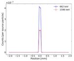
Fig. 9. LSF for collimator of length 100 mm which shows the distribution of
full-energy counts at 662 and 1596-keV photon energies in the perpendicular
reduced efficiency of the individual segment by collecting direction of a line source. The error bars represent the one-sigma estimated
uncertainties in the values from the MCNP results.
data in six channels in parallel. For the particular case of
662-keV photons, the segmented HPGe detector would be
3.7 = (6 × 14/23) times faster in comparison to an unseg-
mented HPGe detector of the same overall dimensions, there-
fore more than compensating for the lower efficiency with the
larger number of detection elements.
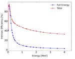
The energy efficiency response curve for scattering segment
number 3 at different incident photon energies was obtained
in separate simulations and is shown in Fig. 8. Both total and
full-energy intrinsic efficiencies were plotted as a function of
energy.
B. LSF and the MTF Curve
Two sets of LSFs were obtained for the two different col-
limator lengths, 100 and 150 mm, according to Section IV-B.
The LSFs for the two cases were plotted and are shown in
Figs. 9 and 10, respectively. The MTF curves for the two
cases were obtained by taking the Fourier transforms of the Fig. 10. LSF for collimator of length 150 mm. The error bars represent the
LSFs which were plotted versus line pairs per millimeter as one-sigma estimated uncertainties in the values from the MCNP results.
shown in Figs. 11 and 12 for the collimator of lengths 100 and
150 mm, respectively. frequency is measured in line pairs per millimeter, the spatial
For the shorter collimator (100 mm), the LSF at higher pho- resolution was obtained as 1/(2 × spatial frequency). The
ton energy (1596 keV) has tails that asymptotically approach spatial resolution for the 100-mm-long collimator at 30%
nonzero values (Fig. 9), meaning that some of the high-energy contrast was thus obtained as 0.1 and 0.16 mm for 662 and
gamma rays penetrate the collimator bulk. The effect of this 1596 keV photons, respectively. The spatial resolution for the
on the MTF can also be noted as shown in Fig. 11, where 150-mm-long collimator was obtained almost equal (0.1 mm)
the 100-mm collimator shows much reduced performance for at both photon energies.
the 1596-keV photons when compared with 662-keV photons.
On the other hand, for the 150-mm-long collimator, these tails
of the LSF are almost zero also at 1596 keV, and a better C. Test Object Reconstructions
contrast was thus obtained in the MTF. The projection data for test object reconstructions were
The limiting resolution of the collimated detector system obtained by analyzing the spectra from each scattering seg-
was obtained from the MTF curve at MTF30, which corre- ment. For test object-1, the single projection at projection
sponds to the line frequency where 30% contrast is transferred. angle 0◦ was plotted and is shown in Fig. 13(a). In total,
The spatial frequencies at MTF30 for both the collimators 150 (25 positions × 6 detection elements) lateral positions
and at both photon energies, 662 and 1596 keV, were obtained were simulated, and the full-energy counts per source particle
and used to quantify the spatial resolution. Since the spatial were obtained.RATHORE et al.: CALCULATION OF SPATIAL RESPONSE OF A COLLIMATED SEGMENTED HPGe DETECTOR FOR GET 719
Fig. 11. MTF curve for a collimator of length 100 mm. The MTF30 value Fig. 13. Test object reconstructions. (a) Single projection plot at 0◦ for
corresponds to the spatial resolution at 30% contrast transfer. object-1. (b) Reconstructed image of object-1. (c) Reconstructed image with
added Poisson counting noise to the projection matrix before reconstruction.
Fig. 12. MTF curve for a collimator of length 150 mm.
Fig. 14. Test object reconstructions. (a) Single projection plot at 0◦ for
object-2, step size = 0.2 mm, and slit width = 0.2 mm. (b) Reconstructed
image of object-2. (c) Reconstructed image with added Poisson counting noise
It should be noted that from the simulated spectra one just to the projection matrix before reconstruction.
obtains the probability of full-energy deposition per source
particle, and therefore, to get the total counts at each position image shown in Fig. 13(c), Poisson noise was added to the
we have multiplied the projection data by a factor such that at sonogram, and then the image was reconstructed.
the central scan position approximately 1000 counts should be In the first scan of test object-2, a total of 150 lateral
obtained after multiplication. It was assumed that a minimum positions were scanned with 0.2-mm-wide slits with a step
of 1000 counts (based on a previous tomographic reconstruc- size of 0.2 mm. The same procedure (as used in the test
tion experience of nuclear fuel object [17]) at this position object-1 case) was used to make the reconstructions, and
would be required to have a satisfactory reconstruction; also the reconstructed images are shown in Fig. 14(b) and (c) as
assuming the counts as Poisson-distributed, this would result obtained without and with added Poisson noise, respectively.
in uncertainty of ±3%. This was done for all the reconstruction In the second scan of test object-2, the step size and the
cases. slit width were decreased from 0.2 to 0.1 mm. Therefore,
The images were reconstructed using the FBP algorithm in total 300 (50 positions × 6 detection elements) lateral
with a ramp filter from the scikit image processing [18] positions were scanned, and the full energy counts per source
module in Python. The reconstructed images are shown in particle were obtained. The reconstructions are shown in
Fig. 13(b) and (c). The reconstructed image in Fig. 13(b) was Fig. 15(b) and (c) as obtained without and with added Poisson
obtained by repeating the same projection data at 150 equi- noise, respectively. As can be seen in Fig. 16, which is the plot
angular rotational positions distributed in 2π and making the of the central slices of the reconstructed images from the two
reconstruction with the sinogram thus obtained. To obtain the scans, the features are more resolved in the second scan for the720 IEEE TRANSACTIONS ON NUCLEAR SCIENCE, VOL. 69, NO. 4, APRIL 2022
TABLE II
ROUGH E STIMATE OF THE T OTAL R EQUIRED T IME FOR AN A XIAL S CAN
the optical-field-of-view [19] is four times smaller, leading to
an increase in the total scan time by a factor of 16, when
compared with a factor of 14.3, which was obtained using the
MCNP simulation count rates instead.
VI. C ONCLUSION
The spatial response of a collimated segmented HPGe
Fig. 15. Test object reconstructions. (a) Single projection plot at 0◦ for
detector was obtained from the simulation study using par-
object-2, step size = 0.1 mm, and slit width = 0.1 mm. (b) Reconstructed ticle transport code MCNP6.2. The LSF and the MTF were
image of object-2. (c) Reconstructed image with added Poisson counting noise obtained for two different lengths of the multislit collimator.
to the projection matrix before reconstruction.
The limiting spatial resolution obtained for 30% contrast is
approximately the same as the width of the slit (0.1 mm) for
662-keV photons and 0.16 mm for 1596-keV photons with a
100-mm-long collimator. The values of the limiting resolution
improve with a 150-mm-long collimator, especially for 1596-
keV photons. For a shorter collimator, the transmission of
high-energy photons through the collimator material and slit
corners deteriorates the LSF and subsequently the MTF.
Test object reconstructions were made using projection data
obtained from the simulations of simplified symmetrical test
objects. These simplifications were used to save the compu-
tation time; still, these reconstructions exhibit the viability
and performance of the segmented HPGe detector for GET
measurements. However, it should be noted that in realistic
measurements on nuclear fuel in test reactors, the activity of
the sample may vary by many orders of magnitude, depending
Fig. 16. (a) Central slice plot of the reconstructed image [Fig. 14(b)]. on whether high-burnup fuel is studied, or on the opposite
(b) Central slice plot of the reconstructed image [Fig. 15(b)]. extreme, fresh fuel exposed only to the reactor pulse of in-pile
transient tests. Collimator selection and experimental settings
such as step size and measurement time per position should
quite obvious reason of having more projection data. However, thus be adapted to the activity of the sample. Development of
more time needs to be spent during an experimental measure- methods to facilitate such adaptions, experiment design, and
ment to acquire this amount of projection data as illustrated planning is ongoing.
below. The obtainable resolution may thus be limited by time The detector is currently in the manufacturing phase and is
constraints. likely to be installed in early 2022, and the experimental val-
For example, simulations were performed to roughly esti- idation of the results will be carried out after the installation.
mate the required time to complete a 2π scan of a 30-mm Eventually, the detector will be available for the postirradiation
diameter UO2 object with around 2.2 × 10¹¹ photons/cm/s examination of irradiation-tested fuel samples.
initial activity (of 137 Cs, 662 keV, from 60 MWd/kg burnup) at
one axial location with different collimator slits and step sizes.
Again, the total time was calculated by assuming a minimum VII. O UTLOOK AND D ISCUSSION
of 1000 counts at the central scan position. The number of The choice of a collimator is dictated by the spatial
angular rotation steps of the object was assumed equal to resolution requirements at the photon energies of interest;
the number of lateral measuring positions. A 100-mm-long using a longer collimator certainly helps in achieving a better
collimator was used in all the simulations, and for comparison, spatial resolution but it is not always possible as it can
the required time for one case is given in Table II. The required prolong the measurement duration, due to lowering the count
time increases manifold as the slit size and the step size are rate. Using the two different lengths (100 and 150 mm) in
halved as given in the table. This increase in time is reasonable the simulations, we have shown that the obtainable spatial
since the measuring positions are fourfold, and in addition resolution is similar for the 662-keV case but greatly improvesRATHORE et al.: CALCULATION OF SPATIAL RESPONSE OF A COLLIMATED SEGMENTED HPGe DETECTOR FOR GET 721
for the 1596-keV case. The final choice of the collimator will R EFERENCES
depend on the source activity, isotope of interest, and time for [1] S. Akkoyun et al., “AGATA—Advanced gamma tracking array,” Nucl.
measurement. Thus, if interested only in the 662-keV photons Instrum. Methods Phys. Res. A, Accel. Spectrom. Detect. Assoc. Equip.,
in a long-cooled fuel that lacks the 1596-keV peak, we might vol. 668, pp. 26–58, Mar. 2012, doi: 10.1016/j.nima.2011.11.081.
[2] S. Paschalis et al., “The performance of the gamma-ray energy tracking
be better off using the shorter collimator, which obtains a in-beam nuclear array GRETINA,” Nucl. Instrum. Methods Phys. Res. A,
higher count rate. Depending on the circumstances, different Accel. Spectrom. Detect. Assoc. Equip., vol. 709, pp. 44–55, May 2013,
collimators can be used, and for that matter, the results from doi: 10.1016/j.nima.2013.01.009.
the simulations will be crucial to design the experiments. [3] P. Andersson et al., “Simulation of the response of a segmented high-
purity germanium detector for gamma emission tomography of nuclear
In our study, we have considered spatial resolution only fuel,” Social Netw. Appl. Sci., vol. 2, no. 2, p. 271, Jan. 2020, doi:
in the lateral direction considering many fuel objects may 10.1007/s42452-020-2053-4.
have some symmetry in the axial direction (thus the height [4] V. Rathore, L. Senis, E. A. Sundén, P. Jansson, A. Håkansson,
and P. Andersson, “Geometrical optimisation of a segmented HPGe
of the collimator slit can be taken larger than the width), detector for spectroscopic gamma emission tomography—A simula-
but it is also worth mentioning that the spatial resolution tion study,” Nucl. Instrum. Methods Phys. Res. A, Accel. Spectrom.
in the axial direction may also be important particularly in Detect. Assoc. Equip., vol. 998, May 2021, Art. no. 165164, doi:
10.1016/j.nima.2021.165164.
cases where the fuel inside the cladding is fragmented and [5] P. Andersson, S. Holcombe, and T. Tverberg. (Jul. 2016). Inspection of a
may not have axial symmetry. In such cases, slit height needs LOCA Test Rod at the Halden Reactor Project Using Gamma Emission
to be adjusted according to the measurement requirements. Tomography. [Online]. Available: https://www.osti.gov/biblio/22764099
[6] B. Biard, “Quantitative analysis of the fission product distribution in
For the test objects’ reconstructions, only two representative a damaged fuel assembly using gamma-spectrometry and computed
photon energies were used, but it should also be noted that tomography for the Phébus FPT3 test,” Nucl. Eng. Des., vol. 262,
the spent fuel emits photons of many different energies. pp. 469–483, Sep. 2013, doi: 10.1016/j.nucengdes.2013.05.019.
[7] J. Schulthess et al., “Non-destructive post-irradiation examina-
In such cases, peaks may overlap or scatter background may tion results of the first modern fueled experiments in TREAT,”
hinder the determination of some peaks, but considering the J. Nucl. Mater., vol. 541, Dec. 2020, Art. no. 152442, doi:
excellent energy resolution of HPGes and availability of fast 10.1016/j.jnucmat.2020.152442.
data acquisition systems, this is not expected to be a significant [8] B. Juste, R. Miró, P. Monasor, and G. Verdú, “Monte Carlo cal-
culation of the spatial response (modulated transfer function) of a
issue. scintillation flat panel and comparison with experimental results,”
In this work, the required time to complete the scan was Radiat. Phys. Chem., vol. 116, pp. 181–185, Nov. 2015, doi:
calculated for a representative burnup case of 60 MWd/kg 10.1016/j.radphyschem.2015.01.005.
[9] J. Diaz, T. Kim, V. Petrov, and A. Manera, “X-ray and gamma-ray
and two representative slit dimensions. The estimated time tomographic imaging of fuel relocation inside sodium fast reactor test
of 257 h to complete an axial scan with a 0.1-mm-wide slit assemblies during severe accidents,” J. Nucl. Mater., vol. 543, Jan. 2021,
is indeed a long time, and if one has to scan many axial Art. no. 152567, doi: 10.1016/j.jnucmat.2020.152567.
[10] C. J. Werner, Ed., “MCNP6.2 release notes,” Los Alamos Nat. Lab.,
positions (e.g., in scanning a fragmented fuel that may not have Los Alamos, NM, USA, Tech. Rep. LA-UR-18-20808, 2018.
axial symmetry) then it would certainly be very time-expensive [11] A. Gopal and S. S. Samant, “Validity of the line-pair bar-pattern
although not impossible. However, a reasonable compromise method in the measurement of the modulation transfer function (MTF)
in megavoltage imaging,” Med. Phys., vol. 35, no. 1, pp. 270–279,
can be obtained using a 0.2-mm-wide slit that can complete Jan. 2008, doi: 10.1118/1.2816108.
one axial scan in less than 24 h. It can be noted that the [12] C. J. Werner, Ed., “MCNP users manual—Code version 6.2,” Los
real measurement time will also be affected by factors such Alamos Nat. Lab., Los Alamos, NM, USA, Tech. Rep. LA-UR-17-
29981, 2017.
as detector dead time and time losses for movement of the
[13] S. Holcombe, S. Jacobsson Svärd, and L. Hallstadius, “A novel
detector relative to the fuel object. Therefore, in the future, all gamma emission tomography instrument for enhanced fuel
the factors which introduce uncertainty in the total scan time characterization capabilities within the OECD Halden reactor
required need to be considered and analyzed. Furthermore, project,” Ann. Nucl. Energy, vol. 85, pp. 837–845, Nov. 2015, doi:
10.1016/j.anucene.2015.06.043.
it is also planned for using different reconstruction methods [14] S. Caruso, M. Murphy, F. Jatuff, and R. Chawla, “Validation of 134 Cs,
137 Cs and 154 Eu single ratios as burnup monitors for ultra-high burnup
to analyze the effect on the quality of the reconstructed image.
Even though the demonstration scaled-down detector ver- UO2 fuel,” Ann. Nucl. Energy, vol. 34, nos. 1–2, pp. 28–35, Jan. 2007,
doi: 10.1016/j.anucene.2006.11.009.
sion has only six scattering segments and the primary objective [15] P. Jansson, S. J. Svärd, A. Håkansson, and A. Bäcklin, “A device for
of using it is to evaluate the capabilities of the novel segmented nondestructive experimental determination of the power distribution in
HPGe detector concept for spent fuel measurements, we also a nuclear fuel assembly,” Nucl. Sci. Eng., vol. 152, no. 1, pp. 76–86,
Jan. 2006, doi: 10.13182/NSE06-A2565.
envision some use cases of the device. In particular, fuel [16] C. Willman, A. Håkansson, O. Osifo, A. Bäcklin, and S. J. Svärd,
objects of up to 30-mm width can be covered with a single “Nondestructive assay of spent nuclear fuel with gamma-ray spec-
5-mm sweep of the detector, by utilization of the six segments. troscopy,” Ann. Nucl. Energy, vol. 33, no. 5, pp. 427–438, Mar. 2006,
doi: 10.1016/j.anucene.2005.12.005.
This may be useful, for example, in the inspection of transient [17] P. Andersson and S. Holcombe, “A computerized method (UPPREC)
fuel test rods, often with the relocation of fuel causing a larger for quantitative analysis of irradiated nuclear fuel assemblies with
size than nominal fuel rod diameter. Inspections of irradiated gamma emission tomography at the Halden reactor,” Ann. Nucl. Energy,
vol. 110, pp. 88–97, Dec. 2017, doi: 10.1016/j.anucene.2017.06.025.
fuel assemblies (of larger size) would require repeating several
[18] S. van der Walt et al., “Scikit-image: Image processing in Python,”
30-mm scans, each made by moving the segmented detector PeerJ, vol. 2, p. e453, Jun. 2014, doi: 10.7717/peerj.453.
5 mm. Even in this case, the segmented detector can speed up [19] L. Senis et al., “Evaluation of gamma-ray transmission through
data collection or produce data with higher spatial sampling rectangular collimator slits for application in nuclear fuel spec-
trometry,” Nucl. Instrum. Methods Phys. Res. A, Accel. Spectrom.
frequency at the same time, compared with an unsegmented Detect. Assoc. Equip., vol. 1014, Oct. 2021, Art. no. 165698, doi:
detector. 10.1016/j.nima.2021.165698.You can also read


