BTLA-HVEM Couple in Health and Diseases: Insights for Immunotherapy in Lung Cancer - Frontiers
←
→
Page content transcription
If your browser does not render page correctly, please read the page content below
MINI REVIEW
                                                                                                                                          published: 31 August 2021
                                                                                                                                     doi: 10.3389/fonc.2021.682007

                                              BTLA-HVEM Couple in Health
                                              and Diseases: Insights for
                                              Immunotherapy in Lung Cancer
                                              Clemence Demerlé , Laurent Gorvel * and Daniel Olive *

                                              Cancer Research Center in Marseille (CRCM), INSERM U1068, CNRS U7258, Aix Marseille University (AMU), Paoli Calmette
                                              Institute (IPC), Marseille, France

                                              Lung cancer is the leading cause of cancer deaths worldwide. Immunotherapies (IT) have
                                              been rapidly approved for lung cancer treatment after the spectacular results in
                                              melanoma. Responses to the currently used checkpoint inhibitors are strikingly good
                                              especially in metastatic diseases. However, durable responses are observed in only 25%
                                              of cases. Consequently, there is an urgent need for new immunotherapy targets. Among
                                              the multiple checkpoints involved in the tumor immune escape, the BTLA-HVEM couple
                          Edited by:
                            Jin Hou,
                                              appears to be a promising target. BTLA (B- and T- Lymphocyte Attenuator) is a co-
   Second Military Medical University,        inhibitory receptor mainly expressed by B and T cells, repressing the activation signal
                               China
                                              transduction. BTLA shares similarities with other immune checkpoints such as PD-1 and
                       Reviewed by:
                                              CTLA-4 which are the targets of the currently used immunotherapies. Furthermore, BTLA
                  Marieke F. Fransen,
           Academic Medical Center,           expression points out terminally exhausted and dysfunctional lymphocytes, and correlates
                          Netherlands         with lung cancer progression. The ligand of BTLA is HVEM (Herpes Virus Entry Mediator)
                    Tatsuro Okamoto,
National Hospital Organization Kyushu
                                              which belongs to the TNF receptor family. Often described as a molecular switch, HVEM is
                Cancer Center, Japan          constitutively expressed by many cells, including cells from tumor and healthy tissues. In
                  *Correspondence:            addition, HVEM seems to be involved in tumor immuno-evasion, especially in lung tumors
                        Laurent Gorvel
                                              lacking PD-L1 expression. Here, we propose to review the role of BTLA-HVEM in
              laurent.gorvel@inserm.fr
                            Daniel Olive      immuno-escape in order to highlight its potential for designing new immunotherapies.
                 daniel.olive@inserm.fr
                                              Keywords: immune escape, lung cancer, T cell exhaustion, HVEM/TNFRSF14, BTLA, immunotherapy

                   Specialty section:
         This article was submitted to
                    Thoracic Oncology,
               a section of the journal
                                              INTRODUCTION
                 Frontiers in Oncology
                                              For decades, tumors have been directly targeted by chemotherapy, radiotherapy, or resected when
          Received: 17 March 2021             possible. The association of these treatments often leads to tumor eradication. However, collateral
         Accepted: 09 August 2021             effects on the non-tumor cells are not negligible. Mesenchymal and immune cells in the tumor
         Published: 31 August 2021
                                              environment are either resident or recruited, and promote or inhibit tumor growth. On the one
                            Citation:         hand, the infiltration of the tumor by immunosuppressive cells such as regulatory T cells (Tregs),
     Demerlé C, Gorvel L and Olive D
                                              myeloid derived suppressor cells (MDSCs), and tumor associated macrophages (TAM) is
       (2021) BTLA-HVEM Couple in
     Health and Diseases: Insights for
                                              prejudicial to tumor immune control and lead to an unfavourable prognosis (1). On the other
     Immunotherapy in Lung Cancer.            hand, M1 macrophages, Natural Killer cells, T CD8+, and T gd lymphocytes are crucial for anti-
            Front. Oncol. 11:682007.          tumor immunity. In addition to divert key resources from their environment, tumor cells developed
     doi: 10.3389/fonc.2021.682007            mechanisms to evade immune recognition and switch cytotoxic cells off (2). Among these

Frontiers in Oncology | www.frontiersin.org                                        1                                     August 2021 | Volume 11 | Article 682007
Demerlé et al. BTLA-HVEM Mediated Immuno-Escape
mechanisms, one of the most studied is lymphocytes exhaustion Entry Mediator). Although BTLA shares similarities with PD-1
through co-inhibitory molecules signalling. Co-inhibitory and and CTLA-4, they differ in terms of expression and functions.
co-stimulating receptors expressed by T lymphocytes are known HVEM is widely expressed among cell types and participate to
as immune checkpoints (3). The balance between the signals immune homeostasis. In tumors, HVEM upregulation was largely
received through these receptors determines lymphocytes reported (Table 1). Here, we propose to review the implication of
activation. Tumors are able to escape immune response by BTLA-HVEM in tumor immune-evasion and its potential for
reducing the expression of costimulatory ligands or developing new IT to treat lung cancer.
upregulating the co-inhibitory molecules. For example, the co-
inhibitory receptor Programmed cell Death 1 (PD-1) and its
ligands PD-L1 and PD-L2 are often overexpressed by tumors to
inhibit T cells activation (4). BTLA AND HVEM IMPLICATION IN
Immunotherapies (IT) have changed the paradigm in cancer IMMUNE HOMEOSTASIS
treatments. Instead of the direct tumor killing by chemotherapy or
radiation, IT acts on immune cells to turn them into in situ BTLA
weapons to eliminate tumor cells. Current ITs are antagonistic B and T cell attenuator (BTLA) was discovered after PD-1 and
antibodies, which block co-inhibitory signalling such as the CTLA-4 almost 20 years ago (18). BTLA belongs to the CD28
CTLA-4/CD80-CD86 and PD-1/PD-L1-PD-L2 pathways. After family and shares structural similarities with PD-1 and CTLA-4.
the promising results in metastatic melanoma, anti-PD-1 IT It exhibits an extracellular immunoglobulin domain, an
(Pembrolizumab and Nivolumab) were rapidly approved for immunoreceptor tyrosine inhibitory motif (ITIM) as well as an
lung cancer treatment 6 years ago (5). Recently, anti-PD-L1 IT immunoreceptor tyrosine-based switch motif (ITSM). BTLA
(Atezoliumab and Durvalumab) were also approved in this signal transduction consists in the phosphorylation of ITIMs
context. Anti-CTLA-4 efficiency remains unclear in lung cancer. and Src homology 2 (SH2) domain–containing phosphatase 1
FDA approval was only given in 2020 in combination with anti- (SHP-1)/SHP-2 association, which leads to the repression of T
PD-1 for metastatic NSCLC expressing PD-L1 (6). Biomarkers cell proliferation and cytokine production (19). The inhibitory
were studied to predict responses, including CD8+ T lymphocyte function of BTLA was confirmed in mice through a BTLA
infiltration, PD-L1 tumor expression, or tumor mutational burden deficient model showing an enhanced sensitivity to auto-
at diagnosis (7). These indicators about the local immune context immune encephalomyelitis (18). In vitro, BTLA deficient T
remain weakly reliable to predict patient response to IT or who cells show an increased TCR-induced proliferation compared
will suffer from hyper-progression (8). Altogether, current ITs for to normal T cells. In healthy humans, BTLA expression is high
NSCLC show a long-term efficiency in 20%–30% of treated on naïve CD4+ and T CD8+ T cells from peripheral blood. BTLA
patients. Therefore, new IT strategies are needed to propose expression remains high during CD4+ differentiation whereas
alternative treatments for advanced lung cancer patients who are BTLA is downregulated during CD8+ T cell differentiation (20).
not responding or are relapsing under anti-PD-1/PD-L1 IT. Similar results were observed on gd-T cells, which is another
BTLA (B and T Lymphocyte Attenuator) is another important cytotoxic population (21). BTLA is highly expressed on resting
co-inhibitory receptor which ligand is HVEM (Herpes Virus Vg9Vd2 cells, the major gd-T-cell subset in human peripheral
TABLE 1 | Review of HVEM upregulation in solid cancers.
Cancer Number of HVEM positivity PD-L1 status Disease Progression Prognosis Year of Reference
patients publication
Melanoma 116 98.3% Mainly Mutual n.a bad 2019 (9)
exclusive
Colorectal cancer 234 94,9% n.a more advanced tumor status and bad 2015 (10)
pathological stage
Gastric cancer 136 89.0% n.a Lymph node metastasis bad 2017 (11)
and depth of invasion
Glioblastoma 34 72.7% n.a n.a bad 2019 (12)
hepatocellular carcinoma 150 Only low/high n.a Intra and extra hepatic recurrences bad 2015 (13)
HVEM status
clear cell renal carcinoma 140 Only low/high n.a n.a bad 2019 (14)
HVEM status
human oesophageal squamous 103 Only low/high n.a Depth of tumor invasion and lymph bad 2013 (15)
cell carcinoma HVEM status node metastasis
breast cancer 1005 16.5% Negative High grade and advanced bad 2017 (16)
correlation pathological stage
Non-small cell-lung cancer 527 18.6% Negative lymph node N2 metastasis Not 2018 (17)
correlation significant
This table reviews the published studies on HVEM expression and correlation with disease progression and prognosis. (n.a, not assessed).
Frontiers in Oncology | www.frontiersin.org 2 August 2021 | Volume 11 | Article 682007Demerlé et al. BTLA-HVEM Mediated Immuno-Escape
blood, and is downregulated during Vg9Vd2 differentiation. moderate expression in the heart, placenta, skeletal muscles, and
Serriari et al. (20) showed that BTLA expression is increased pancreas, and very low in the brain. Among immune cells,
on CMV-specific CD8+ T cells and decreased on memory CD8+ HVEM is strongly expressed by resting T and B cells, NK cells,
T cell subsets when CMV infection is controlled. Furthermore, Tregs, monocytes, and DCs (25). Mesenchymal cells and
the authors demonstrated that in vitro BTLA blockade enhanced epithelial cells also express HVEM (26). In epithelial cells,
CD8+ T cell proliferation, suggesting that BTLA is a promising HVEM expression plays a critical role in innate mucosal
target to improve the control of viral infections. defence against pathogenic bacteria (27). In a mouse model of
Originally described on B and T lymphocytes, BTLA expression acute enteropathogenic infection, authors found that HVEM-/-
was more recently observed on murine type 1 conventional mice present a severe infection with an increased inflammation,
dendritic cells (cDC1) (22). BTLA-positive DCs take part in bacterial dissemination, and reduced survival. Similar results
peripheral Treg induction in an acute encephalomyelitis mouse were found in another mice model of lung infection by
model. This beneficial tolerance mechanism was not observed with Streptococcus pneumoniae, showing that epithelial innate
BTLA- DCs. Zhang et al. reported that BTLA+DCs promoted Treg immunity is impaired in HVEM-/- condition.
and Th2 polarization of T cells in human lung tuberculosis (23). HVEM interaction with BTLA was the first ligation between
Altogether, these data highlight the importance of BTLA in an Ig structure and TNFR family to be described (28). HVEM has
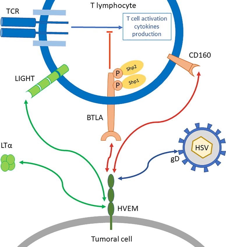
immune homeostasis. other ligands outside of BTLA (Figure 1). Indeed, HVEM binds
to CD160, LIGHT, Lymphotoxin-a (LTa), and herpes simplex
HVEM virus glycoprotein D. When HVEM is engaged with LIGHT or
HVEM, or TNFRSF14, is a TNF-receptor family member. It was LTa, a co-stimulatory signal is delivered, while HVEM binding
discovered in 1996 for its role in the entry of Herpes Simplex to BTLA or CD160 triggers a co-inhibitory signal. Thus, HVEM
Virus (HSV) into cells (24). HVEM expression is observed in is often described as a molecular switch depending on the
tissues, with a higher expression in the lung, kidney, and liver, engaged ligand (29). However, HVEM-/- mice show increased
FIGURE 1 | BTLA/HVEM network. BTLA is a co-inhibitory receptor that represses TCR signal transduction in T lymphocytes through the recruitment of Shp1 and
Shp2. Its ligand HVEM can also bind other molecules such as LIGHT or Lymphotoxin-a (LTa) which triggers co-stimulatory signals (green arrows), or CD160 another
co-inhibitory receptor (red arrows) on distinct binding sites. HVEM is also binding to glycoprotein D from Herpes Simplex Virus (HSV).
Frontiers in Oncology | www.frontiersin.org 3 August 2021 | Volume 11 | Article 682007Demerlé et al. BTLA-HVEM Mediated Immuno-Escape
responses to T cell stimulation and enhanced susceptibility to carcinoma (13), clear cell renal carcinoma (14), human
auto-immune disorders (30). This suggests that HVEM co- esophageal squamous cell carcinoma (15), and breast cancer
inhibitory signal overcomes its HVEM co-stimulation counterpart. (16) (Table 1). In the field of hematological malignancies, a
higher HVEM expression in follicular lymphoma (33) and
HVEM-BTLA Binding chronic lymphocytic leukemia (34) was correlated to a
BTLA-HVEM binding sites were investigated by crystallography. poorer prognosis.
Compared to other CD28 receptors such as PD-1 and CTLA-4, Thus, the numerous data in hematological and solid tumors
BTLA differs by its extracellular domain lacking a C’’ strand. The concerning HVEM role in tumor immune escape provide a
crystal structure of BTLA-HVEM interaction indicates that strong rationale for the study of HVEM in lung cancer and
BTLA binds to a N-terminal cysteine-rich domain of HVEM assess its potential as a major candidate for IT.
(31). Moreover, this cysteine-rich domain (CRD1) is also the
binding site for CD160, the other inhibitory ligand of HVEM, HVEM Dysregulation in Lung Cancer
but not for the costimulatory ligand LIGHT (32). Therefore, the In 2018, Ren et al. (17) published the first study dedicated to
inhibitory and stimulatory signals are received by HVEM on HVEM in lung cancer. They analysed the HVEM expression in
distinct structural binding sites. This suggests that inhibitory 415 NSCLC biopsies and 56 NSCLC cell lines. HVEM expression
signals could be blocked without altering the binding of was positive in 18.6% (77/415) of the biopsies and 48.2% (27/56)
stimulatory ligands. These important structural aspects of the cell lines. PD-L1 expression was also evaluated by IHC in
highlight the importance of modulation rather than complete 491/527 patients, and 31% (152/491) were positive.
inhibition of HVEM, which has a complex molecular network. First, the authors showed that a higher HVEM expression was
significantly associated with lymph node (N2) metastasis. A
higher HVEM expression was also evidenced in the advanced
stage group (stages III–IV) without reaching statistical
HVEM ROLE IN TUMOR significance. However, HVEM expression was not predictive of
the overall survival. Noteworthy, HVEM status was not linked to
HVEM Dysregulation in Tumors
age, gender, smoking status, oncogenic status, pathology, and
HVEM upregulation represents another immune escape
ethnicity. Thus, the upregulation of HVEM would be a tumor-
mechanism, which is similar to the overexpression of PD-L1 or
driven mechanism of immune escape that occurs during tumor
PD-L2 by tumor cells to engage PD-1 on immune cells. In
growth and disease progression. Second, they found a negative
melanoma, HVEM was shown to be overexpressed in some
correlation between PD-L1 and HVEM expression suggesting
tumors and contiguous to BTLA positive T cells (9). Malissen
that the underlying mechanisms involved in PD-L1 or HVEM
et al. identified an “HVEM signature” gathering genes involved
upregulation are different. So far, this is the first and only study
in melanoma proliferation and aggressiveness. Surprisingly, this
which demonstrates the importance of HVEM in immune
signature was not correlated with a classical IFN-g signature.
evasion in lung cancer, especially when PD-L1 is lacking.
HVEM expression was not linked to PD-L1 expression status.
Altogether, these data show that HVEM upregulation is
Moreover, HVEM high expression in their cohort was associated
closely linked to tumor progression and aggressiveness in many
to a significantly poorer prognosis, which was confirmed in The
solid cancers, including lung cancer, and hematological malignancies.
Cancer Genome Atlas (TCGA) melanoma cohort.
In colorectal cancer, HVEM expression was upregulated in
malignant lesions (10). High HVEM expression was associated to
the tumor status and pathological stage. HVEM status was an BTLA ROLE IN TUMORS
independent prognostic value. In another study on 136 gastric
cancer biopsies, an increased HVEM expression was associated BTLA Expression in Tumors
to disease progression and poorer overall survival (11). In melanoma, HVEM+ tumor cells were found to be contiguous
Interestingly, a recent study in glioblastoma highlighted to BTLA+ tumor infiltrating lymphocytes (TILs) (9). Gertner-
similar results. HVEM expression was increased in aggressive Dardenne et al. showed that HVEM-positive lymphoma cells in
subtypes of glioma, and was associated to a poorer prognosis contact with gd- T cells polarize the distribution of BTLA and
(12). Assessed by IHC on 34 glioma tissues, HVEM expression was Vd2-TCR to the immunological synapse (21). In gastric cancer,
localized to the peri-necrotic zone and areas of microvascular Lan et al. (11) reported an upregulation of HVEM in malignant
proliferation. Based on transcriptomic analysis, HVEM expression lesion as discussed previously, but they also evaluated BTLA
was related to immune cell infiltration and stromal cells of expression in the same biopsies. As HVEM, BTLA is more
the microenvironment. HVEM expression was also linked to the expressed in malignant tissues compared to normal tissues.
expression of PD-1, PD-L1, CTLA-4, LAG3, and VISTA, leading Indeed, a higher BTLA expression was positively correlated
to the conclusion that HVEM is crucially involved in the with a higher HVEM expression. Moreover, BTLA expression
modulation of the immune and inflammatory responses, impacted the prognosis, with a 5-year overall survival (OS) rate
especially T cell activation. at 48.3% for the low BTLA expression group, falling to 17.9%
The negative prognosis impact of HVEM high expression was when BTLA was highly expressed. A higher BTLA expression
described in many others cancers such as hepatocellular was also associated to lymph node metastasis.
Frontiers in Oncology | www.frontiersin.org 4 August 2021 | Volume 11 | Article 682007Demerlé et al. BTLA-HVEM Mediated Immuno-Escape
BTLA is downregulated during physiological or virally-induced Therefore, these data demonstrate that CD8+ T cell exhaustion
T cells differentiation (20, 21, 35), whereas in tumor conditions, evolves during tumor progression. To measure T cell dysfunction,
BTLA expression follows a different pattern. Derré et al. (35) the authors set an in vitro assay based on CD3/CD28 T cells
analyzed PBMC from melanoma patients, and showed that BTLA activation with CD25 induction, granzyme-B expression, and
expression remains high on Melan-AMART-1–specific lymphocytes cytokine production readouts. Results were rather heterogeneous
despite effector cell differentiation. This phenotype was reversed by among patients, ranging from highly dysfunctional state, which
conventional vaccination with Melan-AMART-1 peptide, which corresponds to a weak response after polyclonal activation, to
leads to a progressive BTLA downregulation on vaccine-specific lowly affected CD8+ T cells, which strongly respond to stimulation.
CD8+ T cells. Furthermore, IFN-g production is also restored Interestingly, the dysfunctional state was positively correlated with
showing that BTLA-triggered inhibition can be overcome. the IR score. Finally, authors demonstrated that PD-1 blockade
BTLA is related to other co-inhibitory receptors. For this can partially rescue T cell function depending on the PD-1
reason, BTLA was analyzed along with other co-inhibitory expression. In fact, only CD8+ T cells with an intermediate PD-
receptor expression. In advanced melanoma, Fourcade et al. 1 expression benefit from the anti-PD-1 treatment to restore their
(36) demonstrated that 42% of NY-ESO-1-specific CD8+ T function. This suggests that PD-1high CD8+ T cells are too
lymphocytes co-expressed BTLA and PD-1. These cells have a exhausted for their function to be restored by PD-1 blockade
partial dysfunctional phenotype compared to the PD1+ BTLA+ alone. To note, the PD-1high subset also expresses higher amounts
TIM-3+ subset, which is reported as a highly dysfunctional of TIM-3, CTLA-4, LAG-3, and BTLA compared to the PD-1int
subset. TIM-3 and PD-1 are upregulated when NY-ESO-1- subset. Thus, a combined strategy to complete PD-1 blockade is an
specific CD8+ T lymphocytes received a prolonged stimulation interesting strategy to explore. So far, this major publication
with a cognate antigen. BTLA expression followed a different studying BTLA involvement in human lung cancer raises new
pattern. This suggests that BTLA upregulation depends on insights for T cells dysfunction and tumor progression.
different conditions, rather than a functional exhaustion driven Noteworthy, Lou et al. investigated the correlation between
by a high antigen load. Also, BTLA blockade by anti-BTLA epithelial-mesenchymal transition (EMT) and immune
antibody enhanced IFN-g, TNFa, and IL-2 production by NY- activation in lung cancer (39). They established that EMT was
ESO-1-specific CD8+ T cells. Interestingly, a synergistic effect closely linked to an inflammatory transcriptomic signature.
was observed in functional assays when anti-BTLA was Moreover, in tumors displaying EMT, the transcriptomic
combined with anti-PD-1 IT. expression of immune checkpoints, including BTLA, was
increased and associated with regulatory T cells recruitment.
BTLA: The Keystone of T Cell Exhaustion Precisely, BTLA was found to be increased in mesenchymal
in Lung Cancer tissue, suggesting that EMT could be modulated by the
In lung cancer, BTLA expression and function is understudied. inflammatory micro-environment through a BTLA-dependent
However, some critical data have been reported. Indeed, in a mechanism. This study opens a new field of research exploring
mouse model of subcutaneous lung tumor implantation, Mittal BTLA expression on non-immune cells.
et al. showed that BTLA frequency on CD4+ and CD8+ T cells
was increased, along with other co-inhibitory markers such as
PD-1 and 2B4 (37). They concluded that T cell exhaustion was BTLA Expression on Tumor Cells
driven by the tumor implantation and modification of the TME. BTLA expression on tumor cells was reported last year in lung
This study was confirmed and extended in the human lung cancer. Li et al. (40) found a positive BTLA expression in 35 on
tumor samples by Thommen et al. (38). Indeed, Thommen et al. 87 lung adenocarcinoma biopsies. These 35 patients presented a
described a progressive increase of dysfunctional T cells shorter relapse-free survival compared to the BTLA negative
correlated to the expression of multiple co-inhibitory receptors group. BTLA expression on TILs was moderate. So far, BTLA
and also to disease progression. Authors performed a detailed tumor expression was assessed in one study. Feng et al. (41)
immunophenotyping of TILs from NSCLC biopsies (n = 25). described a positive BTLA expression on tumor cells in gastric
They found that the expression of PD-1 and Tim-3 was increased cancer. Both studies on lung and gastric cancers assessed the
on the infiltrating CD8+ T cell subset in advanced tumor stages. BTLA expression by IHC with polyclonal anti-BTLA antibodies.
The percentage of BTLA positive cells was rather low, but Further analyses are required to explore the BTLA expression on
following the same pattern as PD-1 and TIM-3, without non-immune cells. To note, BTLA upregulation have been
reaching statistical significance. BTLA+ CD8+ T cells also reported in non-Hodgkin lymphoma (42). However, since
highly expressed other co-inhibitory receptors, suggesting that BTLA is expressed by non-malignant B cells, the comparison
BTLA was upregulated during the late stages of T cell exhaustion. with solid tumors is delicate.
According to the authors, the sequential expression of co-
inhibitory receptors started from PD-1 expression, then TIM-3,
CTLA-4, and LAG-3, and finally, BTLA. To summarize the DISCUSSION
expression of all co-inhibitory receptors, the authors designed
an inhibitory receptor (IR) scoring system. The IR score increased Immuno-evasion through BTLA/HVEM was studied in many
in patients with lymph node invasion and advanced tumor stages. haematological malignancies and solid tumors. In lung cancer,
However, it did not correlate with the primary tumor size. data remain scarce but promising.
Frontiers in Oncology | www.frontiersin.org 5 August 2021 | Volume 11 | Article 682007Demerlé et al. BTLA-HVEM Mediated Immuno-Escape
As we reviewed, HVEM can be overexpressed in tumor To conclude, BTLA-HVEM couple is truly involved in
conditions. This upregulation is directly linked to tumor immune escape. Indeed, HVEM upregulation by tumor cells
aggressiveness and correlated to a poorer prognosis. In lung dampens anti-tumor immunity through BTLA engagement,
cancer, Ren et al. (17) reported that HVEM expression was resulting in disease progression and a poorer prognosis. The
associated to disease progression, and was distinct from PD-L1 inhibitory signal triggered by BTLA can be overcome, as shown
expression. However, in this study, HVEM expression was not in melanoma (35, 36). In addition, combining blockade of BTLA
correlated with the overall survival. In this cohort, 75% of and PD-1 has demonstrated an interesting synergistic effect.
patients were classified as early stage (I–II), moreover, Therefore, targeting BTLA or HVEM represents a promising
molecular classification was not complete (only the EGFR and new IT that remains to be tested in lung cancer.
KRAS mutations status available). Finally, only 27 biopsies were
HVEM positive in the advanced stage group (III–IV). Therefore,
we hypothesize that HVEM is predictive of the OS only for a
subset of patients that has yet to be defined. The small number of
patients in the advanced-stage group is another limitation to AUTHOR CONTRIBUTIONS
assess the prognosis impact of HVEM for these patients. Further
CD designed and wrote the review, and designed the figures. LG
studies are required to evaluate the correlation between HVEM
proofread and wrote the manuscript. DO provided the fundings
expression and clinical parameters in lung cancer.
and proofread the manuscript. All authors contributed to the
Instead of PD-1, BTLA is only expressed on a small subset of
article and approved the submitted version.
T cells. Nonetheless, these BTLA+ lymphocytes, which are also
PD-1+, TIM3+, CTLA-4+, and LAG-3+, seem to be terminally
exhausted and poorly functional, and are associated to disease
progression in lung cancer (38). These data are coherent with
previous publications in other malignancies such as gastric ACKNOWLEDGMENTS
cancer (11) or melanoma (35, 36). Recently, BTLA expression
was described on tumor cells (40), raising questions about the We are deeply grateful to Transcan17 Digest and RHU
role of BTLA on non-immune cells. PIONEER (ANR-17-RHU5-0007) for their support.
Prognosis. Onco Targets Ther (2017) 10:919–26. doi: 10.2147/
REFERENCES OTT.S128825
1. Fridman WH, Zitvogel L, Sautès–Fridman C, Kroemer G. The Immune 12. Han M-Z, Wang S, Zhao W-B, Ni S-L, Yang N, Kong Y, et al. Immune
Contexture in Cancer Prognosis and Treatment. Nat Rev Clin Oncol (2017) Checkpoint Molecule Herpes Virus Entry Mediator is Overexpressed and
14:717–34. doi: 10.1038/nrclinonc.2017.101 Associated With Poor Prognosis in Human Glioblastoma. EBioMedicine
2. Hanahan D, Weinberg RA. Hallmarks of Cancer: The Next Generation. Cell (2019) 43:159–70. doi: 10.1016/j.ebiom.2019.04.002
(2011) 144:646–74. doi: 10.1016/j.cell.2011.02.013 13. Hokuto D, Sho M, Yamato I, Yasuda S, Obara S, Nomi T, et al. Clinical Impact
3. Chen L, Flies DB. Molecular Mechanisms of T Cell Co-Stimulation and Co- of Herpesvirus Entry Mediator Expression in Human Hepatocellular
Inhibition. Nat Rev Immunol (2013) 13:227–42. doi: 10.1038/nri3405 Carcinoma. Eur J Cancer Oxf Engl 1990 (2015) 51:157–65. doi: 10.1016/
4. Iwai Y, Ishida M, Tanaka Y, Okazaki T, Honjo T, Minato N, et al. j.ejca.2014.11.004
Involvement of PD-L1 on Tumor Cells in the Escape From Host Immune 14. Tang M, Cao X, Li Y, Li G-Q, He Q-H, Li S-J, et al. High Expression of Herpes
System and Tumor Immunotherapy by PD-L1 Blockade. Proc Natl Acad Sci Virus Entry Mediator Is Associated With Poor Prognosis in Clear Cell Renal
USA (2002) 99:12293–7. doi: 10.1073/pnas.192461099 Cell Carcinoma. Am J Cancer Res (2019) 9:975–87.
5. Garon EB, Rizvi NA, Hui R, Leighl N, Balmanoukian AS, Eder JP, et al. 15. Migita K, Sho M, Shimada K, Yasuda S, Yamato I, Takayama T, et al. Significant
Pembrolizumab for the Treatment of Non-Small-Cell Lung Cancer. N Engl J Involvement of Herpesvirus Entry Mediator in Human Esophageal Squamous
Med (2015) 372:2018–28. doi: 10.1056/NEJMoa1501824 Cell Carcinoma. Cancer (2014) 120:808–17. doi: 10.1002/cncr.28491
6. Hellmann MD, Paz-Ares L, Bernabe Caro R, Zurawski B, Kim S-W, Carcereny 16. Tsang JYS, Chan K-W, Ni Y-B, Hlaing T, Hu J, Chan S-K, et al. Expression
Costa E, et al. Nivolumab Plus Ipilimumab in Advanced Non-Small-Cell Lung and Clinical Significance of Herpes Virus Entry Mediator (HVEM) in Breast
Cancer. N Engl J Med (2019) 381:2020–31. doi: 10.1056/NEJMoa1910231 Cancer. Ann Surg Oncol (2017) 24:4042–50. doi: 10.1245/s10434-017-5924-1
7. Bodor JN, Boumber Y, Borghaei H. Biomarkers for Immune Checkpoint 17. Ren S, Tian Q, Amar N, Yu H, Rivard CJ, Caldwell C, et al. The Immune
Inhibition in Non-Small Cell Lung Cancer (NSCLC). Cancer (2020) 126:260– Checkpoint, HVEM may Contribute to Immune Escape in Non-Small Cell
70. doi: 10.1002/cncr.32468 Lung Cancer Lacking PD-L1 Expression. Lung Cancer (2018) 125:115–20.
8. Wang L, Hu Y, Wang S, Shen J, Wang X. Biomarkers of Immunotherapy in doi: 10.1016/j.lungcan.2018.09.004
Non-Small Cell Lung Cancer. Oncol Lett (2020) 20:139. doi: 10.3892/ 18. Watanabe N, Gavrieli M, Sedy JR, Yang J, Fallarino F, Loftin SK, et al. BTLA Is
ol.2020.11999 a Lymphocyte Inhibitory Receptor With Similarities to CTLA-4 and PD-1.
9. Malissen N, Macagno N, Granjeaud S, Granier C, Moutardier V, Gaudy- Nat Immunol (2003) 4:670–9. doi: 10.1038/ni944
Marqueste C, et al. HVEM Has a Broader Expression Than PD-L1 and 19. Chemnitz JM, Lanfranco AR, Braunstein I, Riley JL. B and T Lymphocyte
Constitutes a Negative Prognostic Marker and Potential Treatment Target for Attenuator-Mediated Signal Transduction Provides a Potent Inhibitory Signal
Melanoma. Oncoimmunology (2019) 8:e1665976. doi: 10.1080/2162402X. to Primary Human CD4 T Cells That Can Be Initiated by Multiple
2019.1665976 Phosphotyrosine Motifs. J Immunol Baltim Md 1950 (2006) 176:6603–14.
10. Inoue T, Sho M, Yasuda S, Nishiwada S, Nakamura S, Ueda T, et al. HVEM doi: 10.4049/jimmunol.176.11.6603
Expression Contributes to Tumor Progression and Prognosis in Human 20. Serriari N-E, Gondois-Rey F, Guillaume Y, Remmerswaal EBM, Pastor S,
Colorectal Cancer. Anticancer Res (2015) 35:1361–7. Messal N, et al. B and T Lymphocyte Attenuator Is Highly Expressed on
11. Lan X, Li S, Gao H, Nanding A, Quan L, Yang C, et al. Increased BTLA and CMV-Specific T Cells During Infection and Regulates Their Function.
HVEM in Gastric Cancer are Associated With Progression and Poor J Immunol (2010) 185:3140–8. doi: 10.4049/jimmunol.0902487
Frontiers in Oncology | www.frontiersin.org 6 August 2021 | Volume 11 | Article 682007Demerlé et al. BTLA-HVEM Mediated Immuno-Escape
21. Gertner-Dardenne J, Fauriat C, Orlanducci F, Thibult M-L, Pastor S, Fitzgibbon Partially Reversed by Vaccination. J Clin Invest (2010) 120:157–67.
J, et al. The Co-Receptor BTLA Negatively Regulates Human Vg9vd2 T-Cell doi: 10.1172/JCI40070
Proliferation: A Potential Way of Immune Escape for Lymphoma Cells. Blood 36. Fourcade J, Sun Z, Pagliano O, Guillaume P, Luescher IF, Sander C, et al.
(2013) 122:922–31. doi: 10.1182/blood-2012-11-464685 CD8(+) T Cells Specific for Tumor Antigens Can Be Rendered Dysfunctional
22. Jones A, Bourque J, Kuehm L, Opejin A, Teague RM, Gross C, et al. by the Tumor Microenvironment Through Upregulation of the Inhibitory
Immunomodulatory Functions of BTLA and HVEM Govern Induction of Receptors BTLA and PD-1. Cancer Res (2012) 72:887–96. doi: 10.1158/0008-
Extrathymic Regulatory T Cells and Tolerance by Dendritic Cells. Immunity 5472.CAN-11-2637
(2016) 45:1066–77. doi: 10.1016/j.immuni.2016.10.008 37. Mittal R, Chen C-W, Lyons JD, Margoles LM, Liang Z, Coopersmith CM,
23. Zhang J-A, Lu Y-B, Wang W-D, Liu G-B, Chen C, Shen J, et al. BTLA- et al. Murine Lung Cancer Induces Generalized T Cell Exhaustion. J Surg Res
Expressing Dendritic Cells in Patients With Tuberculosis Exhibit Reduced (2015) 195:541–9. doi: 10.1016/j.jss.2015.02.004
Production of IL-12/IFN-a and Increased Production of IL-4 and TGF-b, 38. Thommen DS, Schreiner J, Muller P, Herzig P, Roller A, Belousov A, et al.
Favoring Th2 and Foxp3+ Treg Polarization. Front Immunol (2020) 11:518. Progression of Lung Cancer Is Associated With Increased Dysfunction
doi: 10.3389/fimmu.2020.00518 of T Cells Defined by Coexpression of Multiple Inhibitory Receptors. Cancer
24. Montgomery RI, Warner MS, Lum BJ, Spear PG. Herpes Simplex Virus-1 Immunol Res (2015) 3:1344–55. doi: 10.1158/2326-6066.CIR-15-0097
Entry Into Cells Mediated by a Novel Member of the TNF/NGF Receptor 39. Lou Y, Diao L, Cuentas ERP, Denning WL, Chen L, Fan YH, et al. Epithelial-
Family. Cell (1996) 87:427–36. doi: 10.1016/S0092-8674(00)81363-X Mesenchymal Transition Is Associated With a Distinct Tumor
25. Murphy KM, Nelson CA, Šedý JR. Balancing Co-Stimulation and Inhibition With Microenvironment Including Elevation of Inflammatory Signals and
BTLA and HVEM. Nat Rev Immunol (2006) 6:671–81. doi: 10.1038/nri1917 Multiple Immune Checkpoints in Lung Adenocarcinoma. Clin Cancer Res
26. Guo H, Pang K, Wei Y, Yi C, Wu X. Herpes Virus Entry Mediator in Human (2016) 22:3630–42. doi: 10.1158/1078-0432.CCR-15-1434
Corneal Epithelial Cells Modulates the Production of Inflammatory Cytokines 40. Li X, Xu Z, Cui G, Yu L, Zhang X. BTLA Expression in Stage I-III Non-
in Response to HSV Type 1 Challenge. Ophthalmic Res (2015) 54:128–34. Small-Cell Lung Cancer and Its Correlation With PD-1/PD-L1 and
doi: 10.1159/000437209 Clinical Outcomes. Onco Targets Ther (2020) 13:215–24. doi: 10.2147/
27. Shui J-W, Larange A, Kim G, Vela JL, Zahner S, Cheroutre H, et al. HVEM OTT.S232234
Signalling at Mucosal Barriers Provides Host Defence Against Pathogenic 41. Feng X-Y, Wen X-Z, Tan X-J, Hou J-H, Ding Y, Wang K-F, et al. Ectopic
Bacteria. Nature (2012) 488:222–5. doi: 10.1038/nature11242 Expression of B and T Lymphocyte Attenuator in Gastric Cancer: A Potential
28. Sedy JR, Gavrieli M, Potter KG, Hurchla MA, Lindsley RC, Hildner K, et al. B Independent Prognostic Factor in Patients With Gastric Cancer. Mol Med Rep
and T Lymphocyte Attenuator Regulates T Cell Activation Through (2015) 11:658–64. doi: 10.3892/mmr.2014.2699
Interaction With Herpesvirus Entry Mediator. Nat Immunol (2005) 6:90–8. 42. M’Hidi H, Thibult M-L, Chetaille B, Rey F, Bouadallah R, Nicollas R, et al.
doi: 10.1038/ni1144 High Expression of the Inhibitory Receptor BTLA in T-Follicular Helper Cells
29. Cai G, Freeman GJ. The CD160, BTLA, LIGHT/HVEM Pathway: A and in B-Cell Small Lymphocytic Lymphoma/Chronic Lymphocytic
Bidirectional Switch Regulating T-Cell Activation. Immunol Rev (2009) Leukemia. Am J Clin Pathol (2009) 132:589–96. doi: 10.1309/AJCPPHKG
229:244–58. doi: 10.1111/j.1600-065X.2009.00783.x YYGGL39C
30. Wang Y, Subudhi SK, Anders RA, Lo J, Sun Y, Blink S, et al. The Role of
Herpesvirus Entry Mediator as a Negative Regulator of T Cell–Mediated Conflict of Interest: DO is a co-founder and shareholder of ImCheck
Responses. J Clin Invest (2005) 115:711–7. doi: 10.1172/JCI200522982 Therapeutics, Alderaan Biotechnology and Emergence Therapeutics and has
31. Compaan DM, Gonzalez LC, Tom I, Loyet KM, Eaton D, Hymowitz SG, et al. research funds from ImCheck Therapeutics, Alderaan Biotechnology, Cellectis
Attenuating Lymphocyte Activity: The Crystal Structure of the BTLA-HVEM and Emergence Therapeutics. The other authors declare no conflicts of interest.
Complex. J Biol Chem (2005) 280:39553–61. doi: 10.1074/jbc.M507629200 The funders had no role in the design of the study, in the writing of the
32. Liu W, Garrett SC, Fedorov EV, Ramagopal UA, Garforth SJ, Bonanno JB, manuscript, or in the decision to publish.
et al. Structural Basis of CD160:HVEM Recognition. Struct Lond Engl 1993
(2019) 27:1286–95.e4. doi: 10.1016/j.str.2019.05.010 Publisher’s Note: All claims expressed in this article are solely those of the authors
33. Carreras J, Lopez-Guillermo A, Kikuti YY, Itoh J, Masashi M, Ikoma H, et al. and do not necessarily represent those of their affiliated organizations, or those of
High TNFRSF14 and Low BTLA Are Associated With Poor Prognosis in the publisher, the editors and the reviewers. Any product that may be evaluated in
Follicular Lymphoma and in Diffuse Large B-Cell Lymphoma this article, or claim that may be made by its manufacturer, is not guaranteed or
Transformation. J Clin Exp Hematop JCEH (2019) 59:1–16. doi: 10.3960/ endorsed by the publisher.
jslrt.19003
34. Sordo-Bahamonde C, Lorenzo-Herrero S, Gonzalez-Rodriguez AP, Payer R, Copyright © 2021 Demerle,́ Gorvel and Olive. This is an open-access article distributed
Gonzá lez-Garcı́a Á , Ló pez-Soto E, et al. BTLA/HVEM Axis Induces NK Cell under the terms of the Creative Commons Attribution License (CC BY). The use,
Immunosuppression and Poor Outcome in Chronic Lymphocytic Leukemia. distribution or reproduction in other forums is permitted, provided the original
Cancers (2021) 13:1766. doi: 10.3390/cancers13081766 author(s) and the copyright owner(s) are credited and that the original publication
35. Derré L, Rivals J-P, Jandus C, Pastor S, Rimoldi D, Romero P, et al. BTLA in this journal is cited, in accordance with accepted academic practice. No use,
Mediates Inhibition of Human Tumor-Specific CD8+ T Cells That Can Be distribution or reproduction is permitted which does not comply with these terms.
Frontiers in Oncology | www.frontiersin.org 7 August 2021 | Volume 11 | Article 682007You can also read