MiR-212-5p inhibits nasopharyngeal carcinoma progression by targeting METTL3
←
→
Page content transcription
If your browser does not render page correctly, please read the page content below
Open Medicine 2022; 17: 1241–1251
Research Article
Hongyu Zhou#, Nana Zhang*#
miR-212-5p inhibits nasopharyngeal carcinoma
progression by targeting METTL3
https://doi.org/10.1515/med-2022-0515 and promoted apoptosis of NPC cells; miR-212-5p inhibi-
received December 16, 2021; accepted June 3, 2022 tion functioned oppositely. Mechanistically, miR-212-5p
Abstract: This study was conducted to investigate the inhibited the proliferation and promoted apoptosis of
effect of microRNA-212-5p (miR-212-5p) on the proliferation NPC cells via suppressing METTL3 expression. miR-212-
and apoptosis of nasopharyngeal carcinoma (NPC) cells. 5p/METTL3 was associated with processes of RNA trans-
Microarray datasets (EXP00394 and EXP00660) were port and cell cycle. In conclusion, miR-212-5p inhibits the
downloaded from the dbDEMC database, and the differ- progression of NPC by targeting METTL3.
entially expressed microRNAs between high-grade and Keywords: NPC, miR-212-5p, METTL3, proliferation, apoptosis
low-grade NPC were analyzed. miR-212-5p and methyl-
transferase like 3 (METTL3) expression levels in NPC
tissues and cells were determined by the quantitative
real-time polymerase chain reaction and Western blot. 1 Introduction
Besides, the relationship between miR-212-5p expression
and clinicopathological characteristics of patients was Nasopharyngeal carcinoma (NPC) is a tumor derived from
analyzed by the Chi-square test. Cell counting kit-8 assay, the nasopharyngeal epithelium and has a higher mor-
5-ethynyl-2-deoxyuridine (EdU) assay, and flow cytometry bidity and mortality among head and neck malignancies
were adopted to detect the effect of miR-212-5p on the cell [1,2]. Despite NPC’s sensitiveness to radiotherapy, the
proliferation and apoptosis. Kyoto Encyclopedia of Genes prognosis of NPC patients is poor, and the 5-year survival
and Genomes and Gene Ontology analysis were performed rate of NPC patients is reported to be less than 50% [3,4].
to explore the potential biological functions and the It is, therefore, pivotal to elaborate on the molecular
signal pathways related to the target genes of miR-212- mechanism of the progression of NPC to explore new
5p. Bioinformatics prediction and dual luciferase reporter therapeutic targets for NPC.
gene assay were used to verify the relationship between MicroRNAs (miRNAs, miRs) are a category of non-
miR-212-5p and METTL3 3′ untranslated region. Besides, coding RNAs that induce the degradation or inhibit the
western blot was adopted to detect the expression of translation of mRNAs by interacting with the 3′ untrans-
METTL3. Gene set enrichment analysis was performed to lated region (3′UTR) of these mRNAs [5]. miRNAs can act
analyze the downstream pathways in which METTL3 was as tumor promotors or inhibitors to regulate the prolifera-
enriched. It was found that miR-212-5p was downregulated tion, differentiation, and apoptosis of cells [6,7]. For
in NPC tissues, and the low miR-212-5p expression was example, miR-4321-5p expression is reduced in NPC,
associated with lymph node metastasis and poor differen- and miR-4321-5p overexpression represses the migration
tiation. miR-212-5p overexpression inhibited the growth and invasion of NPC cells via targeting N-myristoyltrans-
ferase 1 [8]. miR-124-3p expression is inhibited in NPC.
miR-124-3p overexpression curbs the activation of phos-
phatidylinositol 3-kinase/AKT/mammalian rapamycin target
# Contributed equally. signal pathway by targeting PCDH8 and inhibits the viability
and colony formation of NPC cells [9]. As reported, as one
of the important members of the miR family, miR-212-5p is
* Corresponding author: Nana Zhang, Department of dysregulated in a variety of human tumors, such as breast
Otorhinolaryngology Head and Neck Surgery, Wuhan Fourth
cancer, colorectal cancer (CRC), and clear cell renal cell
Hospital, Wuhan 430033, Hubei, China,
e-mail: mchxznnh6522@163.com
carcinoma cells, and is associated with the growth and
Hongyu Zhou: Department of Otorhinolaryngology Head and Neck metastasis of tumor cells [10–12]. Here, bioinformatics pre-
Surgery, Wuhan Fourth Hospital, Wuhan 430033, Hubei, China dicted that miR-212-5p expression is inhibited in high-
Open Access. © 2022 Hongyu Zhou and Nana Zhang, published by De Gruyter. This work is licensed under the Creative Commons Attribution
4.0 International License.
1242 Hongyu Zhou and Nana Zhang
grade tumor tissues of NPC patients; however, how miR- Park Memorial Institute-1640 medium with 10% fetal
212-5p functions in NPC is unclear. bovine serum (Gibco, Carlsbad, CA), 100 U/mL penicillin
Previous studies have shown that methyltransferase (Gibco), and 0.1 mg/mL streptomycin (Gibco) at 37°C in
like 3 (METTL3) promotes the malignant biological beha- 5% CO2 and 95% relative humidity, with the medium
viors of NPC cells [13,14]. In this study, bioinformatics changed every 3 days. When cells reached 80–90% con-
showed that METTL3 may be a target of miR-212-5p. fluence, cells were trypsinized with 0.25% trypsin (Roche,
This study focused on how miR-212-5p functions on the Basel, Swizerland). Then, the cells were inoculated into
malignant phenotype of NPC cells and its downstream six-well plates at the density of 1 × 10⁵ cells/ml and incu-
mechanisms. bated for 24 h at 37°C in 5% CO2. miR-212-5p mimic (miR-
212-5p: 3′-UCAUUCGUCAGAUCUCGGUUCCA-5′), mimics
control (miR-NC: 5′-UUCUCCGAACGUGUCACGUTT-3′), miR-
212-5p inhibitor (miR-212-5p-in: 3′-UGGAACCGAGAUCUGACG
2 Materials and methods AAUGA-5′), inhibitors control (miR-in: 5′-UUGUACUACACAA
AAGUACUG-3′), empty vector (NC), METTL3 overexpression
plasmid (METTL3), small interfering RNA (siRNA) targeting
2.1 Bioinformatics analysis
METTL3 (si-METTL3), and siRNA normal control (si-NC) from
GenePharma (Shanghai, China) were subsequently trans-
Microarray datasets available from dbDEMC database were
fected into 5-8F and 6-10B cells with LipofectamineTM 2000
selected according to following selection criteria. Inclusion
kit (Invitrogen; Carlsbad, CA). A final concentration of 50 nM
criteria were expressed as follows: human case and control
of miRNA mimic/inhibitor and inhibitors was used.
study, miRNA expression profile, and complete raw data.
EXP00394 and EXP00660 were obtained and analyzed in
this study. Differentially expressed miRNA analysis was per-
formed with the “limma” package in R language (version
2.4 Quantitative reverse transcription-
3.5.2). The adjusted P < 0.05 and |log2fold change| > 1 were
chosen as the cutoff value. With the data in LinkedOmics polymerase chain reaction
database, gene set enrichment analysis (GSEA) for METTL3
was conducted to predict its biological functions in NPC. Total RNA was extracted by TRIzol reagent (Invitrogen),
Pathways with NES > 1, nominal P value < 0.05, and false and reversely transcribed into cDNA by the miScript Reverse
discovery rate q value < 0.25 were screened out. Transcription Kit (QIAGEN, GmbH, Hilden, Germany), and
then the miScript SYBR Green PCR system (QIAGEN) was
used to perform PCR reactions on a Rotorgene 3000 series
2.2 Collection of tissue samples PCR machine (Corbett Research, Sydney, Australia). The
expression levels of miRNA and mRNA were quantified by
Forty-five patients who admitted to Wuhan Fourth Hospital Rotorgene software. With U6 or GAPDH as internal refer-
from June 2016 to April 2019 and diagnosed with NPC by the ences, the relative expressions of miR-212-5p and METTL3
Department of the Pathology were included, and tumor tis- mRNA were calculated by the 2−ΔΔCT method. The sequences
sues and adjacent noncancerous tissues were collected of primers: miR-212-5p: 5′-ACCTTGGCTCTCTAGACTGCT-3′,
during biopsy. All samples were frozen in liquid nitrogen 5′-GCAGGGTCCGAGGTATTC-3′; U6: 5′-CTCGCTTCGGCAGC
immediately and stored at −80°C for further use. All sub- ACA-3′, 5′-AACGCTTCACGAATTTGCGT-3′; METTL3: 5′-AAG
jects did not receive chemotherapy, radiotherapy, and other CTGCACTTCAGACGAAT-3′, 5′-GGAATCACCTCCGACACTC-3′;
anticancer treatments before the biopsy. This research was GAPDH: 5′-CGGAGTCAACGGATTTGGTCGTAT-3, 5′-AGCCTT
approved by the Ethics Committee of Wuhan Fourth Hospital, CTCCATGGTGGTGAAGAC-3′.
and all participants signed an informed consent form.
2.3 Cell culture and transfection 2.5 Cell counting kit-8 assay
NPC cell lines (CNE1, 5-8F, C666-1, 6-10B) and the normal The transfected cells were inoculated into a 96-well cell
nasopharyngeal epithelial cell line NP69 were available plate (1,000 cells per well). After 24 h, 48 h and 72 h,
from the Cancer Institute of Southern Medical University 10 μL of CCK-8 reagent (Invitrogen, Shanghai, China)
(Guangzhou, China). All cells were cultured in Roswell was added into each well and accordingly incubated for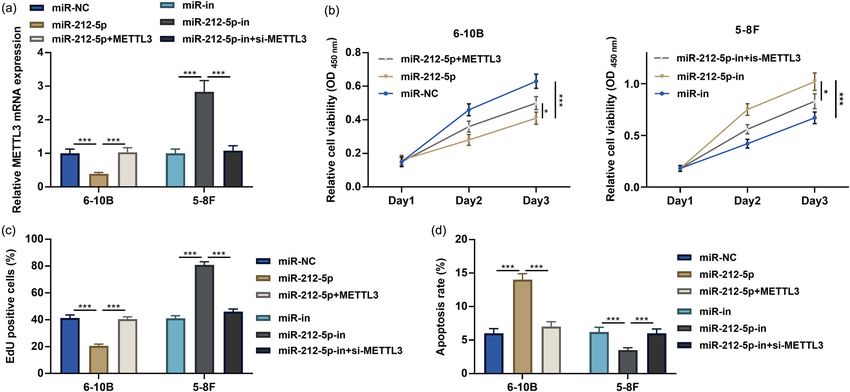
miR-212-5p, M3TTL3, and nasopharyngeal carcinoma 1243
4 h. The values of OD450nm were detected by a microplate luminescence intensity of renilla luciferase to that of firefly
reader. luciferase.
2.9 Western blot
2.6 5-Ethynyl-29-deoxyuridine assay
Transfected cells were collected and lysed with RIPA lysis
6-10B and 5-8F cells were inoculated into 24-well plates, buffer (Pierce, Rockford, IL), and the supernatant was
respectively. The cells were subsequently incubated for collected by high-speed centrifugation. The protein con-
2 h with 5 μmol/L EdU (Beyotime Biotechnology, Shanghai, centrations were measured by the BCA Protein Assay Kit
China) and rinsed with phosphate buffer saline (PBS). The (Rockford, IL). The extracted protein was mixed with the
cells were then fixed with paraformaldehyde for 10 min. loading buffer, and the sample was heated at 95°C for
200 μL of glycine at 2 mg/mL was added, and then the cells 10 min. 30 μg protein was separated on polyacrylamide
were incubated for 5 min. Next, the cells were incubated gel by electrophoresis and transferred to polyvinylidene
with 100 μL of 0.5% TritonX-100, decolorized on a shaker fluoride membranes. The membranes were blocked with
for 10 min, and immersed twice in PBS for 5 min each time. 5% bovine serum albumin for 1 h, then incubated with
Then the cells were incubated with Apollo and stained with primary antibody anti-METTL3 (ab195352, 1:1,000, Abcam
Hoechst. After the cells were washed with PBS, the cells Cambridge, MA) overnight at 4℃. Moreover, the mem-
were observed under a fluorescence microscope. branes were washed and incubated with secondary anti-
body goat anti-rabbit IgG H&L (ab97051, 1:2,000, Abcam
Cambridge) for 1 h at room temperature. Subsequent to
washing membrane three times, the protein bands were
2.7 Flow cytometry visualized by the chemiluminescent reagent ECL (Beyotime,
Shanghai, China), with GAPDH as an internal reference.
For cell cycle analysis, 6-10B and 5-8F cells were fixed in Bio-Rad Gel Dol EZ imager (GEL DOC EZ IMAGER, Bio-
70% cold ethanol overnight at 4°C. After washing with Rad, CA) was adopted to image the proteins, and the gray
PBS twice, the cells were incubated in the binding buffer values were analyzed by Image J software.
containing 50 μg/mL propidium iodide (PI; BD Bioscience,
San Jose, CA), 20 μg/mL RNase A (Sigma) and 0.2% Triton-
X100 for 30 min at room temperature in the dark. Then, 2.10 Statistical analyses
2 × 10⁴ cells were analyzed using a flow cytometer. For cell
apoptosis analysis, the cells were incubated with Annexin V All experiments were performed in triplicate. SPSS 24.0
staining solution and PI staining solution at room tempera- software was used for statistical analysis, and GraphPad
ture for 30 min at room temperature in the darkness. Then, Prism 9 was used to plot the figures of the experimental
2 × 10⁴ cells were analyzed using a flow cytometer. data. Comparisons between two groups were conducted
by Student’s t-test, and differences among multiple groups
were compared by one-way analysis of variance, with
P < 0.05 indicating a statistically significant difference.
2.8 Dual luciferase reporter gene assay
Ethics statement: Our study was approved by the Ethics
METTL3 wild-type sequence containing the miR-212-5p Review Board of Wuhan Fourth Hospital.
binding site and the mutant sequence without the miR-
212-5p binding site were cloned into the pmirGLO vector
(Promega, Madison, WI) to construct a wild-type reporter 3 Results
vector (METTL3-WT) and a mutant reporter vector (METTL3-
MUT). Besides, METTL3-WT or METTL3-MUT and miR-212-
5p mimics or miR-NC were co-transfected into 293 T cells by 3.1 MiR-212-5p expression was
Lipofectamine™ 2,000 kit. 48 h later, the dual luciferase downregulated in NPC
reporter gene assay system (Promega) was used to detect
the luciferase activity. Specifically, the binding intensity of Microarray datasets EXP00394 and EXP00660 were down-
METTL3 to miR-212-5p was expressed by the ratio of the loaded from the dbDEMC database, and the differentially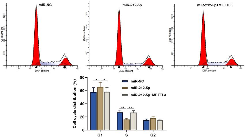
1244 Hongyu Zhou and Nana Zhang
expressed miRNAs between high-grade tumor tissues and cells, and miR-212-5p inhibitors were transfected into 5-8F
low-grade tumor tissues of NPC patients were analyzed, cells. qRT-PCR proved the transfection was successful
and it was revealed that miR-212-5p expression was sig- (Figure 2a). CCK-8 assay, EdU assay, and flow cytometry
nificantly downregulated in high-grade tumor tissues of showed that miR-212-5p overexpression restrained the
NPC patients (Figure 1a). Besides, the results of quanti- growth and promoted the apoptosis of 6-10B cells com-
tative reverse transcription-polymerase chain reaction pared with the control, while its inhibition worked
(qRT-PCR) highlighted that miR-212-5p expression was oppositely on 5-8F cells (Figure 2b–d).
significantly inhibited in NPC tissues compared with
that in adjacent tissues (Figure 1b). In addition, miR-
212-5p was inhibited in four NPC cell lines as against
the NP69 cells (Figure 1c). The Chi-square test indicated 3.3 miR-212-5p specifically represses
that low expression of miR-212-5p was closely associated METTL3
with poorly differentiated tumors and lymph node meta-
stasis in NPC patients (Table 1). miRNA directly binds to the 3′-UTR of a specific mRNA,
leading to mRNA degradation and translational repres-
sion [15]. The downstream targets of miR-212-5p were
predicted by StarBase database and analyzed by Kyoto
3.2 Inhibition of miR-212-5p promoted NPC Encyclopedia of Genes and Genomes (KEGG), and the find-
cell viability and restrained the ings highlighted that the target genes were closely associated
apoptosis with p53 signaling pathway and apoptosis (Figure 3a). Gene
Ontology (GO) analysis showed that these targets were
Subsequently, the function of miR-212-5p in NPC was mainly enriched in the regulation of cell proliferation
explored. miR-212-5p mimics were transfected into 6-10B and protein phosphorylation in biological processes (BPs);
Figure 1: miR-212-5p expression is inhibited in NPC tissues and cells. (a) Volcano plots: microarray datasets EXP00394 and EXP00660 were
downloaded from the dbDEMC database to analyze the differential expression of miRNAs between high-grade and low-grade NPC tissues.
(b) miR-212-5p expression in NPC tissues and adjacent tissues was detected by qRT-PCR. (c) miR-212-5p expression in human normal
nasopharyngeal epithelial cells NP69 cells and NPC cell lines (CNE1, 5-8F, C666-1, 6-10B) was detected by qRT-PCR. ***P < 0.001.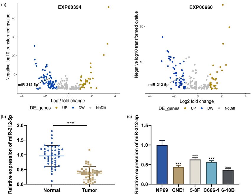
miR-212-5p, M3TTL3, and nasopharyngeal carcinoma 1245
Table 1: The correlation between miR-212-5p expression and clinicopathological characteristics of NPC patients
Clinical and pathological indicators Number miR-212-5p expression Chi-square (math.) P-value
High expression Low expression
Age (years) 45 23 22
≥50 24 12 12 0.025 0.8731246 Hongyu Zhou and Nana Zhang Figure 3: METTL3 is a target of miR-212-5p. (a) The pathway enrichment analysis on the downstream target gene set of miR-212-5p was conducted by the KEGG analysis. (b) The functional enrichment analysis on the downstream target genes of miR-212-5p was conducted by the GO analysis. (c) Binding sequences of METTL3 mRNA 3′UTR wild type or its mutant to miR-212-5p were showed. (d) Dual-luciferase reporter gene assay was used to verify the effect of overexpression of miR-212-5p on the luciferase activity of METTL3-WT and METTL3-MUT. (e and f) The expression of METTL3 mRNA and protein in 6-10B or 5-8F cells transfected with miR-212-5p mimics or inhibitors were detected by qRT-PCR and western blot. (g) The expression of METTL3 mRNA in NPC tissues and adjacent tissues was detected by qRT-PCR. (h) The correlation between METTL3 expression and miR-212-5p expression in NPC tissues was detected by Pearson’s correlation analysis. ***P < 0.001. nucleus and cytoplasm in cellular components; and pro- 293T cells, but that of METTL3-MUT was not significantly tein binding and metal ion binding in molecular functions impacted (Figure 3d). qRT-PCR and western blot showed (Figure 3b). METTL3 was one of these target genes, and that overexpression of miR-212-5p significantly decreased there was a binding site between miR-212-5p and METTL3 METTL3 mRNA and protein expression in 6-10B cells, mRNA 3′-UTR (Figure 3c). Dual-luciferase reporter gene while METTL3 mRNA and protein expressions were greatly assay showed that overexpression of miR-212-5p signifi- increased in 5-8F cells transfected with miR-212-5p inhibi- cantly inhibited the luciferase activity of METTL3-WT in tors (Figure 3e and f). In addition, METTL3 mRNA was
miR-212-5p, M3TTL3, and nasopharyngeal carcinoma 1247
demonstrably highly expressed in NPC tissues as against In addition, flow cytometry showed that miR-212-5p over-
that in adjacent tissues (Figure 3g). The correlation ana- expression induced the cell cycle arrest, while METTLE3
lysis showed that miR-212-5p expression in NPC tissues overexpression accelerated the cell cycle (Figure A1).
was negatively correlated with METTL3 mRNA expression
in NPC tissues (Figure 3h).
3.5 miR-212-5p regulates the expression via
METTL3
3.4 miR-212-5p inhibits the proliferation
and promotes the apoptosis of NPC cells We used GSEA for signal pathway enrichment analysis,
and the findings suggested that the high expression of
by targeting METTL3
METTL3 was associated with the process of RNA transport
and cell cycle (Figure 5a). Western blot showed that miR-
To investigate the function of miR-212-5p/METTL3 axis in
212-5p overexpression repressed cyclin D1 expression,
NPC, we co-transfected miR-212-5p mimics and METTL3
while overexpression of METTL3 reversed these effects;
overexpression plasmids into 6-10B cells, and miR-212-5p
inhibition of miR-212-5p promoted cyclin D1 expression,
inhibitors and METTL3 siRNA into 5-8F cells. qRT-PCR
while knockdown of METTL3 counteracted these effects.
showed that transfection of METTL3 overexpression plasmid
These results further suggested that miR-212-5p inhibited
could reverse the impact of miR-212-5p overexpression on
cell cycle progression of NPC cells by regulating METTL3
METTL3 expression; silencing METTL3 counteracted the
(Figure 5b).
effects of miR-212-5p inhibition on METTL3 expression
(Figure 4a). CCK-8, EdU, and flow cytometry assays proved
that miR-212-5p overexpression inhibited the viability and
accelerated the apoptosis of 6-10B cells compared with 4 Discussion
controls, while overexpression of METTL3 reversed these
effects (Figure 4b–d); inhibition of miR-212-5p accelerated NPC is mainly distributed in the southern region of China,
the growth and inhibited the apoptosis in 5-8F cells, while with obvious geographical and ethnic characteristics;
knockdown of METTL3 impaired these effects (Figure 4b–d). currently, combined radiotherapy and chemotherapy is
Figure 4: miR-212-5p regulates the proliferation and apoptosis of NPC cells by targeting METTL3. (a) miR-212-5p mimics and METTL3
overexpression plasmids were co-transfected into 6-10B cells, and miR-212-5p-in and si-METTL3 were co-transfected into 5-8F cells. The
expression of METTL3 mRNA was detected by qRT-PCR. (b and c) After transfection, the cell proliferation was assessed by the CCK-8 assay
and EdU assay. (d) After transfection, the cell apoptosis was assessed by flow cytometry. *P < 0.05, **P < 0.01, and ***P < 0.001.1248 Hongyu Zhou and Nana Zhang
Figure 5: METTL3 activates the DNA replication, cell cycle signaling pathway. (a) Signaling pathway enrichment analysis was conducted
through GSEA. (b) miR-212-5p and METTL3 overexpression plasmids were co-transfected into 6-10B cells, and miR-212-5p-in and si-METTL3
were transfected into 5-8F cells. The expression of Cyclin D1 was analyzed by western blot.
the main treatment strategy of NPC, and local recurrence malignant biological behaviors of CRC cell by targeting
and distant metastasis are important reasons for the poor SIRT2 [11]. In addition, miR-212-5p expression is also reduced
prognosis of NPC patients [16,17]. Early diagnosis and in renal clear cell carcinoma (RCC), and miR-212-5p overex-
development of treatment therapy are of significance to pression blocks the migration, invasion, and proliferation of
improve the prognosis of NPC patients [18,19]. RCC cells by targeting TBX15 [12]. Here, we demonstrated that
miRNAs are a category of noncoding RNAs with a miR-212-5p expression was demonstrably decreased in NPC
length of 22–24 nt, which can modulate protein expres- tissues and cells, which was related to clinicopathological
sion at posttranscriptional level via combining with the 3′- indexes in NPC patients. Functionally, miR-212-5p overex-
UTR of target mRNAs [20–23]. miRNAs are widely involved pression restrained the growth and promoted the apoptosis
in the regulation of many BP in cancer biology [24]. There of NPC cell; miR-212-5p inhibition exerted the opposite effect.
is increasing evidence that miRNAs are novel biomarkers Given our findings, miR-212-5p may function as a tumor sup-
and promising therapeutic targets of NPC [25]. Here, we pressor in NPC.
focused on miR-212-5p, which, as reported, is reduced in METTL3 is identified as the first m6A methyltrans-
hepatocellular carcinoma (HCC), and miR-212-5p overex- ferase [27,28]. Previous studies have shown that METTL3
pression restrains the migrative and invasive abilities of is functionally complex, and it plays crucial roles in a
HCC cell via targeting the ubiquitin-binding enzyme E2T variety of tissues/cells and is involved in regulating the
[26]. miR-212-5p expression is also reduced in CRC tissues tumorigenesis and cancer progression [29,30]. Report-
and cell lines, and miR-212-5p overexpression represses edly, high expression of METTL3 is associated with poormiR-212-5p, M3TTL3, and nasopharyngeal carcinoma 1249
prognosis of gastric cancer patients; METTL3 overexpres- [4] Ono T, Azuma K, Kawahara A, Sasada T, Matsuo N, Kakuma T,
sion expedites epithelial–mesenchymal transition pro- et al. Prognostic stratification of patients with nasopharyngeal
cess and enhances the migrative and invasive abilities carcinoma based on tumor immune microenvironment. Head
Neck. 2018;40(9):2007–19.
of gastric cancer cells [31]. The upregulated METTL3
[5] Keramati F, Jafarian A, Soltani A, Javandoost E, Mollaei M,
facilitates the metastasis of CRC cells via modulating Fallah P. Circulating miRNAs can serve as potential diagnostic
miR-1246/SPRED2/MAPK signaling pathway [32]. In NPC, biomarkers in chronic myelogenous leukemia patients. Leuk
knockdown of METTL3 inhibits the expression levels of Res Rep. 2021;16:100257.
β-catenin/transcription factor 7-like 2 target genes vimentin [6] Rupaimoole R, Slack FJ. MicroRNA therapeutics: towards a new
era for the management of cancer and other diseases. Nat Rev
and N-cadherin and blocks the migration and invasion of
Drug Discov. 2017;16(3):203–22.
NPC cells, implying the oncogenic roles of METTL3 in NPC [7] Wang Q, Liu J, Zeng J, Yang Z, Ran F, Wu L, et al. Determination
[13]. Another study reports that METTL3 promotes the of miRNA derived from exosomes of prostate cancer via toe-
migratory and invasive ability of NPC cells by regulating hold-aided cyclic amplification combined with HRP enzyme
Snail [33]. Also, METTL3 accelerates the progression of catalysis and magnetic nanoparticles. Anal Biochem.
2021;630:114336.
NPC through mediating m6A modification of EZH2 [14].
[8] Ma X, Yuan Y, Lu J, Li M, Yu Y, Liu J, et al. Long noncoding RNA
In addition, METTL3 exerts pro-cancer effects in tumors
ANCR promotes migration, invasion, EMT progress and stem-
and is also specifically regulated by miRs [34–36]. In the ness of nasopharyngeal carcinoma cells via the miR-4731-5p/
present work, we proved that miR-212-5p could target NMT1 axis. Pathol Res Pract. 2021;224:153540.
METTL3 to suppress the viability and facilitate the apop- [9] Ye J, Liao Q, Zeng X, Liu C, Ding Y, Liu X, et al. MicroRNA-124-3p
tosis of NPC cells. inhibited progression of nasopharyngeal carcinoma by inter-
action with PCDH8 and the inactivation of PI3K/AKT/mTOR
Considering miR-212-5p is associated with tumor dif-
pathway. J Cancer. 2021;12(16):4933–44.
ferentiation and lymph node metastasis in the patients, it [10] Lv ZD, Yang DX, Liu XP, Jin LY, Wang XG, Yang ZC, et al. MiR-
may be used as a biomarker to predict the prognosis of 212-5p suppresses the epithelial-mesenchymal transition in
the patients, and the response to the treatment, and it triple-negative breast cancer by targeting Prrx2. Cell Physiol
may also be a potential therapy target for NPC. There Biochem. 2017;44(5):1785–95.
are several limitations in this work. First, in vivo studies [11] Du F, Li Z, Zhang G, Shaoyan S, Geng D, Tao Z, et al. SIRT2, a
direct target of miR-212-5p, suppresses the proliferation and
are important to further validate the function of miR-212-
metastasis of colorectal cancer cells. J Cell Mol Med.
5p/METTL3 axis in NPC progression. In addition, whether 2020;24(17):9985–98.
miR-212-5p/METTL3 axis can regulate other phenotypes [12] Deng JH, Zheng GY, Li HZ, Ji ZG. MiR-212-5p inhibits the
of NPC cells (such as chemosensitivity and radiosensi- malignant behavior of clear cell renal cell carcinoma cells by
tivity) awaits further investigation. targeting TBX15. Eur Rev Med Pharmacol Sci.
2019;23(24):10699–707.
[13] Liu ZF, Yang J, Wei SP, Luo XG, Jiang QS, Chen T, et al.
Funding information: None. Upregulated METTL3 in nasopharyngeal carcinoma enhances
the motility of cancer cells. Kaohsiung J Med Sci.
Conflict of interest: None. 2020;36(11):895–903.
[14] Meng QZ, Cong CH, Li XJ, Zhu F, Zhao X, Chen FW. METTL3
Data availability statement: The data used to support the promotes the progression of nasopharyngeal carcinoma
through mediating M6A modification of EZH2. Eur Rev Med
findings of this study are available from the corresponding
Pharmacol Sci. 2020;24(8):4328–36.
author upon request. [15] Sárközy M, Kahán Z, Csont T. A myriad of roles of miR-25
in health and disease. Oncotarget. 2018;9(30):
21580–612.
[16] Xiao Y, Li F, Zheng A, Chen Q, Chen F, Cheng X, et al. Ginkgolic
References acid suppresses nasopharyngeal carcinoma growth by indu-
cing apoptosis and inhibiting AKT/NF-κB signaling. J Med
[1] Bossi P, Chan AT, Licitra L, Trama A, Orlandi E, Hui EP, et al. Food. 2021;24:806–16.
Nasopharyngeal carcinoma: ESMO-EURACAN Clinical Practice [17] Peng Z, Wang Y, Wang Y, Fan R, Gao K, Zhang H, et al.
Guidelines for diagnosis, treatment and follow-up(†). Ann Comparing the effectiveness of endoscopic surgeries with
Oncol. 2021;32(4):452–65. intensity-modulated radiotherapy for recurrent rT3 and rT4
[2] Wu L, Li C, Pan L. Nasopharyngeal carcinoma: A review of nasopharyngeal carcinoma: A meta-analysis. Front Oncol.
current updates. Exp Ther Med. 2018;15(4):3687–92. 2021;11:703954.
[3] Zhang B, He X, Ouyang F, Gu D, Dong Y, Zhang L, et al. [18] Prasetyo A, Budiman J, Sadhana U. The relationship between
Radiomic machine-learning classifiers for prognostic biomar- tumor-infiltrating lymphocytes (TILs) and nasopharyngeal
kers of advanced nasopharyngeal carcinoma. Cancer Lett. carcinoma (NPC): A systematic review. Iran J Otorhinolaryngol.
2017;403:21–7. 2021;33(117):191–200.1250 Hongyu Zhou and Nana Zhang
[19] Li W, Zhang H, Lu H, Wang H, Gu Y, Li H, et al. Clinical outcomes [28] Wang X, Feng J, Xue Y, Guan Z, Zhang D, Liu Z, et al.
of salvage endoscopic nasopharyngectomy for patients with Corrigendum: Structural basis of N(6)-adenosine methylation by
advanced recurrent nasopharyngeal carcinoma. Front Oncol. the METTL3-METTL14 complex. Nature. 2017;542(7640):260.
2021;11:716729. [29] Zhao Z, Cai Q, Zhang P, He B, Peng X, Tu G, et al. N6-
[20] Chen X, Zhou C, Wang CC, Zhao Y. Predicting potential small Methyladenosine RNA Methylation Regulator-Related
molecule-miRNA associations based on bounded nuclear norm Alternative Splicing (AS) gene signature predicts non-small
regularization. Brief Bioinform. 2021;22(6):bbab328. cell lung cancer prognosis. Front Mol Biosci. 2021;8:657087.
[21] Fang X, Wang H, Zhuo Z, Tian P, Chen Z, Wang Y, et al. miR-141- [30] Lin S, Choe J, Du P, Triboulet R, Gregory RI. The m(6)A
3p inhibits the activation of astrocytes and the release of Methyltransferase METTL3 promotes translation in human
inflammatory cytokines in bacterial meningitis through down- cancer cells. Mol Cell. 2016;62(3):335–45.
regulating HMGB1. Brain Res. 2021;1770:147611. [31] Yue B, Song C, Yang L, Cui R, Cheng X, Zhang Z, et al. METTL3-
[22] Biswas K, Jolly MK, Ghosh A. First passage time properties of mediated N6-methyladenosine modification is critical for
miRNA-mediated protein translation. J Theor Biol. epithelial-mesenchymal transition and metastasis of gastric
2021;529:110863. cancer. Mol Cancer. 2019;18(1):142.
[23] Modi A, Purohit P, Gadwal A, Ukey S, Roy D, Fernandes S, et al. [32] Peng W, Li J, Chen R, Gu Q, Yang P, Qian W, et al. Upregulated
In-silico analysis of differentially expressed genes and their METTL3 promotes metastasis of colorectal cancer via miR-
regulating microRNA involved in lymph node metastasis in 1246/SPRED2/MAPK signaling pathway. J Exp Clin Cancer Res.
invasive breast carcinoma. Cancer Invest. 2022;40(1):55–72. 2019;38(1):393.
[24] Mollaei H, Safaralizadeh R, Rostami Z. MicroRNA replacement [33] Yu X, Zhao H, Cao Z. The m6A methyltransferase METTL3
therapy in cancer. J Cell Physiol. 2019;234(8):12369–84. aggravates the progression of nasopharyngeal carcinoma
[25] Wang S, Yang T, He Z. Investigations on the role of the through inducing EMT by m6A-modified Snail mRNA. Minerva
MicroRNA-338-5p/Wnt family member 2B (WNT2B) axis in Med. 2022;113(2):309–14.
regulating the pathogenesis of nasopharyngeal carcinoma [34] Chang X, Lin YY, Bai LN, Zhu W. miR-302a-3p suppresses
(NPC). Front Oncol. 2021;11:684462. melanoma cell progression via targeting METTL3. J Chemother.
[26] Ren X, Li A, Ying E, Fang J, Li M, Yu J. Upregulation of ubiquitin- 2022;34(1):55–66.
conjugating enzyme E2T (UBE2T) predicts poor prognosis and [35] Li M, Wang Q, Zhang X, Yan N, Li X. CircPUM1 promotes cell
promotes hepatocellular carcinoma progression. growth and glycolysis in NSCLC via up-regulating METTL3
Bioengineered. 2021;12(1):1530–42. expression through miR-590-5p. Cell Cycle.
[27] Chai RC, Chang YZ, Chang X, Pang B, An SY, Zhang KN, et al. 2021;20(13):1279–94.
YTHDF2 facilitates UBXN1 mRNA decay by recognizing METTL3- [36] Zhang F, Yan Y, Cao X, Zhang J, Li Y, Guo C. Methylation of
mediated m(6)A modification to activate NF-κB and promote microRNA-338-5p by EED promotes METTL3-mediated trans-
the malignant progression of glioma. J Hematol Oncol. lation of oncogene CDCP1 in gastric cancer. Aging (Albany NY).
2021;14(1):109. 2021;13(8):12224–38.miR-212-5p, M3TTL3, and nasopharyngeal carcinoma 1251 Appendix Figure A1: Flow cytometry assay was used to detect the effects of miR-212-5p and METTLE3 on cell cycle progress.
You can also read